Comparative Study
. 2025 Jan;637(8048):1170-1177.
doi: 10.1038/s41586-024-08337-5. Epub 2025 Jan 1.

Structural diversity of axonemes across mammalian motile cilia

Affiliations
Comparative Study

Structural diversity of axonemes across mammalian motile cilia

Miguel Ricardo Leung et al. Nature. 2025 Jan.

Abstract

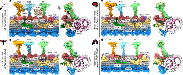
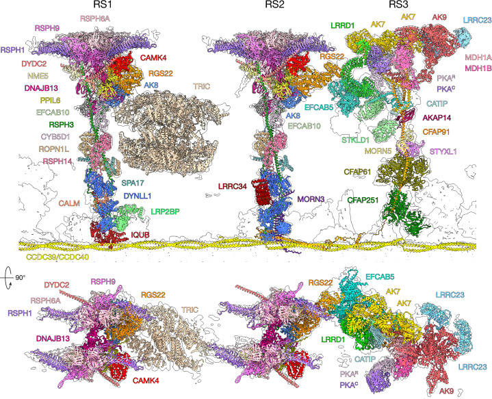
Reproduction, development and homeostasis depend on motile cilia, whose rhythmic beating is powered by a microtubule-based molecular machine called the axoneme. Although an atomic model of the axoneme is available for the alga Chlamydomonas reinhardtii1, structures of mammalian axonemes are incomplete1-5. Furthermore, we do not fully understand how molecular structures of axonemes vary across motile-ciliated cell types in the body. Here we use cryoelectron microscopy, cryoelectron tomography and proteomics to resolve the 96-nm modular repeat of axonemal doublet microtubules (DMTs) from both sperm flagella and epithelial cilia of the oviduct, brain ventricles and respiratory tract. We find that sperm DMTs are the most specialized, with epithelial cilia having only minor differences across tissues. We build a model of the mammalian sperm DMT, defining the positions and interactions of 181 proteins including 34 newly identified proteins. We elucidate the composition of radial spoke 3 and uncover binding sites of kinases associated with regeneration of ATP and regulation of ciliary motility. We discover a sperm-specific, axoneme-tethered T-complex protein ring complex (TRiC) chaperone that may contribute to construction or maintenance of the long flagella of mammalian sperm. We resolve axonemal dyneins in their prestroke states, illuminating conformational changes that occur during ciliary movement. Our results illustrate how elements of chemical and mechanical regulation are embedded within the axoneme, providing valuable resources for understanding the aetiology of ciliopathy and infertility, and exemplifying the discovery power of modern structural biology.


Conflict of interest statement

Competing interests: The authors declare no competing interests.

Figures

Fig. 1
Fig. 1. Cryo-EM reconstructions of the 96-nm axonemal repeat of motile cilia from different mammalian cell types.
Each panel shows a longitudinal and cross-sectional view of a composite cryo-EM map of a 96-nm repeat unit of a doublet microtubule from bovine sperm flagella (a), bovine oviductal cilia (b), porcine brain ventricle cilia (c) and human respiratory cilia (d). The reconstruction in d is EMD-35888 (ref. ). Each major axonemal complex is given a unique colour with the doublet microtubule in grey. IJ, inner junction; MAP, microtubule-associated protein; OJ, outer junction.
Fig. 2
Fig. 2. Newly identified sperm-specific and disease-linked axonemal proteins.
a, Thirty-four newly assigned axonemal proteins are identified in this study, including 21 ‘general’ proteins that are conserved across cell types (blue) and 13 that are sperm specific (pink). Note that CCDC63, DNAH8, DNAH17 and RSPH6A were previously found to be sperm-specific axonemal proteins but are included here for emphasis. b, Proteins implicated in infertility and other motile ciliopathies are colour coded by whether their disruption in humans or model organisms causes defects in sperm flagella (red), other motile cilia (yellow) or both (orange).
Fig. 3
Fig. 3. Structures of RSs in mammalian sperm.
Atomic models of RS proteins (coloured) fitted into the cryo-EM density map from bovine sperm (outline).
Fig. 4
Fig. 4. The sperm-specific barrel is an RS-tethered TRiC chaperone.
a, TRiC is suspended between RS1 and RS2. b, TRiC contacts RS1 by small interfaces with RGS22 and AK8. c, TRiC is tethered to RS2 by a hitherto unidentified linker protein (grey) anchored to DNAJB13 and NME5. d, Assignment of subunit order in the barrel, based on the inherent asymmetries of TRiC, demonstrated by comparison with the structure of yeast TRiC. Asterisk marks an unidentified luminal density of the distal ring (closer to RS2) in RS-tethered TRiC. This density binds at a location different from the binding site of the cochaperone Plp2 in yeast TRiC (grey density).
Fig. 5
Fig. 5. Prestroke state conformation of IDAf.
Left, Molecular surface representations comparing the prestroke state resolved in this study (top) and the poststroke state resolved in EMD-35888 and modelled in PDB 8J07 (ref. ) (bottom). Right, model showing how IDAf would interact with the B-tubule of the neighbouring DMT in pre- and poststroke states. A subtomogram average from intact Tetrahymena thermophila axonemes (EMD-9023) was used to model the position of the neighbouring DMT.
Extended Data Fig. 1
Extended Data Fig. 1. Sample preparation and processing strategies used to resolve the 96-nm repeat of axonemal doublet microtubules (DMTs) from different mammalian motile cilia.
(a) Stitched low-magnification micrographs of a disintegrated bovine sperm cell (left) and corresponding high-magnification image of a DMT with bound radial spokes (RSs) (right). A total of 45,431 such micrographs were processed for this study. (b) Schematic of the isolation of human and porcine oviduct cilia using a syringe inserted into the oviduct. Human cilia were splayed into individual DMTs after demembranating and incubating with ATP to promote DMT-DMT sliding. A similar workflow was used to prepare DMTs from bovine oviducts and porcine brain ventricles. (c) General processing scheme used for single particle analysis (SPA) of DMTs from bovine sperm, bovine/human oviductal cilia, and porcine brain ventricle cilia. All steps were performed in cryoSPARC unless otherwise stated. See Methods and Supplementary Figs. 1–7 for details. (d) Cryo-EM map of the bovine sperm 96-nm repeat with local resolution ranges for individual axonemal complexes indicated. (e) Porcine oviduct cilia subtomogram averaging (STA) workflow. See Methods for details. (f) Fourier shell correlation (FSC) curves for the four segments used to reconstruct the porcine DMT at binning factor 2 by STA. (g) Comparison of oviduct cilia reconstructions obtained by SPA and STA.
Extended Data Fig. 2
Extended Data Fig. 2. Differences in the tektin bundle across mammalian motile cilia.
Cross-sections of doublet microtubules from the indicated cilia types and species. Tektins (TEKT1-5), Tektin-like protein 1 (TEKTL1), tektin-interacting protein 1 (TEKTIP1), and RIBC1/2 are colored according to the legend. Other microtubule-binding proteins are in grey and tubulin is in white. Structures were determined by single-particle analysis (SPA) except for oviductal cilia from Sus scrofa, which was determined by subtomogram averaging (STA).
Extended Data Fig. 3
Extended Data Fig. 3. Comparison of ciliary microtubule associated proteins (CIMAPs) across mammalian motile cilia.
(a) CIMAP1 family proteins are present in all motile cilia examined, but sperm have several additional MAPs including SPMAP1/2, CFAP97D1, EFCAB3, and TSSK. (b) CIMAP2 (only detected in sperm) and CIMAP3 (in all cell types) interact with external axonemal complexes.
Extended Data Fig. 4
Extended Data Fig. 4. Binding sites and interactions of newly identified proteins associated with the nexin-dynein regulatory complex (N-DRC), inner dynein arm f (IDAf), and radial spoke 2 (RS2).
(a) Top-down view of the bovine sperm DMT surface around RS2, N-DRC, and RS3. (b) A complex of ARMH1, CFAP20, and a DNALI1 dimer sits atop CFAP58. This complex represents new binding sites for CFAP20, which is also found at the inner junction and in the central apparatus, and DNALI1, which is also associated with single-headed IDAs. ARMH1 is linked to the N-DRC via unidentified coiled-coils (grey). Comparison with doublet-specific subtomogram averages from mouse sperm (translucent white map) suggests that the ARMH1 complex is asymmetrically distributed around the axoneme. (c) WDR64 binds on top of the CCDC96/113 coiled-coil, where it also interacts with CFAP299, CFAP57, and CFAP58. WDR64 is sperm-specific but may be replaced by WDR49 in other cell types. (d) ANKEF1 and LRRC74A are newly assigned components of the N-DRC distal lobe. ANKEF1 presents a positively charged surface that could bind glutamylated tubulin on the neighboring DMT. (e) LRRC51, which is present in all cilia types studied, binds the DMT surface directly underneath IDAf, between protofilaments A05 and A06 (left panel). AlphaFold Multimer predictions suggest that the elongated density beside LRRC51 could correspond to the C-terminus of CFAP100 (right panel). (f) CFAP206 bridges the inner junction and the base of RS2, thus contributing to the docking of this radial spoke. (g) Domain organization for ARMH1, ANKEF1, CFAP206, LRRC51, LRRC74A, and WDR64.
Extended Data Fig. 5
Extended Data Fig. 5. Structure and composition of sperm radial spoke 3 (RS3).
(a) Atomic models of RS1, RS2, RS3, and RS-tethered TRiC fit into the consensus subtomogram average of the 96-nm repeat from mouse sperm (EMD-27444, Ref. ). (b) Model-in-map fits and (c) domain organization for RS3 proteins assigned in bovine sperm. For CFAP91, the schematic instead indicates regions interacting with other RS3 proteins or axonemal complexes.
Extended Data Fig. 6
Extended Data Fig. 6. Structure of the sperm-specific RS2-RS3 bridge.
(a) The bridge between RS2 and RS3 is formed by the interaction of RGS22, a conserved ciliary protein, with EFCAB5, a sperm-specific protein. The sperm-specific RS3 proteins LRRD1 and STKLD1 interact with EFCAB5 to further support the bridge, along with an unassigned globular density (asterisk). (b) Superposition of RGS22 from RS1 and RS2 showing a large conformational change, potentially induced by binding EFCAB5. (c) Cryo-EM density and corresponding atomic models are shown for the interaction site between RGS22 and EFCAB5.
Extended Data Fig. 7
Extended Data Fig. 7. Structural bases of kinase anchoring to radial spokes.
(a) Overview showing the positions of kinases (colored) in the radial spokes of the bovine sperm DMT. (b) Interactions of adenylate kinase 8 (AK8) with PPIL6 and DNAJB13 stabilize the open conformation of catalytic domain II (left). AK8 is anchored in the neck of RS1 and RS2 via its N-terminal dimerization/docking domain, which interacts with EFCAB10 and packs against RSPH3 (right). (c) AK7 and AK9 are anchored to RS3 via their C-terminal DPY30 domains. The DPY30 domain of AK7-A dimerizes with the DPY30 motif in EFCAB5, while the DPY30 domain of AK7-B interacts with AK9; both pairs of proteins then dock onto CFAP91. (d) CAMK4 interacts with RSPH1, DYDC2, and PPIL6 in the heads of RS1 and RS2. See also the Supplementary Fig. 14. (e) The ciliary PKA holoenzyme is a heterodimer of a catalytic (PKAC) and regulatory (PKAR) subunit. PKAR interacts with a malate dehydrogenase dimer (MDH1A/MDH1B) in the center of the RS3 head.
Extended Data Fig. 8
Extended Data Fig. 8. Structures and conformations of radial spoke-anchored adenylate kinases (AKs).
(a) RS1 and RS2 each have one copy of AK8, while RS3 has two copies of AK7 and one copy of AK9. (b) Models of axonemal AKs with catalytic domains labelled. (c) Catalytic domains of axonemal AKs are observed in either open (PDB 4AKE) or closed (PDB 1AKE) conformations. Densities consistent with small molecules in the putative nucleotide-binding pockets are shown in pink. These densities were colored by aligning PDB 1AKE to the relevant adenylate kinase domain and coloring density within 3 Å of the ligand atoms.
Extended Data Fig. 9
Extended Data Fig. 9. Conformational changes in inner dynein arm f (IDAf) and the tether/tetherhead (T/TH) complex.
(a) Comparison of our atomic models with subtomogram averages of IDAf in the pre- and post-stroke states, obtained from rapidly frozen live, swimming sea urchin sperm (Lin & Nicastro 2018). (b) Composite map of the motor domains of IDAf ( and ), IDAa, and the T/TH complex (CFAP43, CFAP44, and the sperm-specific TEX47). (c) Cryo-EM density for the motor domains of IDAf (DNAH10 and DNAH2). The linker is colored purple and individual AAA domains are colored from blue to red. (d) Changes in the conformation of the linker and stalk domains of the dynein heavy chains (upper panel: pre-stroke state, lower panel: post-stroke state). (e) Relative orientations of the two motors of IDAf change from perpendicular arrangement in the pre-stroke state (upper panels) to a parallel arrangement in the post-stroke state (lower panels). (f) Conformational changes in the T/TH complex, consisting of CFAP43, CFAP44, and the sperm-specific TEX47. (g) Interaction of the motor domain of with CFAP44 and the motor domain of IDAa via CFAP44. The microtubule-binding domain of interacts with the N-DRC (arrows). (h) The motor domain of fβ interacts with CFAP43 and IDAd, while its linker interacts with TEX47 and its stalk with IDAg.
Extended Data Fig. 10
Extended Data Fig. 10. Conformational changes in outer dynein arms (ODAs).
(a) Top panels show molecular surface representations of ODAs in conformations resembling the pre-stroke state (this study) and post-stroke state (PDB 8J07). Bottom panel shows comparison of our atomic models with in situ subtomogram averages of ODAs in the pre- and post-stroke states from rapidly frozen actively swimming sea urchin sperm. (b) Overlaid atomic models showing how transition from post- to pre-stroke state involves rotation of the distal CCDC63/CCDC151 coiled-coil, the IC/LC block, and the linkers of dynein heavy chains in order to accommodate the ~8-nm proximal shift of the motor domains from the neighboring dyneins.

References

    1. Walton, T. et al. Axonemal structures reveal mechanoregulatory and disease mechanisms. Nature618, 625–633 (2023). - PMC - PubMed
    1. Leung, M. R. et al. Structural specializations of the sperm tail. Cell186, 2880–2896 (2023). - PMC - PubMed
    1. Zhou, L. et al. Structures of sperm flagellar doublet microtubules expand the genetic spectrum of male infertility. Cell186, 2897–2910 (2023). - PubMed
    1. Gui, M. et al. De novo identification of mammalian ciliary motility proteins using cryo-EM. Cell184, 5791–5806 (2021). - PMC - PubMed
    1. Gui, M. et al. SPACA9 is a lumenal protein of human ciliary singlet and doublet microtubules. Proc. Natl Acad. Sci. USA119, e2207605119 (2022). - PMC - PubMed

Publication types

MeSH terms

LinkOut - more resources