

Incidence and Management of Splanchnic Vein Thrombosis in Pancreatic Diseases
- PMID: 39743752
- PMCID: PMC11866318
- DOI: 10.1002/ueg2.12744
Incidence and Management of Splanchnic Vein Thrombosis in Pancreatic Diseases
Abstract
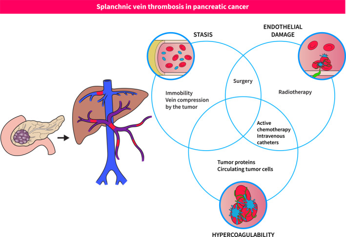
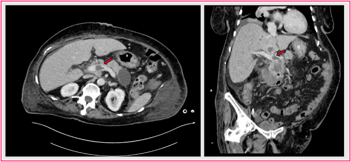
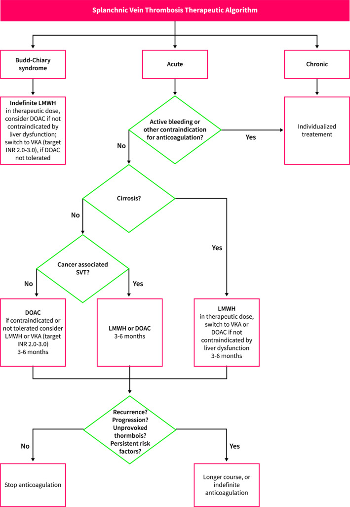
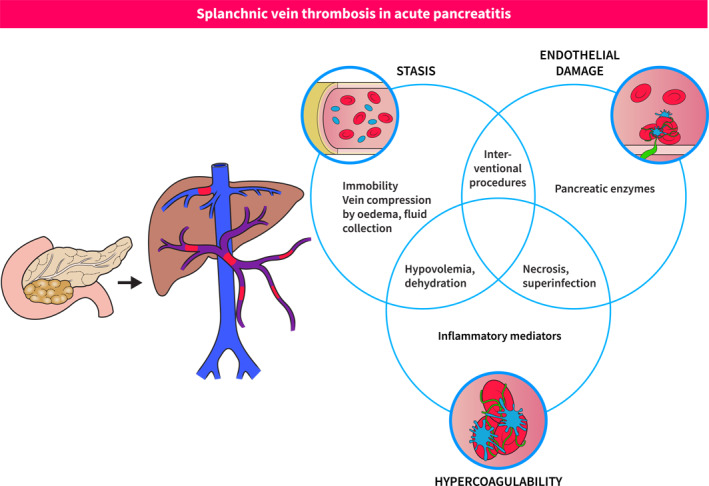
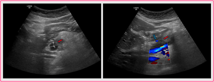
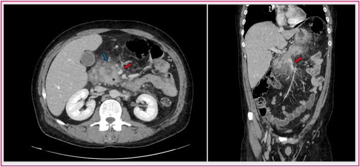
Splanchnic vein thrombosis (SVT) in pancreatic disease has a 20%-30% incidence rate, leading to increased mortality and complication rates. Therefore, the aim of this review is to summarize recent evidence about the incidence, risk factors, and management of pancreatic cancer, pancreatic cystic neoplasm-, and pancreatitis-related SVT. Doppler ultrasound should be the first imaging choice, followed by contrast-enhanced computed tomography or magnetic resonance imaging. Data regarding SVT treatment in acute pancreatitis and pancreatic cancer are scarce; however, for venous thromboembolism treatment, direct oral anticoagulants and low molecular weight heparin have been effective. Further trials must investigate the length of anticoagulant treatment and the need for interventional radiological procedures.
Keywords: cancer; cystic; pancreas; pancreatitis; thrombosis.
© 2025 The Author(s). United European Gastroenterology Journal published by Wiley Periodicals LLC on behalf of United European Gastroenterology.
Conflict of interest statement
The authors declare no conflicts of interest.
Figures
References
-
- Hoekstra J. and Janssen H. L., “Vascular Liver Disorders (I): Diagnosis, Treatment and Prognosis of Budd‐Chiari Syndrome,” Netherlands Journal of Medicine 66, no. 8 (2008): 334–339. - PubMed
Publication types
MeSH terms
Substances
Grants and funding
- BT-ÚNKP-23-3-II-PTE-1996/New National Excellence Program of the Ministry for Innovation and Technology from the source of the National Research, Development and Innovation Fund
- RB-EKÖP-2024-239/New National Excellence Program of the Ministry for Innovation and Technology from the source of the National Research, Development and Innovation Fund
- Innovációs és Technológiai Minisztérium
LinkOut - more resources
Full Text Sources
Medical
