. 2024 Dec 18:12:1450543.
doi: 10.3389/fcell.2024.1450543. eCollection 2024.

Differentiation ability of hematopoietic stem cells and mesenchymal stem cells isolated from human peripheral blood

Affiliations

Differentiation ability of hematopoietic stem cells and mesenchymal stem cells isolated from human peripheral blood

Echambadi Loganathan Samundeshwari et al. Front Cell Dev Biol. .

Abstract

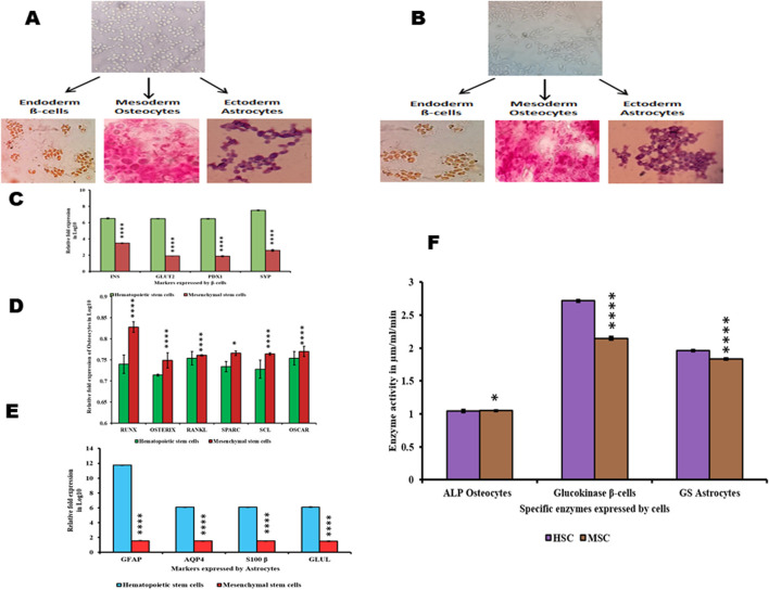
Human hematopoietic stem cells (HSCs) and mesenchymal stem cells (MSCs) are the major stem cells of the bone marrow and are usually isolated from the peripheral blood. In the present study, we isolated these stem cells by an apheresis method from a donor who was administered granulocyte colony-stimulating factor (G-CSF). In vitro propagation of these stem cells showed a plastic-adherence property expressing CD73 and CD105 surface markers, which is a characteristic feature of MSCs. HSCs are non-adherent cells growing as a suspension culture, expressing CD150, CD133, CD34, and CD45 on their surface, which regulate the quiescence nature, and they derive energy from anaerobic glycolysis. The HSCs grow slowly compared to MSCs, are more viable, and survive for long periods under in vitro conditions, which are due to the expression of telomerase, BCL2, and Notch1 genes. The poor viability of MSCs in the culture due to the prominent expression of apoptotic genes BAX, caspase-3, and caspase-9 leads to rapid apoptosis. This was evident even in cells (astrocytes, osteocytes, and beta cells of the islets of Langerhans) differentiated from HSCs and MSCs, thus highlighting the importance of HSCs, the naive stem cells, in regeneration of tissues.

Keywords: differentiation ability; human hematopoietic stem cells; mesenchymal stem cells; peripheral blood stem cells; regenerative medicine.


Conflict of interest statement

The authors declare that the research was conducted in the absence of any commercial or financial relationships that could be construed as a potential conflict of interest.

Figures

FIGURE 1
FIGURE 1
Characterization of HSCs and MSCs isolated from PBSCs (A). Cultured HSCs isolated from PBSCs by the apheresis technique (B). Cultured HSCs were morphologically round with a conspicuous nucleus revealed by Giemsa staining (C). ICC was positive for CD34 anti-human (D). DAPI staining for HSCs (E). Immunofluorescence showing growing HSCs positive for the CD34–FITC conjugate (F). Cultured MSCs isolated from PBSCs (G). Cultured MSCs were spindle-shaped with a nucleus (H). ICC was positive for CD73 anti-human (I). DAPI staining for MSCs (J). Immunofluorescence showing growing MSCs positive for the CD73-PE conjugate (scale bar: 50 µm) (I). Flow cytometry analysis of growing HSCs positive for surface markers (CD34, CD45, and CD133) and MSCs positive for surface markers (CD73 and CD105) (J). Growing HSCs express CD34, CD133, CD45, and CD150 on analysis with RT-PCR (K)- Flow cytometry analysis of growing HSCs and MSCs showing positive for HSCs surface markers (CD34, CD45 and CD133) and MSCs surface markers (CD73 and CD90) (L). Growing MSCs express CD73 and CD105 on analysis with RT-PCR.
FIGURE 2
FIGURE 2
Metabolic and viability characterization of HSCs and MSCs (A). Enzyme activity analysis of glycolytic and TCA-related enzymes in HSCs and MSCs (B). Gene expression analysis of genes involved in the proliferation, viability, and apoptosis of HSCs and MSCs (C). Viability values of HSCs and MSCs obtained by MTT assay at 570 nm (D). Proliferation status of HSCs and MSCs under an in vitro condition. (E,F). Ki-67-PE staining of HSCs and MSCs in the immunofluorescence technique (G,H). Flow cytometry analysis of Ki-67 staining in HSCs and MSCs (I,J). Pimonidazole staining of HSCs and MSCs (K). TRAP assay results: lane 1 shows the smear-like band that represents the presence of telomerase activity obtained from HSCs, and lane 2 shows no band, indicating the absence of telomerase. Two-way ANOVA statistical significance: *p ≤ 0.05, **p ≤ 0.01, ***p ≤ 0.001, and ****p ≤ 0.0001.
FIGURE 3
FIGURE 3
Differentiation of HSCs and MSCs into non-hematopoietic lineages and their characterization (A,B). HSCs and MSCs induced to differentiate into β-cells positive for DTZ staining, osteocytes positive for Alizarin Red staining, and astrocytes were stained with Giemsa (C). Differentiated β-cells expressing INS, GLUT2, PDX1, and SYP (D). Differentiated osteocytes expressing Runx, osterix, RANKL, SPARC, SCL, and OSCAR (E). Differentiated astrocytes expressing GFAP, AQP4, S100β, and GLUL (F). Enzyme activity in differentiated β-cells, osteocytes, and astrocytes. Two-way ANOVA statistical significance: *p ≤ 0.05, **p ≤ 0.01, ***p ≤ 0.001, and ****p ≤ 0.0001.
FIGURE 4
FIGURE 4
Analysis of Ki-67 proliferation marker and telomerase activity in differentiated cells. (A) Immunofluorescence staining of the Ki-67-PE conjugate in differentiated β-cells, osteocytes, and astrocytes. (B) Flow cytometry analysis showing the Ki-67 marker in differentiated cells. (C) TRAP assay results show the presence and absence of telomerase activity in differentiated cells: lane 1, β-cells; lane 2, osteocytes; and lane 3, astrocytes from HSCs; Lane 4, β-cells; lane 2, osteocytes; and lane 3, astrocytes from MSCs.
FIGURE 5
FIGURE 5
Proliferation, viability, and metabolic status of differentiated cells. (A) Proliferation values of differentiated cells. (B) Viability values of differentiated cells. (C–E) Gene expression analysis of differentiated cells from HSCs and MSCs. Two-way ANOVA statistical significance: *p ≤ 0.05, **p ≤ 0.01, ***p ≤ 0.001, and ****p ≤ 0.0001.

Similar articles

References

    1. Ab Kadir R., Zainal Ariffin S. H., Megat Abdul Wahab R., Kermani S., Senafi S. (2012). Characterization of mononucleated human peripheral blood cells. TheScientificWorldJournal 2012, 843843. 10.1100/2012/843843 - DOI - PMC - PubMed
    1. Alenzi F. Q., Alenazi B. Q., Ahmad S. Y., Salem M. L., Al-Jabri A. A., Wyse R. K. (2009). The haemopoietic stem cell: between apoptosis and self renewal. Yale J. Biol. Med. 82 (1), 7–18. - PMC - PubMed
    1. Alhadlaq A., Mao J. J. (2004). Mesenchymal stem cells: isolation and therapeutics. Stem cells Dev. 13 (4), 436–448. 10.1089/scd.2004.13.436 - DOI - PubMed
    1. Baghaei K., Hashemi S. M., Tokhanbigli S., Asadi Rad A., Assadzadeh-Aghdaei H., Sharifian A., et al. (2017). Isolation, differentiation, and characterization of mesenchymal stem cells from human bone marrow. Gastroenterology hepatology bed bench 10 (3), 208–213. - PMC - PubMed
    1. Banerjee P. P., Jagadeesh S. (2009). Non-radioactive assay methods for the assessment of telomerase activity and telomere length. Methods Mol. Biol. Clift. N.J. 523, 383–394. 10.1007/978-1-59745-190-1_25 - DOI - PMC - PubMed

LinkOut - more resources