. 2024 Dec 3;3(4):551-564.
doi: 10.1002/mlf2.12143. eCollection 2024 Dec.

Sodium dodecyl sulfate-coated silver nanoparticles accelerate antimicrobial potentials by targeting amphiphilic membranes

Affiliations

Sodium dodecyl sulfate-coated silver nanoparticles accelerate antimicrobial potentials by targeting amphiphilic membranes

Xiuyan Jin et al. mLife. .

Abstract

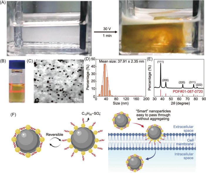
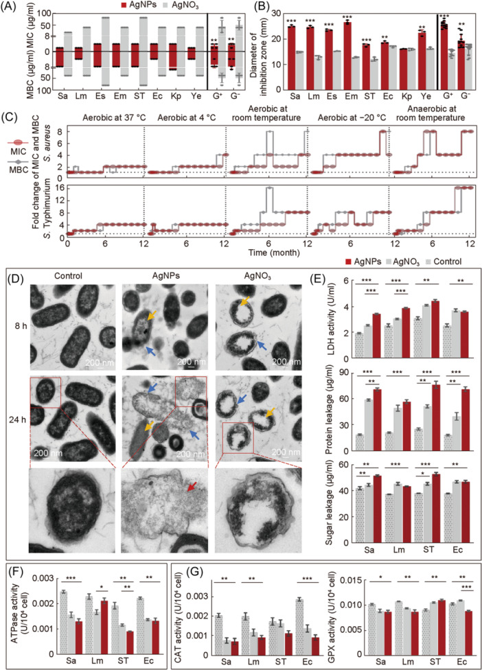
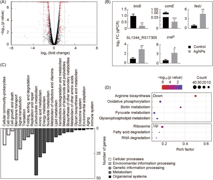
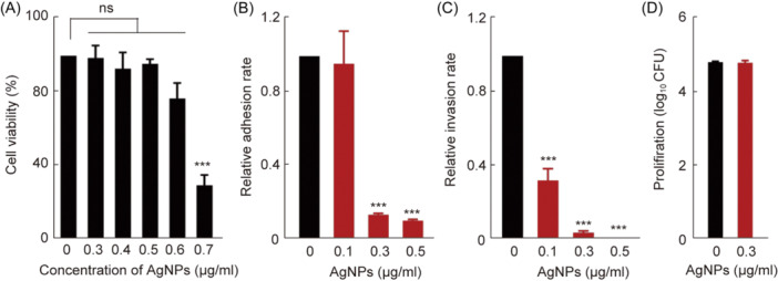
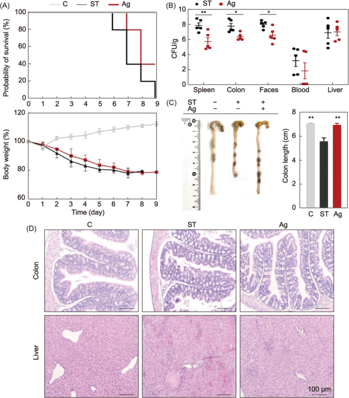
Compelling concerns about antimicrobial resistance and the emergence of multidrug-resistant pathogens call for novel strategies to address these challenges. Nanoparticles show promising antimicrobial activities; however, their actions are hindered primarily by the bacterial hydrophilic-hydrophobic barrier. To overcome this, we developed a method of electrochemically anchoring sodium dodecyl sulfate (SDS) coatings onto silver nanoparticles (AgNPs), resulting in improved antimicrobial potency. We then investigated the antimicrobial mechanisms and developed therapeutic applications. We demonstrated SDS-coated AgNPs with anomalous dispersive properties capable of dispersing in both polar and nonpolar solvents and, further, detected significantly higher bacteriostatic and bactericidal effects compared to silver ions (Ag+). Cellular assays suggested multipotent disruptions targeting the bacterial membrane, evidenced by increasing lactate dehydrogenase, protein and sugar leakage, and consistent with results from the transcriptomic analysis. Notably, the amphiphilic characteristics of the AgNPs maintained robust antibacterial activities for a year at various temperatures, indicating long-term efficacy as a potential disinfectant. In a murine model, the AgNPs showed considerable biocompatibility and could alleviate fatal Salmonella infections. Collectively, by gaining amphiphilic properties from SDS, we offer novel AgNPs against bacterial infections combined with long-term and cost-effective strategies.

Keywords: amphiphilic properties; antimicrobial agents; antimicrobial resistance; feed additive; silver nanoparticles.


Conflict of interest statement

The authors declare no conflict of interests.

Figures

Figure 1
Figure 1
Sustained electrochemical preparation of silver nanoparticles (AgNPs) in electrolyte solution. (A) Production of AgNPs. AgNPs were obtained by electrodeposition in an electrolyte solution of silver nitrate (AgNO3) and sodium dodecyl sulfate (SDS). The AgNPs prepared here were continuously emitted from the electrode interface into the electrolyte solution, resulting in a yellow colloidal solution that formed within 1 min. (B) AgNPs showing the Tyndall effect when dispersed in water and indicating the homogeneity of the synthetic colloidal solution system. (C) Transmission electron microscope analysis of the AgNPs. (D) Particle size distribution of the AgNPs. (E) X‐ray diffraction analysis of the AgNPs. (F) Proposed mechanism for adapting ligands. The reversible orientation change of the surface ligands allowed them to adapt to surrounding liquids and easily pass through the bacterial membrane.
Figure 2
Figure 2
Bactericidal effect of AgNPs in vitro. (A) Minimum inhibitory concentration (MIC) and minimum bactericidal concentration (MBC) of AgNPs and originated derivative AgNO3. (B) Agar well diffusion assay for bacterial inhibition. (C) Long‐term bacterial inhibition and bactericidal efficacy of the AgNPs. The antibacterial effect of the AgNPs against S. aureus and S. Typhimurium under five different preservation conditions (aerobic at 37°C, aerobic at 4°C, aerobic at room temperature, aerobic at −20°C, and anaerobic at room temperature) at different time intervals (0, 0.25, 0.5, 1, 2, 3, 4, 5, 6, 7, 8, 9, 10, 11, and 12 months). (D) Visualization of AgNPs and AgNO3‐treated S. Typhimurium at 8 and 24 h using transmission electron microscopy analysis. The absence of AgNPs or AgNO3 was used as a control. Scale bars: 200 nm. (E) Contents of lactate dehydrogenase (LDH) and the efflux (protein and sugar) of bacterial contents after exposure to AgNPs and AgNO3. (F) ATPase activities after exposure to AgNPs and AgNO3. (G) Bacterial antioxidant enzyme activities after exposure to AgNPs and AgNO3, including glutathione peroxidase (GPX) and catalase (CAT). Red: bacteria treated with AgNPs. Gray: bacteria treated with AgNO3. Gray with points: control. Data were from at least three independent assays: *p < 0.05; **p < 0.01; ***p < 0.001. Sa, Staphylococcus aureus; Lm: Listeria monocytogenes; Es, Enterococcus faecalis; Em, Enterococcus faecium; ST, S. Typhimurium; Ec, Escherichia coli; Kp, Klebsiella pneumoniae; Ye, Yersinia enterocolitica. S. aureus, L. monocytogenes, E. faecalis, and E. faecium are Gam‐positive (G+) bacteria. S. Typhimurium, E. coli, K. pneumoniae, and Y. enterocolitica are Gram‐negative (G) bacteria.
Figure 3
Figure 3
Effect of AgNPs on S. Typhimurium gene expression. (A) Volcanic map of differentially expressed genes (DEGs). DEGs: red point; upregulated genes: X‐axis > 0; downregulated genes: X‐axis < 0. (B) mRNA levels of the DEGs by quantitative reverse transcription polymerase chain reaction (RT‐qPCR). Each scatter represents the expression level of each gene treated with AgNPs. The relative expression level of the target genes was measured with the 2–ΔΔCt method, and 16S rRNA was used as the reference gene. All tests were performed at least three times. (C) Kyoto Encyclopedia of Genes and Genomes (KEGG) pathway classification of the DEGs. (D) KEGG pathway group enrichment of the DEGs. *p < 0.05; **p < 0.01; ***p < 0.001.
Figure 4
Figure 4
In vitro toxicity and protective capacity of AgNPs against Salmonella infections. (A) In vitro biocompatibility effect of AgNPs on the viability of the murine RAW264.7 macrophage cell detected using the CC‐K8 assay. (B–D) The dose effects of AgNPs in bacteria and cell interactions. For S. Typhimurium in the RAW264.7 cells, the geometric means of CFUs per cell were obtained from three independent experiments for cell adhesion, invasion, and proliferation. ***p < 0.001; ns, no significance.
Figure 5
Figure 5
In vivo protective efficacy of AgNPs against Salmonella infections. (A) Comparative analysis of survival and body weight. Changes in the probability of survival and body weight over time after infection were observed from the day of infection. (B) Bacterial tissue load analysis. Amount of Salmonella was detected in the spleen, colon, feces, blood, and liver, but not detected in any control group sample (data not shown). (C) A representative colon length image and a summary of all examined colon length data. (D) Hematoxylin and eosin (H&E) staining analysis of liver and colon sections of each group from the representative samples. *p < 0.05; **p < 0.01. C, control group; ST, Salmonella infection group; Ag, Salmonella infection and AgNP‐treated group. Of 10 mice, five were used for clinical trials, and five were used to observe survival and growth. Samples were collected on Day 5 post infection.
Figure 6
Figure 6
In vivo toxicity of AgNPs. (A) In vivo biocompatibility evaluation of the AgNPs by acute toxicity. AgNPs (0.01, 0.1, 1, 10, and 100 mg/kg) were given orally to mice once daily for 7 days. For the control groups, ddH2O was administered to the animals through the same route as in each test. Within 21 days of acute toxicity, body weight was measured. (B) Tissue pathology. Hematoxylin and eosin (H&E)‐stained liver, kidney, spleen, colon, duodenum, and ileum sections of each group were collected on Day 10 post infection.

Similar articles

Cited by

References

    1. Prestinaci F, Pezzotti P, Pantosti A. Antimicrobial resistance: a global multifaceted phenomenon. Pathog Glob Health. 2015;109:309–318. - PMC - PubMed
    1. Murray C, Ikuta KS, Sharara F, Swetschinski L, Robles Aguilar G, Gray A, et al. Global burden of bacterial antimicrobial resistance in 2019: a systematic analysis. Lancet. 2022;399:629–655. - PMC - PubMed
    1. Jia C, Wang Z, Huang C, Teng L, Zhou H, An H, et al. Mobilome‐driven partitions of the resistome in Salmonella . mSystems. 2023;8:e00883‐23. - PMC - PubMed
    1. Wang Z, Huang C, Liu Y, Chen J, Yin R, Jia C, et al. Salmonellosis outbreak archive in China: data collection and assembly. Sci Data. 2024;11:244. - PMC - PubMed
    1. Wang Z, Zhou H, Liu Y, Huang C, Chen J, Siddique A, et al. Nationwide trends and features of human salmonellosis outbreaks in China. Emerg Microbes Infect. 2024;13:2372364. - PMC - PubMed

LinkOut - more resources