

Pathologic function and therapeutic potential of extracellular vesicle miRNA in sepsis
- PMID: 39744123
- PMCID: PMC11688188
- DOI: 10.3389/fphar.2024.1452276
Pathologic function and therapeutic potential of extracellular vesicle miRNA in sepsis
Abstract
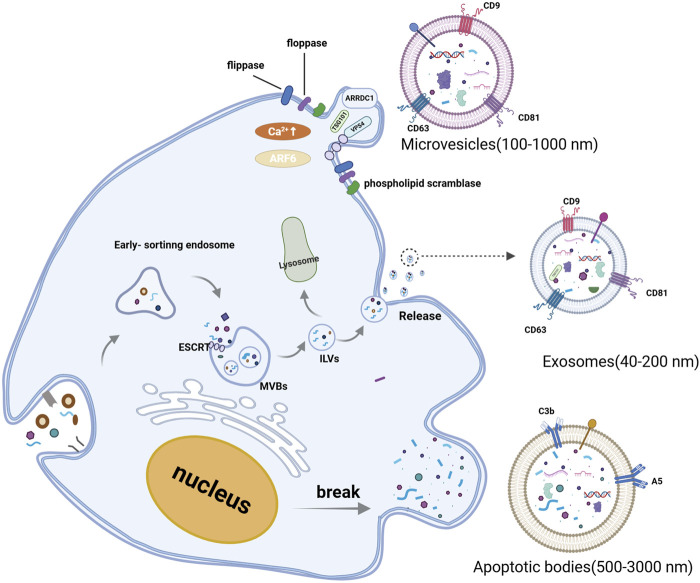
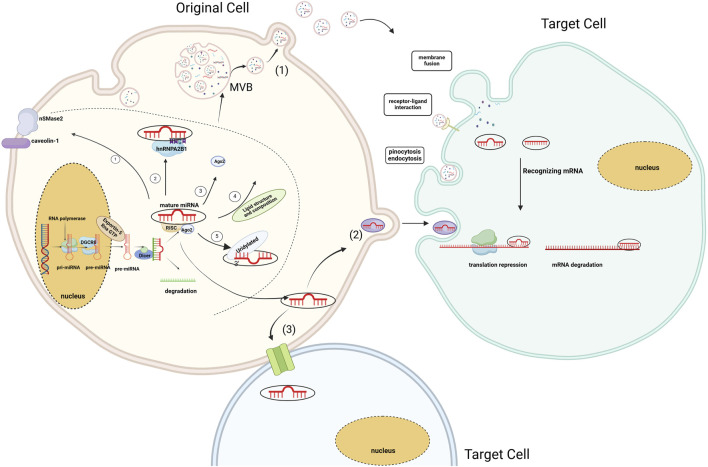
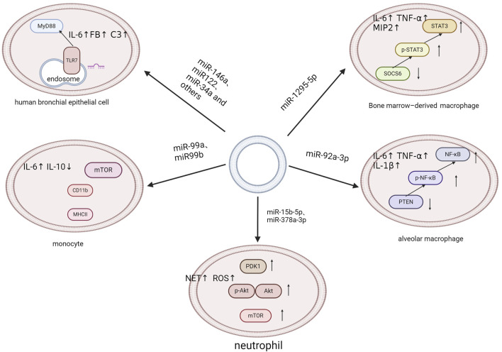
Sepsis is a systemic inflammatory response initiated by an infection, which can lead to multi-organ dysfunction. The pathophysiology of sepsis is complex, and treatment options are limited. Traditional antibiotic therapies have shown limitations, such as promoting the emergence of antibiotic-resistant bacteria and disrupting the natural microbiota. Consequently, there is a pressing need to explore diverse therapeutic approaches for sepsis management. Extracellular vesicles, which play a crucial role in cell-to-cell communication, are released by various cell types throughout the body and possess a membrane structure composed of a lipid bilayer. MicroRNAs may be encapsulated within these structures and can be selectively delivered to target recipient cells through the activation of cell surface receptors or via endocytosis and fusion, thereby modulating the biological functions of target cells. The article examines the pathological alterations that happen as sepsis progresses and the biological control of extracellular vesicles and microRNAs in sepsis. This review focuses on the role of extracellular vesicles and their microRNAs on controlling the inflammatory response, macrophage polarization, programmed cell death, endothelial dysfunction, and microcirculatory changes in sepsis. Furthermore, the obstacles encountered by this novel therapy are also examined.
Keywords: diagnosis; endothelial dysfunction; extracellular vesicle; inflammation; microRNA; programmed cell death; sepsis; therapy.
Copyright © 2024 Deng, Cui, Zhang, Liu, Luo, Liu, Zhang and Fang.
Conflict of interest statement
The authors declare that the research was conducted in the absence of any commercial or financial relationships that could be construed as a potential conflict of interest.
Figures
Similar articles
-
Exosomes and MicroRNAs: key modulators of macrophage polarization in sepsis pathophysiology.Eur J Med Res. 2025 Apr 17;30(1):298. doi: 10.1186/s40001-025-02561-z. Eur J Med Res. 2025. PMID: 40247413 Free PMC article. Review.
-
Role of Extracellular microRNAs in Sepsis-Induced Acute Lung Injury.J Immunol Res. 2023 Jun 19;2023:5509652. doi: 10.1155/2023/5509652. eCollection 2023. J Immunol Res. 2023. PMID: 37378068 Free PMC article. Review.
-
Functional delivery of lncRNA TUG1 by endothelial progenitor cells derived extracellular vesicles confers anti-inflammatory macrophage polarization in sepsis via impairing miR-9-5p-targeted SIRT1 inhibition.Cell Death Dis. 2021 Nov 6;12(11):1056. doi: 10.1038/s41419-021-04117-5. Cell Death Dis. 2021. PMID: 34743197 Free PMC article.
-
Endothelial progenitor cells-secreted extracellular vesicles containing microRNA-93-5p confer protection against sepsis-induced acute kidney injury via the KDM6B/H3K27me3/TNF-α axis.Exp Cell Res. 2020 Oct 15;395(2):112173. doi: 10.1016/j.yexcr.2020.112173. Epub 2020 Jul 15. Exp Cell Res. 2020. PMID: 32679234
-
Aquaporins in sepsis- an update.Front Immunol. 2024 Oct 31;15:1495206. doi: 10.3389/fimmu.2024.1495206. eCollection 2024. Front Immunol. 2024. PMID: 39544938 Free PMC article. Review.
Cited by
-
Circulating extracellular vesicles as potential biomarkers and mediators of acute respiratory distress syndrome in sepsis.Sci Rep. 2025 Feb 14;15(1):5512. doi: 10.1038/s41598-025-89783-7. Sci Rep. 2025. PMID: 39953195 Free PMC article.
References
Publication types
LinkOut - more resources
Full Text Sources