. 2025 Jan 1;22(1):71-86.
doi: 10.7150/ijms.104932. eCollection 2025.

Receptor-Interacting Protein Kinase 3-Mediated Modulation of Endothelial Cell Necroptosis and Mitochondrial Dysfunction through AMPK/Drp1 Signaling Pathway: Insights into the Pathophysiological Mechanisms of Lipopolysaccharide-Induced Acute Lung Injury

Affiliations

Receptor-Interacting Protein Kinase 3-Mediated Modulation of Endothelial Cell Necroptosis and Mitochondrial Dysfunction through AMPK/Drp1 Signaling Pathway: Insights into the Pathophysiological Mechanisms of Lipopolysaccharide-Induced Acute Lung Injury

Zhaoning Zhao et al. Int J Med Sci. .

Abstract

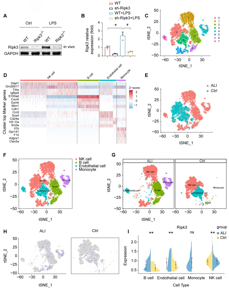
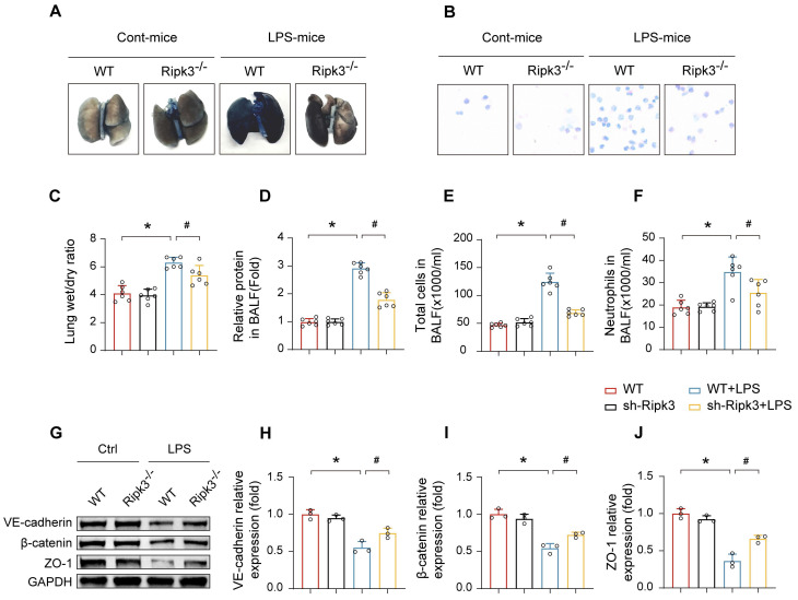
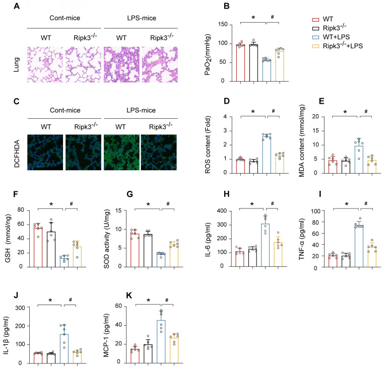
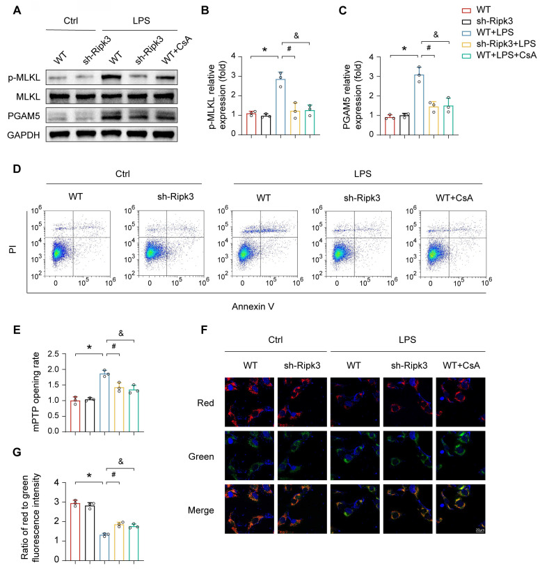
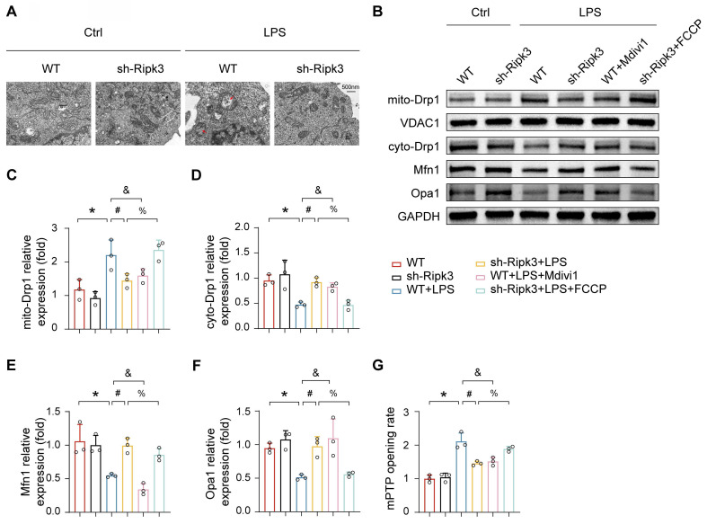
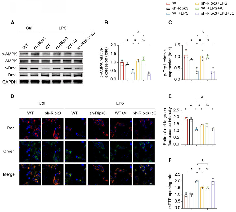
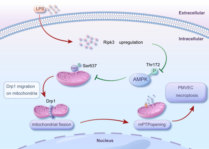
Receptor-interacting protein 3 (Ripk3) plays a crucial part in acute lung injury (ALI) by regulating inflammation-induced endothelial damage in the lung tissue. The precise mechanisms through which Ripk3 contributes to the endothelial injury in ALI still remain uncertain. In the current research, we employed Ripk3-deficient (Ripk3-/-) mice to examine the role of Ripk3 in ALI progression, focusing on its effects on endothelial cells (ECs), mitochondrial damage and necroptosis. Our study observed significant Ripk3 upregulation in lipopolysaccharide- (LPS-) treated lung tissues, as well as in murine pulmonary microvascular endothelial cells (PMVECs). Ripk3 deletion improved lung tissue morphology, reduced inflammation, oxidative stress and endothelial dysfunction under LPS challenge. It also mitigated LPS-induced necroptosis and mitochondrial damage in PMVECs. Ripk3 upregulation suppressed the AMP-activated protein kinase (AMPK) pathway and activated Drp1-mediated mitochondrial fission, increasing mitochondrial permeability transition pore (mPTP) opening and PMVEC necroptosis. Conversely, Ripk3 deletion activated the AMPK/Drp1-mitochondrial fission pathway, preventing mPTP opening and PMVEC necroptosis in ALI. These findings demonstrated that Ripk3 promotes necroptosis through the AMPK/Drp1/mPTP opening pathway, identifying a potential therapeutic target for ALI treatment.

Keywords: Acute lung injury; Cell necroptosis; Mitochondrial damage; Ripk3.


Conflict of interest statement

Competing Interests: The authors have declared that no competing interest exists.

Figures

Figure 1
Figure 1
LPS upregulates Ripk3 in lung ECs. (A-B) The change of Ripk3 expression in vivo was quantified using western blotting. (C-I) Ripk3 expression in different cell types from the lung was determined using single-cell sequencing analysis. Mean ± SEM, ∗p < 0.05 vs. the wild-type (WT) group; #p < 0.05 vs. the LPS group.
Figure 2
Figure 2
Effect of Ripk3 deletion on the alveolar-capillary barrier in LPS-induced ALI mice. (A) Representative images of Evans Blue (EB) extravasation in the lungs. (B-F) The ratio of wet weight to dry weight of the lungs, the protein content, the number of total cells and neutrophils in BALF were analyzed to assess lung permeability. (G-J) The protein levels of VE-cadherin, β-catenin, and ZO-1 in the lung tissues were measured using western blot and quantitative analysis. Mean ± SEM, ∗p < 0.05 vs. the WT group; #p < 0.05 vs. the LPS group.
Figure 3
Figure 3
Ripk3 deletion attenuates LPS-mediated oxidative stress and inflammation response in acute lung injury. (A) Pathological alterations of lung parenchyma observed by HE staining after acute lung injury. (B) Measurement of the partial pressure of arterial oxygen (PaO2). (C-D) DCFHDA staining was used to detect ROS content. (E-G) MDA level, SOD activity, GSH level were also measured in lung tissue. (H-K) Protein levels of IL-6, TNF-α, IL-1β, and MCP-1 were quantified by ELISA. Mean ± SEM, ∗p < 0.05 vs. the WT group; #p < 0.05 vs. the LPS group.
Figure 4
Figure 4
Ripk3 knockdown reduces PMVEC necroptosis by inhibiting mPTP opening. (A-C) Western blots were used to analyze the expression of PGAM5, and p-MLKL. (D) Necroptosis of PMVECs was quantified by flow cytometry with Annexin V/PI staining. The necroptosis group: the percentage of PI+ cells. Cyclosporin A (CsA) was used as the inhibitor to prevent mitochondrial permeability transition pore (mPTP) opening. (E) Evaluation of the rate of mPTP opening. (F-G) Changes of mitochondrial membrane potential were identified using JC-1 staining. Mean ± SEM, ∗p < 0.05 vs. the Ctrl group; #p < 0.05 vs. the LPS group; &p < 0.05 vs. the LPS+CsA group.
Figure 5
Figure 5
Ripk3 knockdown inhibits mPTP opening via Drp1-related mitochondrial fission. (A) Representative transmission electron microscopy (TEM) images depicting changes in mitochondrial morphology. Red arrow: damaged and swollen mitochondria. (B-F) The expression levels of mito-Drp1, cyto-Drp1, Mfn1, and Opa1 were quantified by Western blotting. The mitochondrial fission inhibitor, Mdivi1, was utilized to impede mitochondrial fission in WT cells under LPS administration. The mitochondrial fission activator, FCCP, was employed to promote mitochondrial fission in Ripk3-deficient cells under LPS treatment. (G) The change of mPTP opening time. Mean ± SEM, ∗p < 0.05 vs. the Ctrl group; #p < 0.05 vs. the LPS group; &p < 0.05 vs. the LPS+Mdivi1 group; %p < 0.05 vs. the LPS+ FCCP group.
Figure 6
Figure 6
Ripk3 knockdown alleviates mitochondrial injury through promoting AMPK-mediated Drp1 phosphorylation. (A-C) The phosphorylation of Drp1 and AMPK was measured using western blots. The AMPK pathway activator, AICAR (AI), was employed to activate AMPK pathway in WT cells under LPS treatment. The AMPK pathway inhibitor, compound C (cC), was utilized to block the AMPK pathway in Ripk3-deficient cells under LPS injury. (D-E) Impact of AMPK on mitochondrial membrane potential. (F) The change of mPTP opening time. Mean ± SEM, ∗p < 0.05 vs. the Ctrl group; #p < 0.05 vs. the LPS group; &p < 0.05 vs. the LPS+AI group; %p< 0.05 vs. the LPS+cC group.
Figure 7
Figure 7
Mechanism diagram for the role of Ripk3-AMPK-Drp1-mPTP in LPS-mediated acute lung injury via initiating necroptosis. LPS injury caused upregulation of Ripk3, which resulted in a reduction in the phosphorylation of AMPK. Inactivating AMPK hindered the phosphorylation of Drp1 at Ser637, which triggered fatal mitochondrial fission. Excessive fission caused the opening of mPTP, ultimately leading to the necroptosis of PMVECs. Nevertheless, the recovery of AMPK activity could halt the excessive fission, offering prosurvival effects for the lung in the presence of LPS injury. Our findings revealed a novel role for Ripk3 in LPS-induced acute lung injury through regulating Drp1-mediated mitochondrial fission via the AMPK signaling pathway. These results suggest that targeting Ripk3 could be a promising therapeutic approach for treating acute lung injury in clinical practice.

Similar articles

Cited by

References

    1. Chambers ED, White A, Vang A, Wang Z, Ayala A, Weng T. et al. Blockade of equilibrative nucleoside transporter 1/2 protects against Pseudomonas aeruginosa-induced acute lung injury and NLRP3 inflammasome activation. Faseb j. 2020;34:1516–31. - PMC - PubMed
    1. Liu M, Chen Y, Wang S, Zhou H, Feng D, Wei J. et al. α-Ketoglutarate Modulates Macrophage Polarization Through Regulation of PPARγ Transcription and mTORC1/p70S6K Pathway to Ameliorate ALI/ARDS. Shock. 2020;53:103–13. - PubMed
    1. Hu Q, Zhang S, Yang Y, Yao JQ, Tang WF, Lyon CJ. et al. Extracellular vesicles in the pathogenesis and treatment of acute lung injury. Mil Med Res. 2022;9:61. - PMC - PubMed
    1. Siegel ER, Croze RH, Fang X, Matthay MA, Gotts JE. Inhibition of the lipoxin A4 and resolvin D1 receptor impairs host response to acute lung injury caused by pneumococcal pneumonia in mice. Am J Physiol Lung Cell Mol Physiol. 2021;320:L1085–l92. - PMC - PubMed
    1. Meyer NJ, Gattinoni L, Calfee CS. Acute respiratory distress syndrome. Lancet. 2021;398:622–37. - PMC - PubMed

MeSH terms

Substances

LinkOut - more resources