. 2025 Jan 1;21(1):175-188.
doi: 10.7150/ijbs.103122. eCollection 2025.

Heat acclimation mediates cellular protection via HSP70 stabilization of HIF-1α protein in extreme environments

Affiliations

Heat acclimation mediates cellular protection via HSP70 stabilization of HIF-1α protein in extreme environments

Can Li et al. Int J Biol Sci. .

Abstract

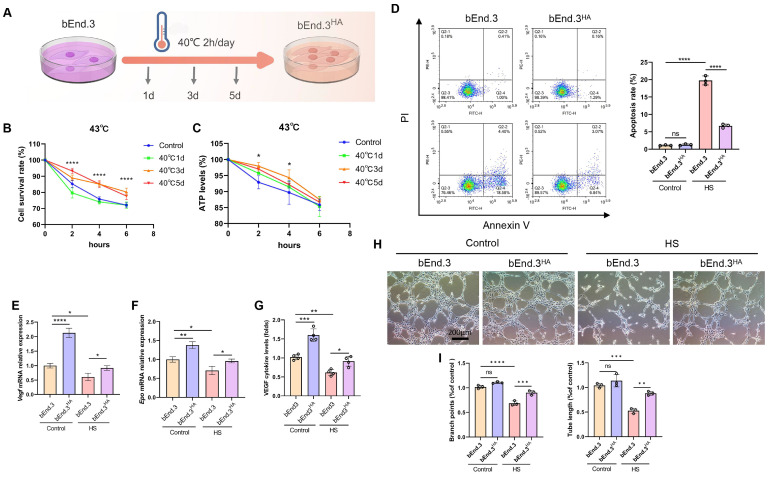
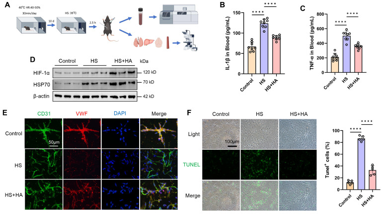
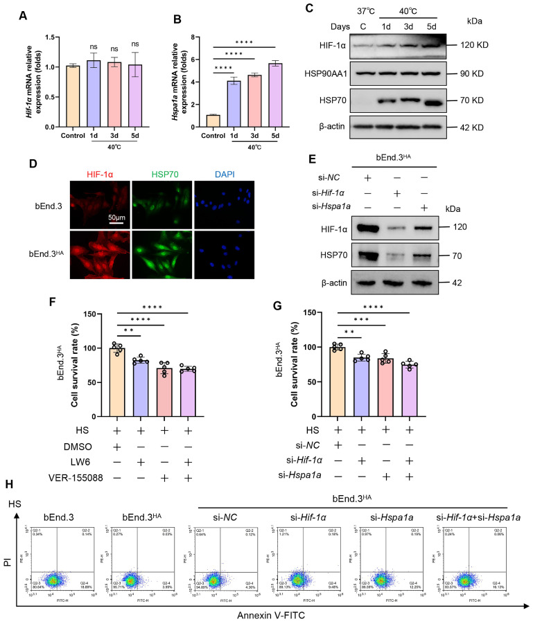
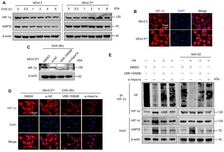
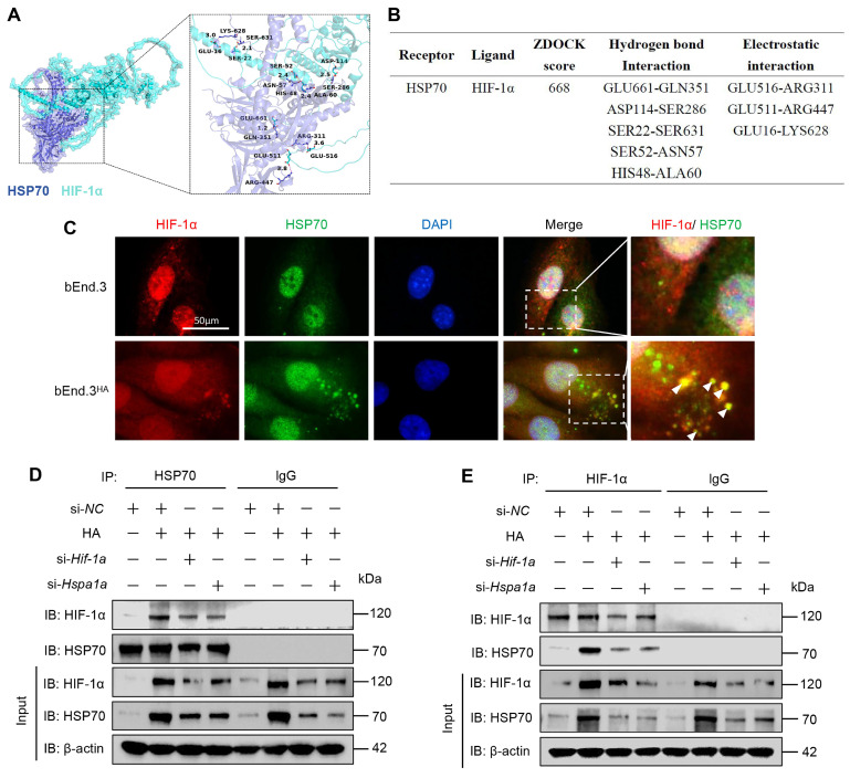
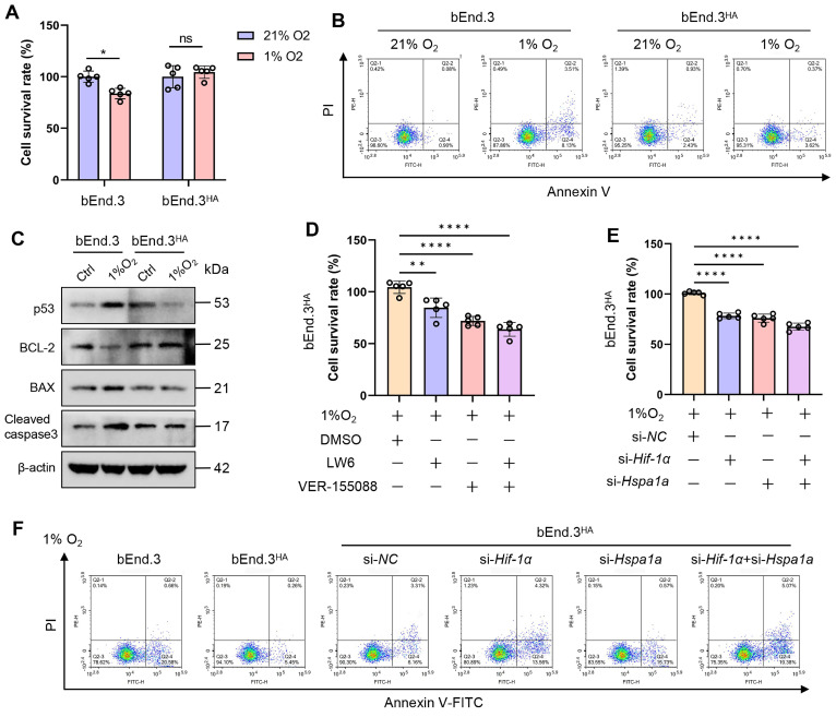
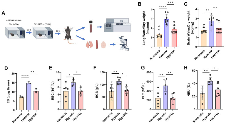
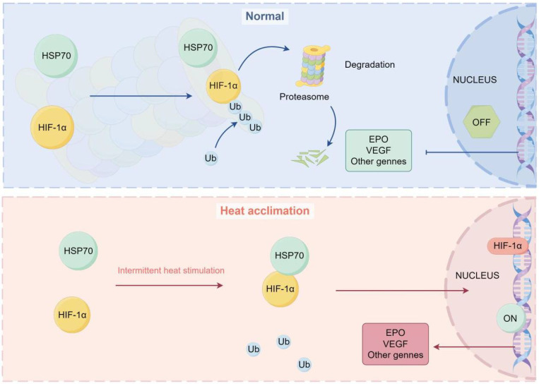
Heat acclimation (HA) is an evolutionarily conserved trait that enhances tolerance to novel stressors by inducing heat shock proteins (HSPs). However, the molecular mechanisms underlying this phenomenon remain elusive. In this study, we established a HA mouse model through intermittent heat stimulation. Subsequently, this model was evaluated using an array of physiological and histological assessments. In vitro, HA cell model with mouse brain microvascular endothelial cells (bEnd.3) was established and analyzed for cell viability and apoptosis markers. We investigated HA-mediated heat and hypoxia tolerance mechanisms using HIF-1α and HSP70 inhibitors and siRNA. Our results demonstrated that HA enhances the tolerance of bEnd.3 cells and mice to both heat and hypoxia, Mechanistically, HA upregulated the expression of HIF-1α and HSP70. However, inhibition of HIF-1α or HSP70 partially attenuated HA-induced tolerance to heat and hypoxia. Additionally, HA significantly decreased the ubiquitination levels of HIF-1α, whereas inhibition of HSP70 increased its ubiquitination. HA also substantially enhanced the interaction between HIF-1α and HSP70. In conclusion, our findings indicate that HA enhances tolerance to heat and hypoxia by stabilizing HIF-1α through increased interaction with HSP70. This discovery elucidates a novel mechanism of cellular protection conferred by HA and provides new strategies and potential targets for human adaptation to extreme environments.

Keywords: Heat acclimation; Heat shock protein 70; Heat stress; Hypoxia; Hypoxia-inducible factor 1-alpha; Ubiquitination.


Conflict of interest statement

Competing Interests: The authors have declared that no competing interest exists.

Figures

Figure 1
Figure 1
Intermittent heat stimulation enhances cellular thermotolerance. A. Schematic diagram of the temperature shift procedure to render bEnd.3 cells into thermoresistant bEnd.3HA (1d, 3d, 5d) cells. B. Cell viability of bEnd.3 and bEnd.3HA (1d, 3d, 5d) cells under HS conditions as determined by CCK-8 assay. C. ATP levels of bEnd.3 and bEnd.3HA (1d, 3d, 5d) cells under HS conditions as determined by CellTiter-LumiTM assay. D. The apoptotic index of bEnd.3 and bEnd.3HA cells under control and HS conditions was determined by flow cytometry. E & F. bEnd.3 and bEnd.3HA cells were treated under control and HS conditions. RT-qPCR analyses of vascular endothelial cell markers Vegf and Epo mRNA. β-actin was used as an internal control. Control: 37℃, HS: 43℃. G. ELISA analyses of VEGF protein secretion in culture medium. H. Representative images of bEnd.3 and bEnd.3HA on matrigel for 6 h. Scale bars = 200 μm. I. Tube length and branch points were quantified. *P < 0.05, **P < 0.01, ***P < 0.001, ****P < 0.0001.
Figure 2
Figure 2
Intermittent heat stimulation training improves high temperature tolerance in mice. A. Flowchart of the experiment to improve heat tolerance in mice through intermittent heat stimulation training. B & C. ELISA was used to analyze the plasma concentrations of cytokines (IL-1β and TNF-ɑ) in control, HS, and HS+HA group mice (n =8) 16 h after HS. D. WB was used to detect the expression of HIF-1α and HSP70 protein. β-actin was used as an internal control. E. IF analysis of isolated microvessels stained with antibodies for each cell component. Endothelial markers (CD31, green channel; VWF, red channel) were detected in all microvessel fragments. Scale bar = 50 μm. F. IF was used to detect apoptosis in isolated microvessels stained with TUNEL. Scale bar = 100 μm. ****P < 0.0001.
Figure 3
Figure 3
HA induces tolerance to high temperatures via the HIF-1α/HSP70 signaling. A & B. The bEnd.3 cells were exposed to 40℃ for 2 h daily, continuously for 1 day, 3 days, and 5 days. RT-qPCR was used to detect the expression of Hif-1α and Hspa1a mRNA. C. WB was used to detect the expression of HIF-1α, HSP90AA1, and HSP70 protein. β-actin was used as an internal control. D. IF was used to detect the expression of HIF-1α and HSP70 protein. E. WB was used to detect the expression of HIF-1α and HSP70 protein. β-actin was used as an internal control. F. bEnd.3HA cells were exposed to HS for 4 h and treated with LW6, VER-155088, or a combination of both. Cell viability was detected using the CCK-8 assay. G. bEnd.3HA cells were exposed to HS for 4 h and treated with si-Hif-1α, si-Hspa1a, or a combination of both. Cell viability was detected using the CCK-8 assay. H. Flow cytometry with Annexin V and PI staining was used to determine the apoptotic index of cells. **P < 0.01, ***P < 0.001, ****P < 0.0001.
Figure 4
Figure 4
HA enhances HIF-1α protein stability via HSP70 in bEnd.3 cells. A. bEnd.3 and bEnd.3HA cells were treated with CHX for the indicated times and analyzed by WB. B. IF analysis of HIF-1α subcellular localization. Nuclei are stained with DAPI (blue). n = 5 per group, scar bar = 50 μm. C. bEnd.3HA cells were treated with DMSO, si-NC, VER-155008, or si-Hspa1a and with CHX for 6 h. WB was used to detect the HIF-1α protein expression. D. IF was used to detect the HIF-1α protein expression. Nuclei are stained with DAPI (blue). n=5 per group, scar bar = 50 μm. E. bEnd.3 and bEnd.3HA cells were treated with si-NC, VER-155008, or si-Hspa1a. The extracts were immunoprecipitated with anti-HIF-1α antibodies and immunoblotted with anti-ubiquitin, anti-HIF-1α, and anti-HSP70 antibodies. β-actin was used as an internal control.
Figure 5
Figure 5
HA promotes the interaction between HIF-1α and HSP70 proteins in bEnd.3 cells. A. ZDOCK prediction was used to model the predicted interaction between HSP70 and HIF-1α. B. Predicted ZDOCK binding score, hydrogen bonds, and electrostatic interactions with amino acid sites between HSP70 and HIF-1α. C. Colocalization of HIF-1α (red) with HSP70 (green) in bEnd.3 and bEnd.3HA cells. Nuclei are stained with DAPI (blue). n=5 per group, scar bar = 50 μm. D & E. bEnd.3 and bEnd.3HA cells were treated with si-NC, si-Hif-1α, or si-Hspa1a. The extracts were immunoprecipitated with anti-HIF-1α or anti-HSP70 antibodies and immunoblotted with anti-HIF-1α and anti-HSP70 antibodies.
Figure 6
Figure 6
HA induces tolerance to hypoxia via the HIF-1α/HSP70 signaling. A. bEnd.3 and bEnd.3HA cells were treated at 1% O2 concentration for 12 h. Cell viability was detected using the CCK-8 assay. B. Flow cytometry with Annexin V and PI staining was used to determine the apoptotic index of cells. C. WB was used to detect the expression of p53, Bcl-2, Bax, and cleaved caspase3 protein. β-actin was used as an internal control. D. bEnd.3HA cells were treated at 1% O2 concentration for 12 h and with LW6, VER-155008, or LW6+VER-155008. Cell viability was detected using the CCK-8 assay. E. bEnd.3HA cells were treated at 1% O2 concentration for 12 h and with si-Hif-1α, si-Hspa1a, or si-Hif-1α+si-Hspa1a. Cell viability was detected using the CCK-8 assay. F. Flow cytometry with Annexin V and PI staining was used to determine the apoptotic index of cells. *P < 0.05, **P < 0.01, ***P < 0.001.
Figure 7
Figure 7
Intermittent heat stimulation training improves hypoxia tolerance in mice. A. Flowchart of the experiment to improve hypobaric hypoxia tolerance in mice through intermittent heat stimulation training. B & C. The water content in the brain and lung of mice (n = 8). D. The EB extravasation assay was used to evaluate the BBB permeability (n = 3). E-H. The levels of red blood cells (RBC), hemoglobin (HGB), platelet (PLT), and neutrophil (NEU) in mice (n = 6). *P < 0.05, **P < 0.01, ***P < 0.001, ****P < 0.0001.
Figure 8
Figure 8
Diagram representing the potential mechanism by which HA regulates stabilization of HIF-1α protein via HSP70 in bEnd.3 cells. Under normal conditions, HIF-1α protein is ubiquitinated and degraded by the proteasome. Under HA conditions, HSP70 interacts with the HIF-1α protein, inhibiting its ubiquitination, stabilizing the HIF-1α protein, and promoting the transcription of downstream genes in the nucleus.

Similar articles

References

    1. Burtscher M, Gatterer H, Burtscher J, Mairbäurl H. Extreme Terrestrial Environments: Life in Thermal Stress and Hypoxia. A Narrative Review. Front Physiol. 2018;9:572. - PMC - PubMed
    1. Le Roy B, Martin-Krumm C, Pinol N, Dutheil F, Trousselard M. Human challenges to adaptation to extreme professional environments: A systematic review. Neurosci Biobehav Rev. 2023;146:105054. - PubMed
    1. Oksala NK, Ekmekçi FG, Ozsoy E, Kirankaya S, Kokkola T, Emecen G. et al. Natural thermal adaptation increases heat shock protein levels and decreases oxidative stress. Redox Biol. 2014;3:25–28. - PMC - PubMed
    1. Sherwood SC, Huber M. An adaptability limit to climate change due to heat stress. Proc Natl Acad Sci U S A. 2010;107:9552–9555. - PMC - PubMed
    1. Gibson OR, Taylor L, Watt PW, Maxwell NS. Cross-Adaptation: Heat and Cold Adaptation to Improve Physiological and Cellular Responses to Hypoxia. Sports Med. 2017;47:1751–1768. - PMC - PubMed

Substances

LinkOut - more resources