. 2025 Jan 1;21(1):95-108.
doi: 10.7150/ijbs.95568. eCollection 2025.

The modulation of calcium and chloride channels induces cardiomyocytes from human pluripotent stem cells

Affiliations

The modulation of calcium and chloride channels induces cardiomyocytes from human pluripotent stem cells

Ya Meng et al. Int J Biol Sci. .

Abstract

Ion channels play a crucial role in cardiac functions, and their activities exhibit dynamic changes during heart development. However, the precise function of ion channels in human heart development remains elusive. In this study, we utilized human embryonic stem cells (hESCs) as a model to mimic the process of human embryonic heart development. During hESCs differentiation into cardiomyocytes, we observed differential expression of ion channel genes, including upregulation of ryanodine receptor 2 (RYR2), which encodes a calcium release channel. Subsequently, we discovered that Suramin, an activator of RyR2, efficiently promoted cardiac differentiation even in the absence of conventional WNT inhibitors. Furthermore, various modulators targeting sodium channels, potassium channels or chloride channels were examined under chemically defined conditions during cardiac differentiation. We found that DIDS, a chloride transport inhibitor, also enhanced hESCs differentiation into cardiomyocytes. Both Suramin and DIDS partially inhibited WNT signaling pathway, and RYR2 knockdown attenuated cardiac differentiation induced by WNT inhibitor treatment, or Suramin or DIDS administration. The resulting cardiomyocytes induced by these ion modulators exhibited specific expression patterns of cardiac genes and displayed typical electrophysiological signals. Notably, compared to WNT inhibitor treatment group, both Suramin and DIDS led to increased generation of atrial-like cardiomyocytes suggesting their potential as alternative inducers for specific cardiomyocyte lineage commitment during human cardiomyocyte induction processes. This study demonstrates that regulation of ion channels plays a crucial role in determining the fate of cardiac cells, providing an effective approach for inducing cardiomyocytes from hPSCs and highlighting their critical involvement in human heart development.

Keywords: calcium channel; cardiomyocyte derivation; chloride channel; human pluripotent stem cells; ryanodine receptor 2.


Conflict of interest statement

Competing Interests: YM and GC filed a patent application based on cardiomyocyte differentiation that is induced by chloride channel regulators. HELP Stem Cell Innovations Co. Ltd. performed the electrophysiological analysis of hESC-derived cardiomyocytes.

Figures

Figure 1
Figure 1
Gene expressions of ion channels show dynamical changes during cardiac differentiation. A Diagram of differentiation strategies for cardiac differentiation. B Gene expressions of hESCs, mesoendoderm, cardiac progenitor, and cardiomyocyte markers at day 0, day 3, day 6, and day 10 of differentiation. C-F Expression of genes encoded calcium channels (C), sodium channels (D), potassium channels (E), and chloride channels (F) in the process of cardiac differentiation.
Figure 2
Figure 2
Calcium channel modulators regulated cardiomyocyte derivation from hESCs. A The schematic diagram showed the strategies for cardiomyocyte derivation. hESCs were treated with CHIR99021 for 1 day, and after one day, the cells were subjected to IWP-2 or calcium channel modulators from day 2 to day 5. B Gene expressions of ITPR2, ITPR3, RYR1, RYR2, and RYR3 during cardiomyocyte derivation. C Gene expressions of ITPR2, ITPR3, RYR1, RYR2, and RYR3 in control (Ctrl) and IWP-2 group were compared by Q-PCR at day 10 of cardiac differentiation. Blue, Ctrl; Red, IWP-2 group. Data shown are mean ± SD of three independent experiments (*p < 0.05 compared with Ctrl). D Calcium channel regulators were added into basal medium from day 2 to day 5 of differentiation. The expressions of the indicated genes were measured by Q-PCR, and normalized by GAPDH. 4-CMC, 4-Chloro-m-cresol; 2-APB, 2-aminoethoxydiphenyl borate; IP3, Inositol 1,4,5-triphosphate.
Figure 3
Figure 3
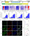
Suramin promoted cardiomyocyte derivation from hESCs. A Suramin exhibits a dose-dependent effect on the gene expression of NKX2-5 and TNNT2 during cardiac differentiation. Data shown are mean ± SD of three independent experiments (*p < 0.05 compared with suramin 0). B Timing effect of suramin on the gene expression of NKX2-5 and TNNT2 during cardiac differentiation. Suramin at a concentration of 50 μM was added at the indicated time of differentiation. Data shown are mean ± SD of three independent experiments (*p < 0.05 compared with none-treated control). C Flow cytometry analysis showed the TNNT2-positive population after 12 days of differentiation. IWP-2 3 μM, suramin 25 μM, or suramin 50 μM was subjected to basal medium from day 2 to day 5 of differentiation. D Immunostaining images showed the levels of TNNT2 (green), NKX2-5 (red) and DAPI (blue) in the differentiated cells. IWP-2 (3 μM) or suramin (50 μM) was added to basal medium from day 2 to day 5 of differentiation. Scale bar, 50 μm.
Figure 4
Figure 4
Chloride channel modulator DIDS induced cardiomyocytes from hESCs. A The schematic diagram of cardiomyocyte derivation. The cells were subjected to IWP-2 or the indicated ion channel modulators from day 2 to day 5. B Effect of Sodium, potassium, and chloride channels on cardiac differentiation. The indicated ion channel modulators were applied from day 2 to day 5 of cardiac differentiation. Gene expressions of TNNT2 and NKX2-5 were determined by Q-PCR at day 10. The scale bar indicated the log2 fold change compared with Ctrl. The doses of ion channel modulators were listed in supplemental table. C Dose-dependent effect of DIDS on the gene expression of NKX2-5 and TNNT2 during cardiomyocyte derivation. Data shown are mean ± SD of three independent experiments (*p < 0.05 compared with DIDS 0). D Timing effect of DIDS on the gene expression of NKX2-5 and TNNT2 during cardiac differentiation. DIDS at a concentration of 4 μM was added at the indicated time of differentiation. Data shown are mean ± SD of three independent experiments (*p < 0.05 compared with Ctrl). E Immunostaining images showed the levels of TNNT2 (green), NKX2-5 (red) and DAPI (blue) in the differentiated cells after 12 days of differentiation. IWP-2 (3 μM) or DIDS (4 μM) was added to basal medium from day 2 to day 5 of differentiation. Scale bar, 50 μm. F Flow cytometry analysis showed the TNNT2-positive population after 12 days of differentiation. IWP-2 (3 μM) or DIDS (4 μM) was added to basal medium from day 2 to day 5 of differentiation.
Figure 5
Figure 5
The induction of cardiac differentiation by Suramin and DIDS was partially reliant on WNT inhibition and RYR2. A Effect of Suramin and DIDS on WNTs expression. hESCs were treated with CHIR99021 for 1 day, and then subjected to IWP-2 (3 μM), suramin (50 μM), or DIDS (4 μM) for 24 hours. Gene expression of WNTs were analyzed by Q-PCR. Data shown are mean ± SD of three independent experiments (*p < 0.05, **p < 0.01 compared with Ctrl). B Immunostaining images of β-Catenin (Green) and DAPI (Blue) in the indicated groups. Scale bar, 20 μm. C Suramin or DIDS promoted IWP-2 induced cardiac differentiation. hESCs were treated with IWP-2, suramin, DIDS, suramin+IWP-2, DIDS+IWP-2 at day 2 to day 5 of differentiation. Gene expression of TNNT2 and NKX2-5 were analyzed by Q-PCR. Data shown are mean ± SD of three independent experiments (*p < 0.05 compared with IWP-2). D Flow cytometry analysis showed the TNNT2-positive population after 12 days of differentiation in the indicated groups. E RYR2 knockdown resulted in decreased gene expression of NKX2-5, TNNT2 and RYR2 in cardiomyocytes induced by IWP-2 (3 μM), suramin (50 μM), or DIDS (4 μM) on day 10 of differentiation. Data shown are mean ± SD of three independent experiments (*p < 0.05 compared to the indicated group).
Figure 6
Figure 6
Analysis of gene expression of cardiomyocytes induced by ion channel modulators. A Cluster analysis of RNA-sequencing data. IWR-1 (5 μM), DIDS (4 μM) or suramin (50 μM) was added to basal medium for 3 days. Samples were harvested for RNA-sequencing after 10 days of differentiation. B Venn diagram showing the differentially expressed genes (DEGs) up-regulated by suramin, DIDS or IWR-1. C Gene ontology analysis of the overlapped 1206 genes up-regulated by suramin, DIDS, or IWR-1 group. D Comparison of the down-regulated DEGs by suramin, DIDS, or IWR-1 group VS non-treated control. E Gene ontology analysis of the overlapped 1191 genes down-regulated by suramin, DIDS, or IWR-1 group. F-G Heatmaps showing the expressions of genes related to different cardiomyocyte subtypes (F) and calcium channels (G).
Figure 7
Figure 7
Electrophysiological analysis of suramin, DIDS or WNT inhibitors-induced cardiomyocytes. A The action potential of ventricular-, atrial- and nodal-like cardiomyocytes in the indicated groups. IWP-2 (3 μM), IWR-1 (5 μM), suramin (50 μM), or DIDS (4 μM) was added to basal medium for 3 days. The action potential was recorded at day 40-50. B-D Quantification of action potential duration (APD)90, APD50, Frequency from the electrophysiology analysis. N=24-25 for each group. Data shown are mean ± SD, *p < 0.05 compared with the indicated group. E Percentage of ventricular-like, nodal-like, and atrial-like action potentials were quantified in the cardiomyocytes induced by IWP-2, IWR-1, suramin, or DIDS. N=24-25 for each group. F Gene expressions of cardiomyocyte subtype markers were determined by Q-PCR at day 40-50 of differentiation. Data shown are mean ± SD of three independent experiments (*p < 0.05 compared with IWP-2 group). G Immunostaining images represented the level of MLC2A (red) and DAPI (blue) in the differentiated cells at day 40-50 of differentiation. Scale bar, 50 μm.

Similar articles

References

    1. Davies MP, An RH, Doevendans P, Kubalak S, Chien KR, Kass RS. Developmental changes in ionic channel activity in the embryonic murine heart. Circ Res. 1996;78:15–25. - PubMed
    1. Conforti L, Tohse N, Sperelakis N. Tetrodotoxin-sensitive sodium current in rat fetal ventricular myocytes-contribution to the plateau phase of action potential. J Mol Cell Cardiol. 1993;25:159–73. - PubMed
    1. Boczek NJ, Ye D, Jin F, Tester DJ, Huseby A, Bos JM. et al. Identification and Functional Characterization of a Novel CACNA1C-Mediated Cardiac Disorder Characterized by Prolonged QT Intervals With Hypertrophic Cardiomyopathy, Congenital Heart Defects, and Sudden Cardiac Death. Circ Arrhythm Electrophysiol. 2015;8:1122–32. - PMC - PubMed
    1. Izarzugaza JMG, Ellesoe SG, Doganli C, Ehlers NS, Dalgaard MD, Audain E. et al. Systems genetics analysis identifies calcium-signaling defects as novel cause of congenital heart disease. Genome Med. 2020;12:76. - PMC - PubMed
    1. Naito AT, Shiojima I, Akazawa H, Hidaka K, Morisaki T, Kikuchi A. et al. Developmental stage-specific biphasic roles of Wnt/beta-catenin signaling in cardiomyogenesis and hematopoiesis. Proc Natl Acad Sci U S A. 2006;103:19812–7. - PMC - PubMed

MeSH terms

Substances

LinkOut - more resources