. 2025 Jan 1;21(1):17-39.
doi: 10.7150/ijbs.105702. eCollection 2025.

Advanced Lung-on-a-Chip Technology: Mimicking the Complex Human Lung Microenvironment

Affiliations

Advanced Lung-on-a-Chip Technology: Mimicking the Complex Human Lung Microenvironment

Eun-Kyung Min et al. Int J Biol Sci. .

Abstract

Intricate crosstalk among various lung cell types is crucial for orchestrating diverse physiological processes. Traditional two-dimensional and recent three-dimensional (3D) assay platforms fail to precisely replicate these complex communications. Many in vitro lung models do not effectively reflect the multicellular complexity of lung tissue. Here, we fabricated an advanced multicellular 3D lung-on-a-chip system that properly replicates the dynamic pulmonary microenvironment and its intricate microarchitecture. Diverse lung cells were incorporated into a microstructure formed from a mixture of natural polymers, including collagen and hyaluronic acid, and blood coagulation factors acting as natural crosslinking agents. The system accurately reflects the complex 3D architecture of the lung. Biomarkers demonstrate more rapid and sensitive responses to toxic substances than functional indicators, such as cell proliferation and apoptosis. SERPINB2 was identified as a biomarker of lung toxicity; it was activated in small airway epithelial cells exposed to various toxic substances. We then developed a fluorescence-linked toxicity biomarker screening platform that enables both intuitive and quantitative evaluation of lung toxicity by measuring the converted fluorescent signal strength. This fluorescent tagging system was incorporated into small airway epithelial cells within a fabricated chip platform; enabling lung-on-a-chip enabled evaluation of the lung toxicity of prospective drug candidates.

Keywords: Fluorescence Screening; Lung Toxicity; Lung-on-a-Chip; Natural Polymers; SERPINB2.


Conflict of interest statement

Competing Interests: The authors have declared that no competing interest exists.

Figures
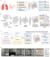
Figure 1
Figure 1
Basic concept of human lung-on-a-chip that accurately mimics structural and multicellular complexities of lung tissue. The figure details the core concept of the multi-compartmentalized lung-on-a-chip platform, illustrating the interactive multicellular communication between the respiratory airways and the adjacent supporting stromal chambers. This platform integrates various cellular elements and 3D structural complexities composed of natural polymers. A thin porous barrier embedded with vascular endothelial cells links the respiratory airway chamber with the supporting stromal chambers, enabling bidirectional cellular communication through the exchange of growth factors and cytokines. Furthermore, to accurately mimic the intricate multicellular interactions within lung tissue microenvironments, the two chambers are encompassed by media channels lined with human vascular cells (A). The 3D mold for the human lung-on-a-chip system, designed to accurately replicate the structural characteristics of the lung tissue. The mold was produced using PLA-based 3D printing technology. Polydimethylsiloxane (PDMS) was subsequently injected into the 3D printed mold, and the resulting chip platform was extracted following polymerization process (B). To replicate the physiological characteristics and multicellular diversity of lung tissue, the PDMS-based lung-on-a-chip platform was populated with multiple human lung cell types, including small airway epithelial cells, stromal cells, vascular endothelial cells, and macrophages. These cells were combined with a natural polymer mixture (hyaluronic acid and collagen), along with blood coagulating factors (thrombin and fibrinogen). The spindle-shaped respiratory airway chamber, which spans the entire chip, is populated with human small airway epithelial cells. This airway chamber is encircled by adjacent stromal chambers that house human stromal cells and macrophages, facilitating a comprehensive representation of the lung tissue microenvironment (C). The PDMS-based lung-on-a-chip platform was designed in a rectangular shape, measuring 100 mm in length, 70 mm in central diameter, and 7 mm in height. The chip platform was fabricated to replicate the microenvironment of the lung tissue and facilitate multicellular communication between the respiratory airways and adjacent supporting stromal chambers (D).
Figure 2
Figure 2
Fabrication of natural polymers-based lung tissue constructs and examination of its physical properties. The diverse cellular constituents constituting lung tissue were integrated with a combination of natural polymer, including hyaluronic acid and collagen, along with non-toxic blood coagulating factors thrombin and fibrinogen (A). The synthesized tissue architecture exhibited a refined surface, soft texture, and white coloration (B). Cross-sectional and lateral microstructural images of the tissue constructs examined by scanning electron microscopy (SEM). SEM images revealed a homogeneously dispersed porous matrix, with pores ranging from approximately 50 to 100 μm in diameter, resulting from the crosslinking of natural polymers combined with blood coagulating factors (C). The mechanical characteristics of the fabricated tissue architecture were evaluated using a universal test machine by administering single-axis compression forces. The tissue samples, each measuring 10 mm in diameter and 10 mm in height, were subjected to varying levels of uniaxial compressive stress to determine their mechanical behavior. To determine the precise failure stress of the fabricated samples, uniaxial compressive force was applied at a constant loading rate of 5 mm/min until the samples fractured (D). The dynamic viscosity of the fabricated tissue architecture was assessed across a range of shear rates from 1 to 10/s. The viscosity of the fabricated tissue architecture progressively decreased from approximately 1000 to 0 Pa-sec (E). To evaluate the swelling dynamics of tissue structures, fabricated samples were immersed in both distilled water and PBS (pH 7.4) at 37°C for 24 h. After the hydration period, the liquid was carefully removed. The tissue architectures were subsequently weighed to assess their water uptake capacity (F). All experiments were performed in triplicate. Significant differences are indicated as follows: *, p < 0.05; **, p < 0.005; and ***, p < 0.001 (two-sample t-test).
Figure 3
Figure 3
Examination of the sustained viability and metabolic functions of multiple cells embedded within each segment of the lung-on-a-chip model. The spatial arrangement of diverse cells integrated into each section of the chip was investigated by first cultivating the tissue structures in a cell-specific culture medium for 24 h, followed by staining with the DNA-targeting fluorochrome 4′,6-diamidino-2-phenylindole (DAPI). The patterns of cellular distribution across each tissue structure were assessed with by fluorescent microscopy. Different types of lung tissue cells are integrated within each compartment of the lung-on-a-chip, including small airway epithelial cells (A), stromal cells (B), vascular endothelial cells (C), and macrophages (D). Following this, the cells were cultured in specialized medium tailored for each cell type and incubated for 1, 7, 14, 21, or 28 days post-integration. Cell viability was assayed using the live & death analysis, using fluorescent dyes to distinctly mark live (green) and dead (red) cells. The prolonged viability of cells in each chamber was then assessed by fluorescent imaging. Seventy or greater percent of the cells embedded in each compartment of the chip remained viable for 28 days, as indicated by the aforementioned green and red fluorescence. Additionally, each compartment of the chip was incubated in a serum-free setting with CCK-8 solution for 48 h. The metabolic functions of the integrated cells were then evaluated by determining the optical density at 450 nm. All experiments were performed in triplicate. Significant differences are indicated as *, p < 0.05; **, p < 0.005; and ***, p < 0.001 (two-sample t-test).
Figure 4
Figure 4
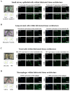
Preservation of distinct cellular characteristics in the natural polymer-based tissue architecture. An assessment was performed to determine if different cells retained their molecular properties following integration into each compartment of the chip. This involved cultivating the cells in tailored medium for a week and subsequently examining them with established biomarkers specific to each cell type. Staining was performed on human small airway epithelial cells for cytokeratin 18 and 19 (A), while lung stromal cells were examined for fibronectin and vimentin (B). Additionally, vascular endothelial cells were labeled for PECAM1 and vWF (C), and macrophages were analyzed using CD11b and CD68 (D). Each experiment was conducted three times. The nuclei in each field were stained with DAPI.
Figure 5
Figure 5
Functional assessments of the respiratory airway and adjacent stromal chambers within the chip: critical protein expression and secretion, and mucin production. To assess whether incorporated cells retained their distinctive properties within respiratory airway chamber in the chip, samples were cultured in a specific culture medium for 7 days before being evaluated with targeted biomarkers (A). Within the natural polymer-based 3D microenvironment of the chip, human small airway epithelial cells exhibited the expression of critical proteins, including MUC5AC, MUC5B, and SPDEF, at both the protein (B) and mRNA (C) levels. Periodic acid-Schiff (PAS) staining to evaluate whether embedded human airway epithelial cells within a chip platform could properly produce glycogen, mucopolysaccharides, and other carbohydrate-rich macromolecules (D). To determine whether the integrated cells preserved their unique characteristics within the stromal compartment of the chip, the samples were maintained in a designated culture medium for a week and subsequently analyzed using specific biomarkers (E). In the chip's 3D microenvironment constructed from natural polymers, human stromal cells showed expression levels of key proteins, such as COL1A1, fibronectin 1, and laminin α1, at the protein (F) and mRNA (G) levels. To evaluate if human stromal cells embedded in a chip platform were capable of adequately secreting essential proteins into the chamber, each cell-loaded and cell-free chamber was cultured in a specific medium tailored for the respective cell types for 7 days. Following this, the medium was switched to a serum-free variant. After 48 h of incubation, the medium was collected and tested for the presence of secreted fibronectin and laminin from the stromal chamber (H). All experiments were performed in triplicate. Significant differences are indicated as *p < 0.05, **p < 0.005, and ***p < 0.001 (two-sample t-test).
Figure 6
Figure 6
Discovery and confirmation of a reliable indicator to anticipating lung toxicity in human small airway epithelial cells. Depiction of the primary stages for the RNA-Seq methodology, comprising the experimental layout, alignment of read, measurement, and graphical representation (A). Comprehensive RNA-seq results displayed in a heatmap that illustrates variations in gene expression between the control groups and those exposed to low (5 ng/ml) and high (7 ng/ml) dioxin doses. The heatmap highlights genes with upregulated (red) or downregulated (green) expression relative to the mRNA levels in the control groups (B). KEGG pathway analyses to identify the potential interconnected pathways and functionalities impacted by toxin exposure (C). Within the array of genes showing differential expression, an observable positive correlation was detected between the marked elevation of SERPINB2 expression and toxic exposure in human small airway epithelial cells (D and E). Using real-time PCR and western blot analysis confirmation of increased SERPINB2 levels following exposure to both low and high doses of the toxin (F). β-Actin was used as the internal protein control, and PPIA was used as the housekeeping gene for real-time PCR. All experiments were performed in triplicate. The data are presented as the means ± SDs. *, p < 0.05; **, p < 0.005; and ***, p < 0.001 (two-sample t-test).
Figure 7
Figure 7
Validating the consistency of the selected indicator (SERPINB2) for lung toxicity in human small airway epithelial cells. Diagrammatic representation outlining how SERPINB2 regulates toxicant-triggered detrimental impacts in small airway epithelial cells (A). Human small airway epithelial cells were transfected with a specific SERPINB2 shRNA and were subsequently exposed or not exposed to dioxin (5 ng/ml) for 72 h. The detrimental impacts on cell growth were evaluated through an MTT assay (B). Depleting SERPINB2 effectively neutralized the detrimental effects of dioxin on the migration of small airway epithelial cells. This outcome was confirmed using a Transwell migration/invasion assay (C) and western blot analysis with antibodies targeting MMP-2 and MMP-9 (D). Human small airway epithelial cells underwent transfection with a targeted SERPINB2 shRNA and were either exposed or not exposed to 5 ng/ml of dioxin. Following this treatment, alterations in DNA fragmentation associated with apoptosis and caspase-3 mediated apoptotic activities were assessed through nuclear staining (E) and western blot analysis (F), respectively. β-actin was used as an internal control. All experiments were performed in triplicate. Data are presented as means ± standard deviations. *, p < 0.05; **, p < 0.005; and ***, p < 0.001 (two-sample t-test).
Figure 8
Figure 8
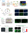
Development of a fluorescent-based detection platform linked with a toxicity detection marker in the respiratory airway compartment of the lung-on-a-chip model. A fluorescent detection platform tagged with SERPINB2 was effectively incorporated into human small airway epithelial cells. Toxin-stimulated SERPINB2 activity was evident as fluorescence of into GFP. Consequently, the lung toxicity of specific drug candidates can be assessed in both qualitative and quantitative manner through the measurement of fluorescent signal strength (A). Human small airway epithelial cells underwent stable transfection with a GFP-tagged SERPINB2 detection vector, which emits a green color. Following exposure to 5 ng/ml dioxin, immunostaining indicated a significant increase in SERPINB2 activity, which was manifest as green fluorescence in these cells (B). This fluorescence reporting platform was implemented in human small airway epithelial cells accurately positioned within the respiratory airway compartment of the lung-on-a-chip. The spatial distribution patterns of the incorporated human small airway epithelial cells within respiratory airway chamber then examined through a fluorescent imaging system (C). Exposure to 5 ng/ml dioxin markedly enhanced SERPINB2 activity, which was evident as green fluorescence within the respiratory airway compartment of the chip (D). The data are presented as the means ± standard deviations. *, p < 0.05; **, p < 0.005; and ***, p < 0.001 (two-sample t-test).
Figure 9
Figure 9
Exposure to different types of toxins triggers the activation of SERPINB2, resulting in emission of green fluorescence within the respiratory airway chamber of the chip. A GFP-tagged SERPINB2 fluorescent detection platform was effectively incorporated into human small airway epithelial cells. Subsequently, these cells were positioned in the appropriate compartments of the chip (A). To validate the consistency of the toxicity assessment marker (SERPINB2)-tagged fluorescent detection platform, toxins were added to the respiratory airway chamber of the chip. The toxins included aristolochic acid I (10 μM), benzidine (10 μM), benzo[a]pyrene (2 μM), semustine (0.5 mM), TPA (5 nM), 1,2-dichloropropane (100 mM), 1,3-butadiene (10 mM), and 4,4'-methylenebis (5 μM). The response of SERPINB2 to various types of toxic exposure was assessed both subjectively and quantitatively through the measurement of the resultant fluorescent signal strength (B). Significant differences are indicated as follows: *, p < 0.05; **, p < 0.005; and ***, p < 0.001 (two-sample t-test).

Similar articles

References

    1. Xie R, Pal V, Yu Y, Lu X, Gao M, Liang S. et al. A comprehensive review on 3D tissue models: Biofabrication technologies and preclinical applications. Biomaterials. 2024;304:122408. - PMC - PubMed
    1. Baptista D, Moreira Teixeira L, Barata D, Tahmasebi Birgani Z, King J, van Riet S. et al. 3D Lung-on-Chip Model Based on Biomimetically Microcurved Culture Membranes. ACS Biomater Sci Eng. 2022;8:2684–99. - PMC - PubMed
    1. Zamprogno P, Schulte J, Ferrari D, Rechberger K, Sengupta A, van Os L. et al. Lung-on-a-Chip Models of the Lung Parenchyma. Adv Exp Med Biol. 2023;1413:191–211. - PubMed
    1. Zamprogno P, Wuthrich S, Achenbach S, Thoma G, Stucki JD, Hobi N. et al. Second-generation lung-on-a-chip with an array of stretchable alveoli made with a biological membrane. Commun Biol. 2021;4:168. - PMC - PubMed
    1. Shah D, Dave B, Chorawala MR, Prajapati BG, Singh S, G ME. et al. An Insight on Microfluidic Organ-on-a-Chip Models for PM(2.5)-Induced Pulmonary Complications. ACS Omega. 2024;9:13534–55. - PMC - PubMed

LinkOut - more resources