

Comparison of sentinel lymph node mapping patterns before and after surgical excision of mast cell tumors in dogs using indirect lymphography: A prospective clinical study
- PMID: 39744465
- PMCID: PMC11665730
Comparison of sentinel lymph node mapping patterns before and after surgical excision of mast cell tumors in dogs using indirect lymphography: A prospective clinical study
Abstract
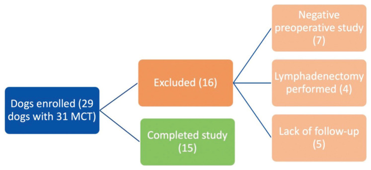
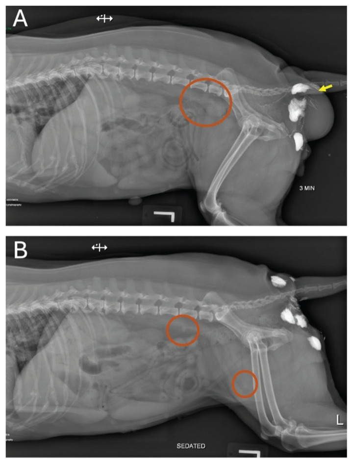
Sentinel lymph node (SLN) mapping has been shown to be important for staging in dogs with mast cell tumors (MCTs). Despite this, many patients are referred to an oncologist after surgical intervention has been carried out. It is unknown whether lymphatic drainage patterns are altered by surgery and whether postoperative SLN mapping can be reliably conducted. The objective of this study was to compare lymphatic drainage patterns from MCT sites before and after surgical removal to determine whether the SLN changes following tumor excision. Twenty-nine client-owned dogs with 31 cytologically diagnosed MCTs were prospectively enrolled, with 14 dogs (N = 15 MCTs) completing the study. Preoperative SLN mapping was conducted using radiographic indirect lymphography (IL). Water-soluble iodinated contrast (WIC) medium was injected peritumorally using a 4-quadrant technique and digital radiography was then used to assess lymphatic drainage patterns. Orthogonal projections were obtained every 1 to 2 min until the SLN was visualized, up to 20 min post-injection. Dogs were re-evaluated 2 to 5 wk postoperatively and radiographic IL was carried out again using the same protocol as previously described with WIC injected around the surgical scar line in a 4-quadrant technique. An SLN was identified for 15 MCTs in 14 dogs preoperatively and in 13/15 MCTs postoperatively. Sixteen dogs with 16 MCTs did not have postoperative lymphography and did not complete the study. Agreement between preoperative and postoperative SLNs was a complete match in 7/15 MCTs, a partial match in 5/15 MCTs, and no match in 3/15 MCTs. A negative IL study was obtained in 2/15 MCTs postoperatively. Complete agreement between preoperative and postoperative SLN identification was detected in 46.7% of cases and there was no agreement in 20% of cases. Surgical intervention did not change the time to SLN identification when carrying out radiographic IL. Thus, surgical removal of MCTs affects lymphatic drainage and can alter the SLN(s) detected. Clinicians should be aware of this finding and interpret results of postoperative lymph node staging with caution.
La cartographie des ganglions sentinelles (SLN) s’est avérée importante pour la stadification chez les chiens atteints de mastocytomes (TMC). Malgré cela, de nombreux patients sont référés à un oncologue après une intervention chirurgicale. On ne sait pas si les schémas de drainage lymphatique sont modifiés par la chirurgie et si la cartographie postopératoire des SLN peut être réalisée de manière fiable. L’objectif de cette étude était de comparer les schémas de drainage lymphatique des sites de TMC avant et après l’ablation chirurgicale afin de déterminer si le SLN change après l’excision de la tumeur. Vingt-neuf chiens appartenant à des clients et présentant 31 TMC diagnostiqués par cytologie ont été recrutés de manière prospective, 14 chiens (N = 15 TMC) ayant terminé l’étude. La cartographie préopératoire des SLN a été réalisée à l’aide d’une lymphographie indirecte radiographique (IL). Un milieu de contraste iodé hydrosoluble (WIC) a été injecté par voie péritumorale à l’aide d’une technique à 4 quadrants et une radiographie numérique a ensuite été utilisée pour évaluer les schémas de drainage lymphatique. Des projections orthogonales ont été obtenues toutes les 1 à 2 minutes jusqu’à ce que le ganglion sentinelle soit visualisé, jusqu’à 20 minutes après l’injection. Les chiens ont été réévalués 2 à 5 semaines après l’opération et une IL radiographique a été réalisée à nouveau en utilisant le même protocole que celui décrit précédemment avec du WIC injecté autour de la ligne de cicatrice chirurgicale dans une technique à 4 quadrants. Un ganglion sentinelle a été identifié pour 15 MCT chez 14 chiens avant l’opération et dans 13/15 MCT après l’opération. Seize chiens avec 16 MCT n’ont pas eu de lymphographie postopératoire et n’ont pas terminé l’étude. La concordance entre les ganglions sentinelles préopératoires et postopératoires était une correspondance complète dans 7/15 MCT, une correspondance partielle dans 5/15 MCT et aucune correspondance dans 3/15 MCT. Une étude IL négative a été obtenue dans 2/15 MCT après l’opération. Une concordance complète entre l’identification préopératoire et postopératoire du ganglion sentinelle a été détectée dans 46,7 % des cas et aucune concordance n’a été constatée dans 20 % des cas. L’intervention chirurgicale n’a pas modifié le délai d’identification du ganglion sentinelle lors de la réalisation d’une IL radiographique. Ainsi, l’ablation chirurgicale des MCT affecte le drainage lymphatique et peut modifier le ou les ganglions sentinelles détectés. Les cliniciens doivent être conscients de ce résultat et interpréter les résultats de la stadification postopératoire des ganglions lymphatiques avec prudence.(Traduit par Docteur Serge Messier).
Copyright and/or publishing rights held by the Canadian Veterinary Medical Association.
Figures
Similar articles
-
Sentinel lymph node mapping in canine mast cell tumours using a preoperative radiographic indirect lymphography: Technique description and results in 138 cases.Vet Comp Oncol. 2023 Sep;21(3):469-481. doi: 10.1111/vco.12906. Epub 2023 May 16. Vet Comp Oncol. 2023. PMID: 37191042
-
Comparison of indirect computed tomographic lymphography and near-infrared fluorescence sentinel lymph node mapping for integumentary canine mast cell tumors.Vet Surg. 2023 Apr;52(3):416-427. doi: 10.1111/vsu.13929. Epub 2022 Dec 27. Vet Surg. 2023. PMID: 36574349
-
Contrast-enhanced ultrasound for sentinel lymph node mapping in the routine staging of canine mast cell tumours: A feasibility study.Vet Comp Oncol. 2021 Sep;19(3):451-462. doi: 10.1111/vco.12647. Epub 2020 Sep 11. Vet Comp Oncol. 2021. PMID: 32840038
-
Nonselective Lymph Node Dissection and Sentinel Lymph Node Mapping and Biopsy.Vet Clin North Am Small Anim Pract. 2019 Sep;49(5):793-807. doi: 10.1016/j.cvsm.2019.04.003. Epub 2019 May 27. Vet Clin North Am Small Anim Pract. 2019. PMID: 31147188 Review.
-
The Use of Sentinel Lymph Node Mapping for Canine Mast Cell Tumors.Animals (Basel). 2024 Apr 3;14(7):1089. doi: 10.3390/ani14071089. Animals (Basel). 2024. PMID: 38612328 Free PMC article. Review.
References
-
- Welle MM, Bley CR, Howard J, Rüfenacht S. Canine mast cell tumours: A review of the pathogenesis, clinical features, pathology and treatment. Vet Dermatol. 2008;19:321–339. - PubMed
-
- Warland J, Amores-Fuster I, Newbury W, Brearley M, Dobson J. The utility of staging in canine mast cell tumours. Vet Comp Oncol. 2014;12:287–298. - PubMed
-
- Krick EL, Billings AP, Shofer FS, Watanabe S, Sorenmo KU. Cytological lymph node evaluation in dogs with mast cell tumours: Association with grade and survival. Vet Comp Oncol. 2009;7:130–138. - PubMed
-
- Stefanello D, Buracco P, Sabattini S, et al. Comparison of 2- and 3-category histologic grading systems for predicting the presence of metastasis at the time of initial evaluation in dogs with cutaneous mast cell tumors: 386 cases (2009–2014) J Am Vet Med Assoc. 2015;246:765–769. - PubMed
-
- Sledge DG, Webster J, Kiupel M. Canine cutaneous mast cell tumors: A combined clinical and pathologic approach to diagnosis, prognosis, and treatment selection. Vet J. 2016;215:43–54. - PubMed
MeSH terms
LinkOut - more resources
Full Text Sources