. 2025 Jan 1;16(2):368-381.
doi: 10.7150/jca.100846. eCollection 2025.

Ultrasound irradiation in the presence of microbubbles may enhance the antitumor effect of chemotherapeutic agents against bladder cancer

Affiliations

Ultrasound irradiation in the presence of microbubbles may enhance the antitumor effect of chemotherapeutic agents against bladder cancer

Takehiro Suzuki et al. J Cancer. .

Abstract

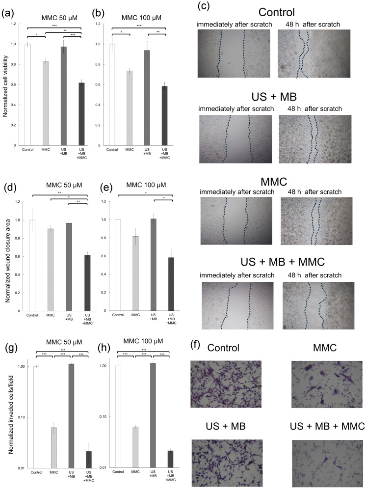
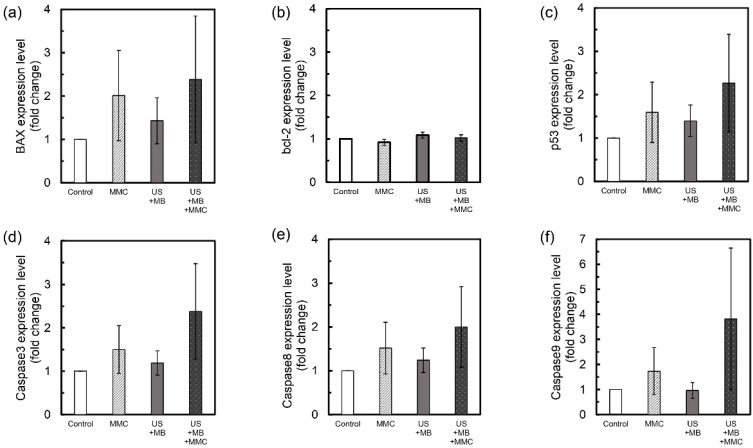
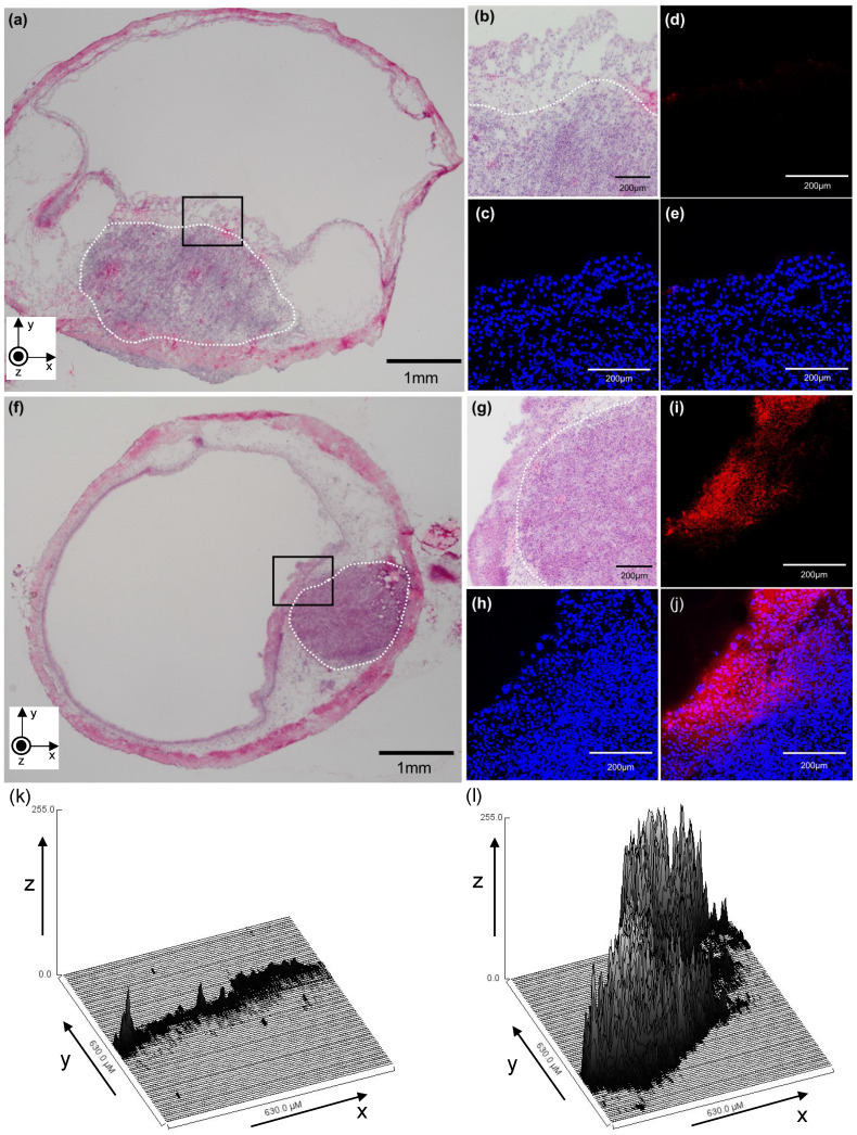
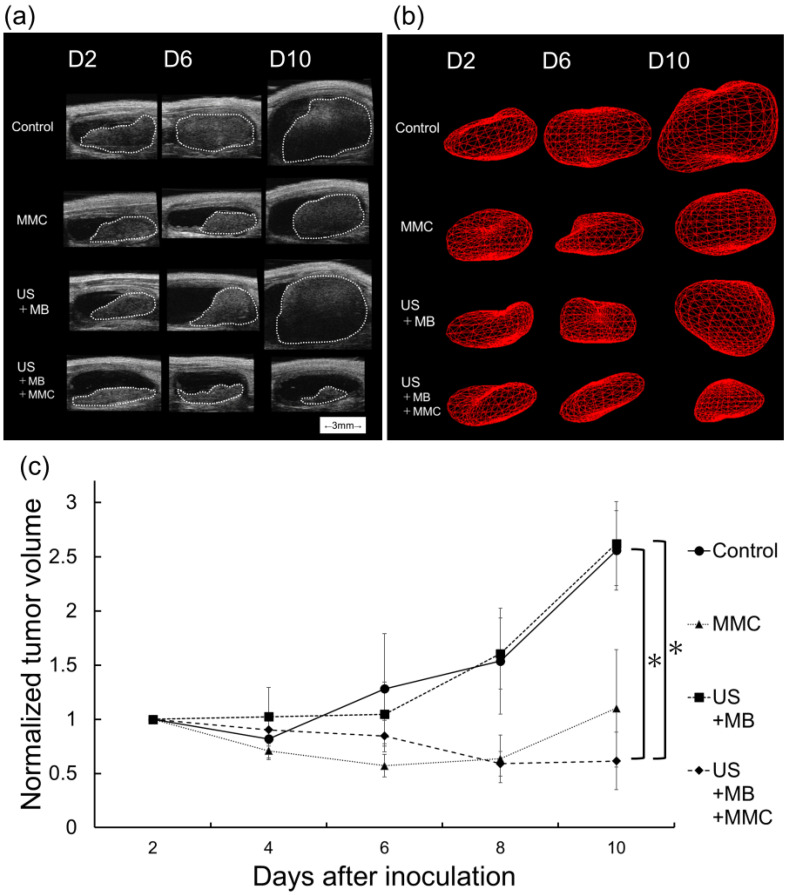
Intravesical instillation of chemotherapy has been performed to reduce the risk of intravesical recurrence of bladder cancer. However, its antitumor effect is not necessarily sufficient, which may be partially due to inadequate delivery of intravesically administered chemotherapeutic agents to bladder tumors. Ultrasound irradiation to target tissues in the presence of microbubbles is a technique to transiently enhance cell membrane permeability and achieve efficient drug delivery to the desired sites without damage to non-target areas; this technique has been used in chemotherapy, immunotherapy, gene therapy, and radiotherapy for the treatment of various cancers. However, the effectiveness of combining intravesical instillation of chemotherapy and this strategy for the treatment of bladder cancer has not been fully investigated. This report shows that mitomycin C combined with ultrasound and microbubbles has a higher antitumor effect than mitomycin C alone against mouse bladder cancer cells. Next, the antitumor effect of intravesical instillation of chemotherapy combined with ultrasound and microbubbles was demonstrated using an orthotopic mouse bladder cancer model. In vivo experiments showed that ultrasound irradiation in the presence of microbubbles enhanced the local delivery of fluorescent molecules and had the potential to enhance the antitumor effect of intravesical instillation of chemotherapy without visible damage to the surrounding normal tissues. The results of the present study demonstrate that intravesical chemotherapy combined with ultrasound and microbubbles is potentially a safe and effective treatment for bladder cancer.

Keywords: bladder cancer; drug delivery; intravesical instillation of chemotherapy; sonoporation.


Conflict of interest statement

Competing Interests: The authors have declared that no competing interest exists.

Figures

Figure 1
Figure 1
In vitro experiments of cell viability, cell migration, and cell invasion. To obtain normalized cell viability, normalized wound closure area (migration), and normalized invaded cells/field (invasion), the value in each group was divided by the mean of the control samples. Panels (a) and (b) represent normalized cell viability of MBT-2 cells measured with the 3-(4,5-dimethylthiazol-2-yl)-2,5-diphenyltetrazolium bromide (MTT) assay, at mitomycin C (MMC) concentrations of 50 μM and 100 μM. Panel (c) displays bright-field images of each group in the migration assay. Images from immediately and at 48 h after the scratch are shown. Panels (d) and (e) display the normalized wound closure area of MBT-2 cells at MMC concentrations of 50 μM and 100 μM. Panel (f) presents microscopic images of each group in the invasion assay. Panels (g) and (h) present normalized invaded cells/field of MBT-2 cells at MMC concentrations of 50 μM and 100 μM. Significant differences in normalized cell viability and normalized wound closure area are found between the MMC and US + MB + MMC groups at MMC concentration of 50 μM. Mean ± SEM values are shown. *P < 0.05. **P < 0.01. ***P < 0.001.
Figure 2
Figure 2
Expression levels of apoptosis-related genes. The mean fold-change in the expression level of each gene, relative to the control group, was measured for the US + MB, MMC, and US + MB + MMC groups (n = 6 for each group). Expression levels of BAX (a), bcl-2 (b), p53 (c), caspase3 (d), caspase8 (e) and caspase9 (f) genes in MBT-2 cells are shown. There is a trend towards higher expression of BAX, p53, caspase3, caspase8, and caspase9 genes in the US + MB + MMC group compared with the other 3 groups. Mean ± SEM values are shown.
Figure 3
Figure 3
A representative confocal image (cross-section) of a bladder in which TOTO-3 fluorescence molecules were delivered using microbubbles (MBs) and ultrasound (US). (a-e) MBs and TOTO-3 without US irradiation. (f-j) MBs and TOTO-3 with US irradiation. (a) A section stained with hematoxylin and eosin (HE). The white dotted line indicates a carcinomatous lesion. (b) An HE-stained section of the bladder (black box) shown in (a) with higher magnification. (c) Nuclei of bladder cells (including MBT-2 cells) were stained with 4′,6-diamidino-2-phenylindole (DAPI). (d) TOTO-3 (red) fluorescence shows the location where the molecules are delivered. (e) Merged image. (f) HE-stained section. The white dotted line indicates a carcinomatous lesion. (g) An HE-stained section of the bladder (black box) shown in (f) with higher magnification. (h) DAPI staining. (i) TOTO-3 (red) fluorescence shows the location where the molecules are delivered. (j) Merged image. (k and l) The distribution of red signal intensity converted into 8-bit grayscale values is shown in a three-dimensional plot, where the x-axis and y-axis correspond to the x-y plane shown in (d) and (i), respectively, and the z-axis shows the mean grayscale intensity of the fluorescent molecule at each point in the region of interest.
Figure 4
Figure 4
Evaluation of treatment efficacy in the experimental bladder cancer model. (a) Bladder imaging from the control, US + MB, MMC, and US + MB + MMC groups acquired using a high-frequency ultrasound imaging system for each group on days, 2, 6, and 10. The white dotted line indicates a carcinomatous lesion. (b) 3D reconstructed image of the bladder tumor. (c) Longitudinal analysis of normalized tumor volume from the four groups described above (n = 6 for each group). Values at different time points were normalized to those on day 2. Mean ± SEM values are shown. *P < 0.05.
Figure 5
Figure 5
Histopathological analysis. Sections from each group stained with hematoxylin and eosin. (a) Control, (b) MMC, (c) US + MB, and (d) US + MB + MMC. Panels (e), (f), (g), and (h) show representative microscopic images with higher magnification of panels (a), (b), (c), and (d) (yellow boxes), respectively. The black dotted lines indicate carcinomatous lesions. In the control and US + MB groups, widespread cancerous lesions are observed, whereas relatively small lesions are observed in sections from the MMC and US + MB + MMC groups.

Similar articles

References

    1. Clark PE, Agarwal N, Biagioli MC. et al. Bladder cancer. J Natl Compr Canc Netw. 2013;11:446–75. - PubMed
    1. van Rhijn BW, Burger M, Lotan Y. et al. Recurrence and progression of disease in non-muscle-invasive bladder cancer: from epidemiology to treatment strategy. Eur Urol. 2009;56:430–42. - PubMed
    1. Lenis AT, Lec PM, Chamie K. et al. Bladder cancer: a review. JAMA. 2020;324:1980–91. - PubMed
    1. Sylvester RJ. Natural history, recurrence, and progression in superficial bladder cancer. Sci World J. 2006;6:2617–25. - PMC - PubMed
    1. Sato T, Sano T, Kawamura S. et al. Improving compliance with guidelines may lead to favorable clinical outcomes for patients with non-muscle-invasive bladder cancer: A retrospective multicenter study. Int J Urol. 2023;12:1155–63. - PMC - PubMed

LinkOut - more resources