. 2024 Nov 6;5(1):1048-1059.
doi: 10.1089/neur.2024.0098. eCollection 2024.

Brain Network Alterations in Chronic Spinal Cord Injury: Multilayer Community Detection Approach

Affiliations

Brain Network Alterations in Chronic Spinal Cord Injury: Multilayer Community Detection Approach

Farzad V Farahani et al. Neurotrauma Rep. .

Abstract

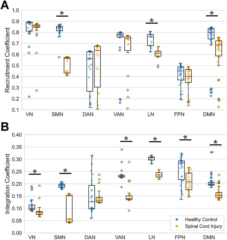
Neurological recovery in individuals with spinal cord injury (SCI) is multifaceted, involving mechanisms such as remyelination and perilesional spinal neuroplasticity, with cortical reorganization being one contributing factor. Cortical reorganization, in particular, can be evaluated through network (graph) analysis of interregional functional connectivity. This study aimed to investigate cortical reorganization patterns in persons with chronic SCI using a multilayer community detection approach on resting-state functional MRI data. Thirty-eight participants with chronic cervical or thoracic SCI and 32 matched healthy controls were examined. Significant alterations in brain community structures were observed in the SCI cohort, particularly within the sensorimotor network (SMN). Importantly, this revealed a pattern of segregation within the SMN, aligning with borders of representations of the upper and lower body and orofacial regions. The SCI cohort showed reduced recruitment and integration coefficients across multiple brain networks, indicating impaired internetwork communication that may underlie sensory and motor deficits in persons with SCI. These findings highlight the impact of SCI on brain connectivity and suggest potential compensatory mechanisms.

Keywords: cortical reorganization; functional connectivity; graph theory; mesoscale; rs-fMRI; spinal cord injury.


Figures

FIG. 1.
FIG. 1.
Flowchart illustrating the data analysis process. The flowchart illustrates the utilization of preprocessed rs-fMRI data to compute mesoscale graph measures. (A) Functional connectivity matrix is computed by partitioning the preprocessed fMRI data into 200 cortical regions assigned to seven networks. (B) Multilayer community detection algorithm is employed to investigate the modular organization of the brain networks. (C–F) A toy example illustrates how integration and recruitment coefficients are derived. The example features nine cortical regions (nodes 1–9) and three functional networks (S1, S2, S3) across t study participants. (C) Multilayer community detection algorithm is used to assign brain regions to three communities (C1, C2, C3) across different study participants (layers defined as study participants: L1, L2, Lt). (D) Co-occurrence matrices are constructed for each participant, where elements indicate shared community labels between nodes. (E) The average of all co-occurrence matrices across participants creates the module allegiance matrix. (F) Integration and recruitment coefficients are calculated to measure the consistency of region inclusion in the same functional network and interaction with other networks, respectively. rs-fMRI, resting-state functional MRI.
FIG. 2.
FIG. 2.
Group differences in module allegiance between healthy controls (HC) and spinal cord injury (SCI) cohorts. Panels illustrate (A) HC cohort module allegiance matrix, (B) SCI cohort module allegiance matrix, (C) significant p values for cohort differences, and (D) detailed view focusing on right-hemisphere nodes in the SCI cohort. The SCI cohort’s module allegiance matrix reveals that the parcels within the sensorimotor network (SMN) segregate into two clusters, marked by decreased module allegiance values between the clusters. These clusters are color-coded and labeled as SMN-1 (cyan) and SMN-2 (lime). L, left hemisphere; R, right hemisphere.
FIG. 3.
FIG. 3.
Recruitment and integration coefficients across networks in healthy controls (HC) and spinal cord injury (SCI) cohorts. (A) Box plot of recruitment coefficients in HC and SCI cohorts, with asterisks denoting statistically significant differences (p < 0.001, FDR corrected) for some networks. (B) Box plot of integration coefficients in HC and SCI cohorts, similarly annotated. Networks represented include visual (VN), sensorimotor (SMN), dorsal attention (DAN), salience/ventral attention (VAN), limbic (LN), frontoparietal (FPN), and default mode (DMN) networks. FDR, false discovery rate.
FIG. 4.
FIG. 4.
Visualization of parcels with significant group differences in recruitment and integration coefficients between healthy controls (HC) and spinal cord injury (SCI) cohorts (A) Visualization of parcels with significantly different recruitment coefficients in the SCI cohort compared to the HC cohort (HC-SCI; p > 0.001, FDR corrected). Circle sizes represent the magnitude of differences. (B) Scatter plot comparing parcels based on SCI and HC recruitment coefficients. The diagonal dotted line indicates the identity line. Parcels below this line show significantly lower recruitment coefficients in the SCI cohort compared to the HC cohort. (C) Visualization of parcels with significant group differences in integration coefficients between the HC and SCI cohorts (HC-SCI; p > 0.001, FDR corrected). (D) Scatter plot comparing parcels based on SCI and HC integration coefficients. Networks represented include visual (VN), sensorimotor (SMN), dorsal attention (DAN), salience/ventral attention (VAN), limbic (LN), frontoparietal (FPN), and default mode (DMN) networks. FDR, false discovery rate.

Similar articles

References

    1. Blight AR. Cellular morphology of chronic spinal cord injury in the cat: Analysis of myelinated axons by line-sampling. Neuroscience 1983;10(2):521–543; doi: 10.1016/0306-4522(83)90150-1 - DOI - PubMed
    1. Nathan PW. Effects on movement of surgical incisions into the human spinal cord. Brain 1994;117(Pt 2):337–346; doi: 10.1093/brain/117.2.337 - DOI - PubMed
    1. Metz GA, Curt A, van de Meent H, et al. . Validation of the weight-drop contusion model in rats: A comparative study of human spinal cord injury. J Neurotrauma 2000;17(1):1–17; doi: 10.1089/neu.2000.17.1 - DOI - PubMed
    1. Basso DM. Neuroanatomical substrates of functional recovery after experimental spinal cord injury: Implications of basic science research for human spinal cord injury. Phys Ther 2000;80(8):808–817. - PubMed
    1. Dietz V. Behavior of spinal neurons deprived of supraspinal input. Nat Rev Neurol 2010;6(3):167–174; doi: 10.1038/nrneurol.2009.227 - DOI - PubMed

LinkOut - more resources