Review
. 2024 Dec 18:15:1485174.
doi: 10.3389/fimmu.2024.1485174. eCollection 2024.

Tumor microenvironment in oral squamous cell carcinoma

Affiliations
Review

Tumor microenvironment in oral squamous cell carcinoma

Chenxi Li et al. Front Immunol. .

Abstract

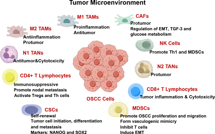
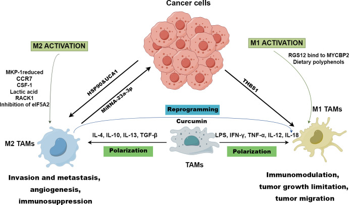
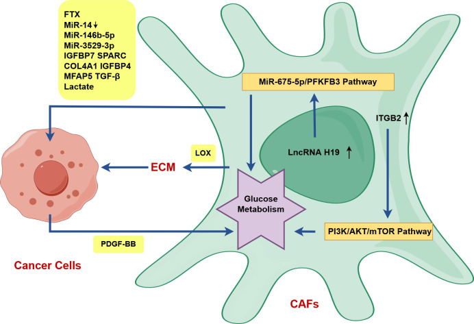
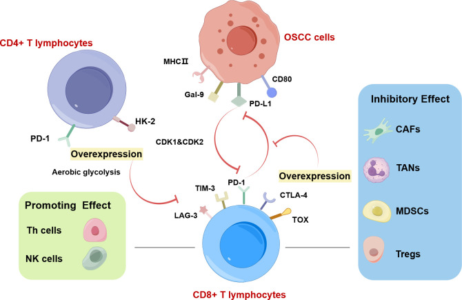
Oral squamous cell carcinoma (OSCC) is a highly aggressive and malignant tumor of oral cavity with a poor prognosis and high mortality due to the limitations of existing therapies. The significant role of tumor microenvironment (TME) in the initiation, development, and progression of OSCC has been widely recognized. Various cells in TME, including tumor-associated macrophages (TAMs), cancer-associated fibroblasts (CAFs), T lymphocytes, tumor-associated neutrophils (TANs), myeloid-derived suppressor cells (MDSCs) and dendritic cells (DCs), form a complicated and important cellular network to modulate OSCC proliferation, invasion, migration, and angiogenesis by secreting RNAs, proteins, cytokines, and metabolites. Understanding the interactions among cells in TME provides the foundation for advanced clinical diagnosis and therapies. This review summarizes the current literature that describes the role of various cellular components and other TME factors in the progression of OSCC, hoping to provide new ideas for the novel OSCC treatment strategies targeting the complicated cellular network and factors that mediate the interactive loops among cells in TME.

Keywords: cellular network; interaction; oral squamous cell carcinoma; tumor microenvironment; tumor-associated macrophages.


Conflict of interest statement

The authors declare that the research was conducted in the absence of any commercial or financial relationships that could be construed as a potential conflict of interest.

Figures

Figure 1
Figure 1
TME in OSCC drawn by Figdraw. Components in TME including cancer-associated fibroblasts (CAFs), myeloid-derived suppressor cells (MDSCs), T lymphocytes, tumor-associated neutrophils (TANs), tumor-associated macrophages (TAMs), and cytokines, are all intricately interconnected with the angiogenesis, proliferation, invasion, and migration of OSCC.
Figure 2
Figure 2
Polarization of TAMs drawn by Figdraw. M1 TAMs polarization is driven by LPS, IFN-γ, TNF-α, IL-12, and IL-18, while IL-4, IL-10, IL-13, and TGF-β promote macrophages towards M2 polarized state. RGS12 binds to MYCBP2 to degrade KIF2A in TAMs driving TAMs to polarize towards the M1 phenotype. However, CCR7, CSF-1, lactic acid, RACK1, reduced MKP-1, and inhibition of eIF5A2 contribute to the genesis of M2 TAMs. M1 TAMs are conducive to immunomodulation, tumor growth limitation, and tumor migration, while M2 TAMs foster invasion and metastasis, angiogenesis, and immunosuppression in OSCC. The TME prompts the polarization of M1 TAMs through cancer-derived exosomal THBS1, while exosomal UCA1 and HSP-90 guide TAMs towards the M2 phenotype polarization. Exosomal miRNA-23a-3p derived from M2 TAMs promotes the proliferation and invasion of OSCC cells and inhibits cancer cell apoptosis. In immunotherapies, and curcumin can reprogram M2 TAMs to M1 TAMs to inhibit OSCC.
Figure 3
Figure 3
CAFs activate OSCC cells in OSCC drawn by Figdraw. ITGB2 expression in CAFs can improve the glycolysis ability of CAFs by activating the PI3K/AKT/mTOR axis, and upregulated lncRNA H19 in CAFs can promote glucose metabolism via the lncRNA H19/miR-675-5p/PFKFB3 signaling cascade. PDGF-BB-induced glucose metabolism reprogramming in CAFs promotes the proliferation, invasion and metastasis of tongue squamous cell carcinoma (TSCC) via the secretion of lactates. CAFs can directly activate OSCC cells and promote OSCC progression by secreting RNAs, proteins, TGF-β and lactate (FTX, decreased miR-14, miR-146b-5p, miR-3529-3p, IGFBP7, SPARC, COL4A1, IGFBP4, MFAP5, TGF-β, lactate). In particular, CAFs indirectly activate OSCC cells via LOX-mediated the remodeling of stromal collagen microenvironment.
Figure 4
Figure 4
CD8+ T lymphocytes in OSCC drawn by Figdraw. Increased PD-1 levels and glycolysis in CD4+ T cells are positively associated with lymph node metastasis of OSCC. The interaction between CD8+ T cells and cancer cells is regulated by immune cells in TME. Among them, CAFs, TANs, MDSCs, and Tregs exhibit inhibitory effects on CD8+ T lymphocytes. Conversely, NK cells and Th cells play a supportive role in promoting CD8+ T lymphocyte function. The highly expressed T lymphocyte proliferation regulator-related genes (RANs) (CDK1 and CDK2) in malignant cells and T cells, can inhibit the function of CD8+ T cells. CD8+ T cells overexpressing PD-1 or TOX in OSCC have low cytotoxicity and weak ability to proliferate, which is associated with aerobic glycolysis. Exhausted CD8+ T cells are characterized by upregulated immunosuppressive checkpoints including PD-1, TIM-3, and CTLA-4.

Similar articles

Cited by

References

    1. Sung H, Ferlay J, Siegel RL, Laversanne M, Soerjomataram I, Jemal A, et al. . Global cancer statistics 2020: GLOBOCAN estimates of incidence and mortality worldwide for 36 cancers in 185 countries. CA Cancer J Clin. (2021) 71:209–49. doi: 10.3322/caac.21660 - DOI - PubMed
    1. Johnson DE, Burtness B, Leemans CR, Lui VWY, Bauman JE, Grandis JR. Head and neck squamous cell carcinoma. Nat Rev Dis Primers. (2020) 6:92. doi: 10.1038/s41572-020-00224-3 - DOI - PMC - PubMed
    1. Shen Y, Chen Y, Lin Y, Li Y, Liu P, Zhang B, et al. . CDK5RAP2 is a Wnt target gene and promotes stemness and progression of oral squamous cell carcinoma. Cell Death Dis. (2023) 14:107. doi: 10.1038/s41419-023-05652-z - DOI - PMC - PubMed
    1. Chandralekha Selvakumar S, Auxzilia Preethi K, Sekar D. MicroRNAs and cancer-associated fibroblasts in the tumour microenvironment of oral squamous cell carcinoma (OSCC). Oral Oncol. (2022) 134:106124. doi: 10.1016/j.oraloncology.2022.106124 - DOI - PubMed
    1. Chen SH, Hsiao SY, Chang KY, Chang JY. New insights into oral squamous cell carcinoma: from clinical aspects to molecular tumorigenesis. Int J Mol Sci. (2021) 22:2252. doi: 10.3390/ijms22052252 - DOI - PMC - PubMed

MeSH terms

LinkOut - more resources