. 2024 Dec 18:18:1481963.
doi: 10.3389/fncel.2024.1481963. eCollection 2024.

Unraveling the SARS-CoV-2 spike protein long-term effect on neuro-PASC

Affiliations

Unraveling the SARS-CoV-2 spike protein long-term effect on neuro-PASC

Filipe Menezes et al. Front Cell Neurosci. .

Abstract

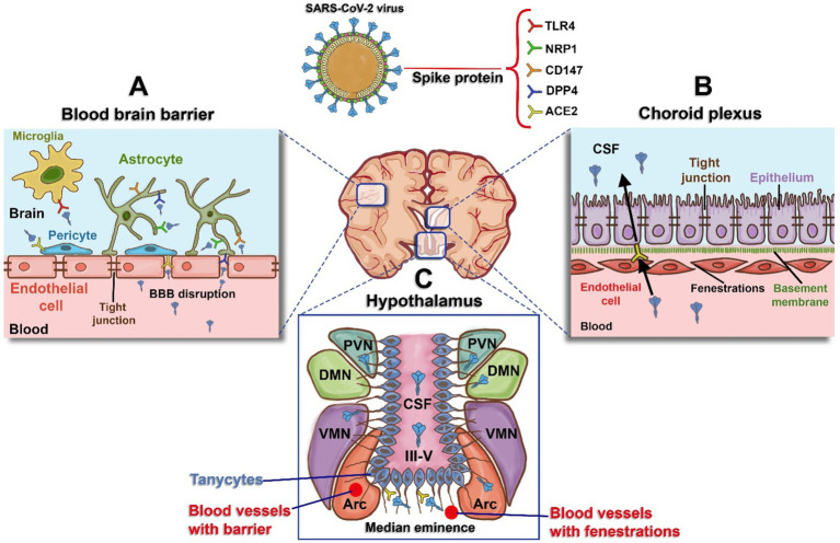
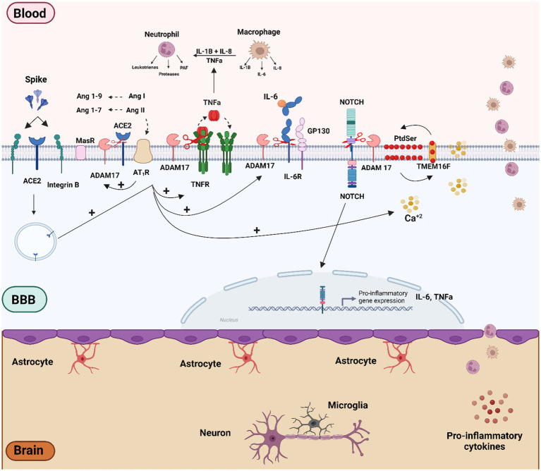
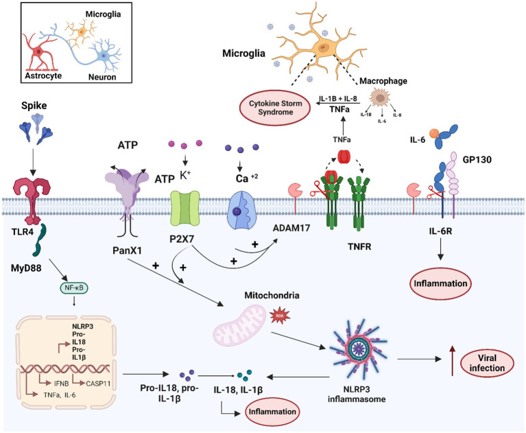
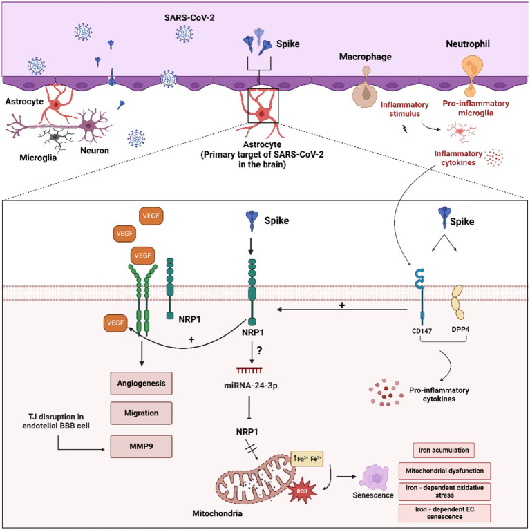
The persistence or emergence of long-term symptoms following resolution of primary SARS-CoV-2 infection is referred to as long COVID or post-acute sequelae of COVID-19 (PASC). PASC predominantly affects the cardiovascular, neurological, respiratory, gastrointestinal, reproductive, and immune systems. Among these, the central nervous system (CNS) is significantly impacted, leading to a spectrum of symptoms, including fatigue, headaches, brain fog, cognitive impairment, anosmia, hypogeusia, neuropsychiatric symptoms, and peripheral neuropathy (neuro-PASC). However, the risk factors and pathogenic mechanisms responsible for neuro-PASC remain unclear. This review hypothesis discusses the leading hypotheses regarding the pathophysiological mechanisms involved in long COVID/PASC, focusing on neuro-PASC. We propose vascular dysfunction mediated by activation of astrocytes and pericytes followed by blood-brain barrier (BBB) disruption as underlying pathophysiological mechanisms of neurological manifestations. Additionally, we provide insights into the role of spike protein at the blood-brain interface. Finally, we explore the potential pathogenic mechanisms initiated by the interaction between the spike protein and cellular receptors at the brain endothelial and tissue levels.

Keywords: SARS-CoV-2 receptors; SARS-CoV-2 spike protein; blood–brain barrier; neuro-PASC; pathophysiology.


Conflict of interest statement

The authors declare that the research was conducted in the absence of any commercial or financial relationships that could be construed as a potential conflict of interest.

Figures

Figure 1
Figure 1
Schematic representation of blood and the brain regions interface and receptors expressed for the spike protein. (A) Blood–Brain Barrier (BBB): The BBB is formed by tight junctions in endothelial cells. The spike protein induces damage to these tight junctions, resulting in BBB disruption, increased permeability, and access to receptors present in astrocytes (human dipeptidyl peptidase - DPP4, Neuropilin-1 - NRP1, and Cluster of Differentiation - CD147), pericytes (Angiotensin-Converting Enzyme 2 - ACE2), and microglia (Toll-like Receptor 4 - TLR4). (B) Choroid Plexus: Specialized tissue located in the wall of the fourth ventricle, composed of endothelial cells that are more permeable than those of the BBB, with gaps known as fenestrations. This enables easier movement for the epithelium on the apical side, which expresses ACE2, and this acts as a potential entry route for spike protein into the cerebrospinal fluid (CSF). (C) Hypothalamus: Hypothalamic tanycytes can be observed around the third ventricle (III-V), expressing ACE2 and transferring spike protein to the regions of the arcuate nucleus (ARC), ventromedial nucleus (VMN), dorsomedial nucleus (DMN), and periventricular nucleus (PVN). Figure created with BioRender.com.
Figure 2
Figure 2
Schematic overview of the molecular mechanisms related to SARS-CoV-2 and S protein effect on vascular dysfunction and BBB disruption. Vascular cells and pericytes of the BBB can be activated by SARS-CoV-2 infection or by the binding of the spike protein to ACE2 and b-integrin receptors. The imbalance of the RAS pathway mediated by ACE2 downmodulation can lead to activation of the Ang II/AT1R axis. Both AT1R activation and viral infection itself can trigger several intracellular signaling, such as Ca2+ influx and activation of TMEM16F-scramblase and externalization of PtdSer to the outer cell membrane, culminating in the activation of ADAM17. The ADAM17 sheddase activity, together with AT1R-mediated RAGE activation may play a preponderant role in the inflammatory process by activating inflammatory factors such as NOTCH and NF-kB proteins, which in turn can lead to the production of pro-inflammatory factors such as TNF-α, IL-6, R-IL6, IL1-β. Figure created with BioRender.com.
Figure 3
Figure 3
Schematic overview of the molecular mechanisms related to microglia activation by the SARS-CoV-2 and S protein. Microglial cells can mainly be activated by SARS-CoV-2/S spike protein through the TLR4-signaling pathway. In addition, the S protein enhances activity of the ATP+ ion channel, and PANX1 and purinergic receptor P2X7/K+, which together with calcium influx can lead to ADAM17 activation and mitochondrial dysfunction. Both TLR4 and PANX1/P2X7 activation can lead to activation of NF-Kb transcription factor and inflammasome system activation. These mechanisms together lead to the production of pro-inflammatory cytokines such as TNF-α, IL-6, RIL-6, IL1-β and IL-18, involved in the cytokine storm and the increase of viral infection. Figure created with BioRender.com.
Figure 4
Figure 4
Schematic overview of the molecular mechanisms related to astrocytes activation by the SARS-CoV-2 and S protein. “Astrogliosis” astrocyte activation can be mediated by interplay between CD147/DPP4 and NRP1 receptors and SARS-CoV-2 and S protein. CD147/DPP4 activation may enhance proinflammatory cytokines, VEGF production and NRP1 activation. Furthermore, VEGF-VEGF-R2/NRP1 is able to induce angiogenesis, migration and disruption of TJ proteins in BBB capillary endothelial cells. In addition, excess VEGF can activate the ADAM9/10 metalloproteinases with the consequent release of the extracellular domain of NRP1 and cytoplasmic tail, inhibiting the VEGF-NRP1 signaling pathway in a negative feedback fashion. Figure created with BioRender.com.

Similar articles

References

    1. Aboudounya M. M., Heads R. J. (2021). COVID-19 and toll-like receptor 4 (TLR4): SARS-CoV-2 May bind and activate TLR4 to increase ACE2 expression, facilitating entry and causing hyperinflammation. Mediat. Inflamm. 2021, 8874339–8874318. doi: 10.1155/2021/8874339, PMID: - DOI - PMC - PubMed
    1. Ackermann M., Verleden S. E., Kuehnel M., Haverich A., Welte T., Laenger F., et al. . (2020). Pulmonary vascular Endothelialitis, thrombosis, and angiogenesis in COVID-19. N. Engl. J. Med. 383, 120–128. doi: 10.1056/NEJMoa2015432, PMID: - DOI - PMC - PubMed
    1. Adingupu D. D., Soroush A., Hansen A., Twomey R., Dunn J. F. (2023). Brain hypoxia, neurocognitive impairment, and quality of life in people post-COVID-19. J. Neurol. 270, 3303–3314. doi: 10.1007/s00415-023-11767-2, PMID: - DOI - PMC - PubMed
    1. Agrawal S., Farfel J. M., Arfanakis K., Al-Harthi L., Shull T., Teppen T. L., et al. . (2022). Brain autopsies of critically ill COVID-19 patients demonstrate heterogeneous profile of acute vascular injury, inflammation and age-linked chronic brain diseases. Acta Neuropathol. Commun. 10:186. doi: 10.1186/s40478-022-01493-7, PMID: - DOI - PMC - PubMed
    1. Akwii R. G., Sajib M. S., Zahra F. T., Mikelis C. M. (2019). Role of Angiopoietin-2 in vascular physiology and pathophysiology. Cells 8:471. doi: 10.3390/cells8050471, PMID: - DOI - PMC - PubMed

LinkOut - more resources