. 2024 Dec 18:11:1483421.
doi: 10.3389/fvets.2024.1483421. eCollection 2024.

Bacterial attachment and junctional transport function in induced apical-out polarized and differentiated canine intestinal organoids

Affiliations

Bacterial attachment and junctional transport function in induced apical-out polarized and differentiated canine intestinal organoids

Shino Yoshida et al. Front Vet Sci. .

Abstract

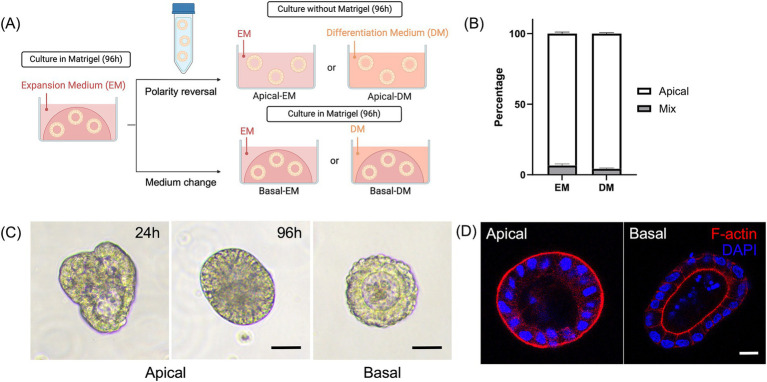
Dogs are increasingly recognized as valuable large animal models for understanding human intestinal diseases, as they naturally develop conditions similar to those in humans, such as Enterohemorrhagic E. coli, Clostridium difficile infection, inflammatory bowel disease, and ulcerative colitis. Given the similarity in gut flora between dogs and humans, canine in vitro intestinal models are ideal for translational research. However, conventional extracellular matrix-embedded organoids present challenges in accessing the lumen, which is critical for gut function. This study aimed to investigate the feasibility of inducing polarity reversal and differentiation in canine apical-out colonic organoids (colonoids), evaluate their barrier integrity, and visualize host-pathogen interactions. Our results demonstrated successful polarity reversal and differentiation induction while maintaining barrier integrity. Polarity reversal allowed for enhanced observation of host-pathogen interactions, facilitating visual assessments and membrane integrity evaluations using both pathogenic and nonpathogenic E. coli. This process led to the downregulation of stem cell marker LGR5 and upregulation of intestinal epithelial cell marker ALPI, indicating differentiation. Further differentiation was observed with the use of a differentiation culture medium, resulting in significant upregulation of ALPI and goblet cell marker MUC2. The findings suggest that apical-out canine colonoids can serve as physiologic and valuable models for studying the pathogenic mechanisms and clinical significance of intestinal diseases in dogs. This model has the potential to advance both canine and human gastrointestinal research, enhancing our understanding of gastrointestinal physiology and pathology and aiding in the development of novel therapeutics.

Keywords: apical-out; canine; differentiation; host-pathogen interaction; intestine; organoid; polarity reversal.


Conflict of interest statement

The authors declare that the research was conducted in the absence of any commercial or financial relationships that could be construed as a potential conflict of interest.

Figures

Figure 1
Figure 1
Development and characterization of the canine apical-out colonoids. (A) A schematic for polarity reversal of colonoids. (B) The percentage of apical-out colonoids (Apical, white box) and colonoids with mixed polarities (Mix, gray box) under confocal microscopy at 96 h (n = 3, each). Data is shown as mean ± standard error of the mean (SEM). 40–60 randomly selected organoids in each biological replicate were counted and classified as apical-out, basal-out, or mixed polarity. (C) Representative images from the phase-contrast microscope at 24 and 96 h. Scale bar = 25 μm. (D) Confocal microscopy images of an apical-out colonoid and Matrigel-embedded colonoid at 96 h with visualization of apical brush borders (F-actin, red) and basal nuclei (DAPI, blue). Scale bar = 10 μm.
Figure 2
Figure 2
Characterization of apical-out colonoids. (A) Comparison of relative gene expression levels of the following markers: LGR5 (stem cells), ALPI (Intestinal epithelial cells), MUC2 (goblet cells), and CHGA (enteroendocrine cells) of apical-out colonoids in EM (white box) versus DM (gray box). Data is shown as mean ± SEM. p < 0.05 was considered statistically significant using the Willcoxon rank-sum test, followed by Bonferroni correction. *p < 0.05; **p < 0.01, ***p < 0.001. (B) Confocal microscopy images of apical-out colonoids at 96 h in DM with visualization of apical brush borders (F-actin, red) and basal nuclei (DAPI, blue), chromogranin A (CHGA, yellow), and Sambucus nigra lectin (SNA, green). Scale bar = 10 μm.
Figure 3
Figure 3
The evaluation of barrier integrity using FITC-dextran assay at 96 h. (A) Representative image of untreated Apical-DM and EDTA-treated Apical-DM. Scale bar = 50 μm. (B) FITC permeability ratio of untreated (Control, white dots) and EDTA-treated Apical-DM (black dots). The median is shown as a horizontal line. For image acquisition, randomly selected 6–15 fields were used in each biological replicate. p < 0.05 was considered statistically significant using the Willcoxon rank-sum test. ***p < 0.001.
Figure 4
Figure 4
Host-pathogen interaction. (A) A representative confocal microscopy image of apical-DM infected with nonpathogenic E. coli (YFP-tagged, green) with visualization of apical brush borders (F-actin, red) and basal nuclei (DAPI, blue). Scale bar = 10 μm. (B) In barrier integrity assay, representative images of apical-out colonoids infected with nonpathogenic E. coli or EHEC. Scale bar = 50 μm. (C) FITC permeability ratio of untreated (Control, white dots), apical-out colonoids infected with nonpathogenic E. coli (gray dots), or EHEC (black dots). The median is shown as a horizontal line. For image acquisition, randomly selected 6–12 fields were used in each biological replicate. p < 0.05 was considered statistically significant using the Willcoxon rank-sum test. ***p < 0.001.

Similar articles

  • Canine intestinal organoids as a platform for studying MHC class II expression in epithelial cells.
    Nakazawa M, Nagao I, Ambrosini YM. Nakazawa M, et al. BMC Mol Cell Biol. 2025 Apr 8;26(1):11. doi: 10.1186/s12860-025-00536-w. BMC Mol Cell Biol. 2025. PMID: 40200149 Free PMC article.
  • Management of urinary stones by experts in stone disease (ESD 2025).
    Papatsoris A, Geavlete B, Radavoi GD, Alameedee M, Almusafer M, Ather MH, Budia A, Cumpanas AA, Kiremi MC, Dellis A, Elhowairis M, Galán-Llopis JA, Geavlete P, Guimerà Garcia J, Isern B, Jinga V, Lopez JM, Mainez JA, Mitsogiannis I, Mora Christian J, Moussa M, Multescu R, Oguz Acar Y, Petkova K, Piñero A, Popov E, Ramos Cebrian M, Rascu S, Siener R, Sountoulides P, Stamatelou K, Syed J, Trinchieri A. Papatsoris A, et al. Arch Ital Urol Androl. 2025 Jun 30;97(2):14085. doi: 10.4081/aiua.2025.14085. Epub 2025 Jun 30. Arch Ital Urol Androl. 2025. PMID: 40583613 Review.
  • Systemic Inflammatory Response Syndrome.
    Baddam S, Burns B. Baddam S, et al. 2025 Jun 20. In: StatPearls [Internet]. Treasure Island (FL): StatPearls Publishing; 2025 Jan–. 2025 Jun 20. In: StatPearls [Internet]. Treasure Island (FL): StatPearls Publishing; 2025 Jan–. PMID: 31613449 Free Books & Documents.
  • Sexual Harassment and Prevention Training.
    Cedeno R, Bohlen J. Cedeno R, et al. 2024 Mar 29. In: StatPearls [Internet]. Treasure Island (FL): StatPearls Publishing; 2025 Jan–. 2024 Mar 29. In: StatPearls [Internet]. Treasure Island (FL): StatPearls Publishing; 2025 Jan–. PMID: 36508513 Free Books & Documents.
  • Signs and symptoms to determine if a patient presenting in primary care or hospital outpatient settings has COVID-19.
    Struyf T, Deeks JJ, Dinnes J, Takwoingi Y, Davenport C, Leeflang MM, Spijker R, Hooft L, Emperador D, Domen J, Tans A, Janssens S, Wickramasinghe D, Lannoy V, Horn SRA, Van den Bruel A; Cochrane COVID-19 Diagnostic Test Accuracy Group. Struyf T, et al. Cochrane Database Syst Rev. 2022 May 20;5(5):CD013665. doi: 10.1002/14651858.CD013665.pub3. Cochrane Database Syst Rev. 2022. PMID: 35593186 Free PMC article.

Cited by

References

    1. Nguyen Y, Sperandio V. Enterohemorrhagic E. coli (EHEC) pathogenesis. Front Cell Infect Microbiol. (2012) 2:90. doi: 10.3389/fcimb.2012.00090, PMID: - DOI - PMC - PubMed
    1. Jonker A, Gouws J, Kapp ER, Henning A. Isolation of a multidrug-resistant Escherichia coli pathotype Stx2:Cnf1:Cnf2:Eae as a potential cause of hemorrhagic diarrhea and secondary septicemia in a dog. JVDI. (2022) 34:339–42. doi: 10.1177/10406387211073816, PMID: - DOI - PMC - PubMed
    1. Sindern N, Suchodolski JS, Leutenegger CM, Gohari IM, Prescott JF, Proksch A, et al. . Prevalence of Clostridium perfringens netE and netF toxin genes in the feces of dogs with acute hemorrhagic diarrhea syndrome. J Vet Intern Med. (2019) 33:100–5. doi: 10.1111/jvim.15361, PMID: - DOI - PMC - PubMed
    1. Starr J. Clostridium difficile associated diarrhoea: diagnosis and treatment. BMJ. (2005) 331:498–501. doi: 10.1136/bmj.331.7515.498 - DOI - PMC - PubMed
    1. German AJ, Hall EJ, Day MJ. Chronic intestinal inflammation and intestinal disease in dogs. J Vet Intern Med. (2003) 17:8–20. doi: 10.1111/j.1939-1676.2003.tb01318.x - DOI - PubMed

LinkOut - more resources