Review
. 2025 Jan 2;135(1):e184134.
doi: 10.1172/JCI184134.

Mechanisms of postischemic cardiac death and protection following myocardial injury

Review

Mechanisms of postischemic cardiac death and protection following myocardial injury

Yusuf Mastoor et al. J Clin Invest. .

Abstract

Acute myocardial infarction (MI) is a leading cause of death worldwide. Although with current treatment, acute mortality from MI is low, the damage and remodeling associated with MI are responsible for subsequent heart failure. Reducing cell death associated with acute MI would decrease the mortality associated with heart failure. Despite considerable study, the precise mechanism by which ischemia and reperfusion (I/R) trigger cell death is still not fully understood. In this Review, we summarize the changes that occur during I/R injury, with emphasis on those that might initiate cell death, such as calcium overload and oxidative stress. We review cell-death pathways and pathway crosstalk and discuss cardioprotective approaches in order to provide insight into mechanisms that could be targeted with therapeutic interventions. Finally, we review cardioprotective clinical trials, with a focus on possible reasons why they were not successful. Cardioprotection has largely focused on inhibiting a single cell-death pathway or one death-trigger mechanism (calcium or ROS). In treatment of other diseases, such as cancer, the benefit of targeting multiple pathways with a "drug cocktail" approach has been demonstrated. Given the crosstalk between cell-death pathways, targeting multiple cardiac death mechanisms should be considered.


Conflict of interest statement

Conflict of interest: The authors have declared that no conflict of interest exists.

Figures

Figure 1
Figure 1. Theoretical relationship between ischemia and infarct size.
The figure illustrates the general relationship between the time of ischemia and infarct size (red). At the start of and during early ischemia, there is little to no cell death if timely reperfusion therapy is administered. As the time of ischemia increases, irreversible myocardial damage occurs until most of the heart is damaged. Applying a cardioprotective strategy along with reperfusion therapy (including PCI or thrombolysis) extends the early window during which most of the undamaged heart tissue can be saved. This shifts the curve to the right (blue line), representing more time to protect. However, it is unlikely that any cardioprotective strategy will completely prevent an infarct if cardioprotection is applied too late.
Figure 2
Figure 2. Molecular changes in the cell during cardiac ischemia.
Cardiac ischemia and the resultant lack of oxygen lead to cessation of aerobic metabolism, transition to anaerobic metabolism, accumulation of glycolytic byproducts such as succinate and lactate, a decrease in intracellular pH, and increases in cytosolic Na+ and Ca2+ (9). Without oxygen to accept electrons from complex IV, the ETC is inhibited, and NADH and FADH2 accumulate. ATP production via complex V (also known as ATP synthase) stops, and the heart must rely on glycolysis as the predominant pathway for ATP generation. During ischemia, approximately 50% of the glycolytically generated ATP is consumed by the reverse mode of the F1F0-ATP synthase and used to maintain mitochondrial membrane potential (Δψ) (10, 199, 200). In the cytosol, glucose is metabolized to pyruvate and subsequently lactate, resulting in acidosis of the cytosol due to retention of protons from degradation of glycolytically generated ATP (9, 198). The increase in cytosolic proton concentration stimulates H+ efflux via the Na+/H+ exchanger (NHE) (120). Na+ that enters is not extruded due to dysfunction of the Na+/K+ pump. The increase in cytosolic Na+ stimulates plasma membrane Na+/Ca2+ exchanger (NCX), leading to an increase in cytosolic Ca2+ (120, 134, 201). An increase in mitochondrial Ca2+ has also been recently shown to occur during ischemia and is thought to lead to cell death through opening of the mitochondrial permeability transition pore (mPTP) (128, 129, 134). Opening of the pore has been shown to be regulated by cyclophilin D (CypD).
Figure 3
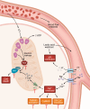
Figure 3. Molecular changes in the cell after reperfusion.
The return of oxygen during reperfusion generates ROS, primarily by mitochondria (10, 11). Damage to the ETC during ischemia leads to increased ROS production on reperfusion. Complex I and complex III are the primary sites of ROS production in the mitochondria (11, 160, 202), but other sites can also contribute (203). The increase in succinate that occurs during ischemia can lead to RET through generation of ROS by complex I. Extracellular pH is rapidly restored, which promotes extrusion of intracellular H+ via NHE, leading to a transient increase in intracellular Na+. As ATP is restored, the Na+-K+ ATPase becomes active and helps to extrude intracellular Na+. Depending on the relative timing of ATP restoration, a sustained increase in cytosolic Na+ can stimulate NCX, leading to a further increase in cytosolic Ca2+ during early reperfusion. ROS can also lead to damage of intracellular proteins such as SERCA and RyR2, leading to altered SR Ca2+ homeostasis. Together, these can lead to greater Ca2+ accumulation in the cytosol and exacerbate reperfusion injury. Any increase in cytosolic Ca2+ present at the start of reperfusion would lead to an increase in mitochondrial Ca2+ accumulation via MCU when the Δψ is restored on reperfusion (204). This further increase in mitochondrial Ca2+ on reperfusion depends on how fast Δψ is restored relative to how quickly cytosolic Ca2+ returns to baseline. Ca2+ overload in the mitochondria is thought to prime the mPTP to open on reperfusion when pH is restored. It is widely cited that mPTP activation is inhibited by the acidic pH induced by ischemia (205, 206) and that upon reperfusion, intracellular and extracellular pH are rapidly corrected, allowing for mPTP opening. However, inhibition of mPTP by acidic pH only occurs in de-energized mitochondria. In energized mitochondria, low pH actually enhances mPTP opening (207). ROS is another activator of mPTP, and it is likely that the increase in ROS that occurs during reperfusion synergizes with the increase in mitochondrial Ca2+ (which may already be there during ischemia) to activate mPTP on reperfusion.
Figure 4
Figure 4. Major contributors of cardiac I/R injury.
Myocardial injury during I/R is triggered by multiple cell-death pathways, all of which contribute to the final infarct size. During ischemia, there is an increase in cytosolic Ca2+; this leads to the activation of calpains, which can trigger plasma membrane rupture, CaMKII, and mitochondrial Ca2+ overload through the MCU. Mitochondrial Ca2+ overload is one of the triggers of mPTP opening. CypD is a regulator of the mPTP. ROS are generated during I/R injury by a variety of mechanisms, including, but not limited to, increased Fe3+ (canonically ferroptosis), electron leak from damage to the ETC, and the accumulation of succinate leading to RET. ROS contribute to mPTP opening and are also implicated in lipid peroxidation, leading to plasma membrane breakage and oxidization of mtDNA (ox-mtDNA) and its leakage into the cytosol. Once in the cytosol, ox-mtDNA can activate caspases, which can either independently cause plasma membrane breakage or activate gasdermins (GSDMs). GSDMs form pores in the plasma membrane. RIPK3 is also activated, leading to MLKL activation, which forms pores in the plasma membrane.

References

    1. Ambrosio G, et al. The relationship between oxygen radical generation and impairment of myocardial energy-metabolism following postischemic reperfusion. J Mol Cell Cardiol. 1991;23(12):1359–1374. doi: 10.1016/0022-2828(91)90183-M. - DOI - PubMed
    1. Bolli R, et al. Direct evidence that oxygen-derived free-radicals contribute to postischemic myocardial dysfunction in the intact dog. Proc Natl Acad Sci U S A. 1989;86(12):4695–4699. doi: 10.1073/pnas.86.12.4695. - DOI - PMC - PubMed
    1. Abdallah Y, et al. Interplay between Ca2+ cycling and mitochondrial permeability transition pores promotes reperfusion-induced injury of cardiac myocytes. J Cell Mol Med. 2011;15(11):2478–2485. doi: 10.1111/j.1582-4934.2010.01249.x. - DOI - PMC - PubMed
    1. Bauer TM, Murphy E. Role of mitochondrial calcium and the permeability transition pore in regulating cell death. Circ Res. 2020;126(2):280–293. doi: 10.1161/CIRCRESAHA.119.316306. - DOI - PMC - PubMed
    1. Halestrap AP, et al. Mitochondrial permeability transition pore opening during myocardial reperfusion: a target for cardioprotection. Cardiovasc Res. 2004;61(3):372–385. doi: 10.1016/S0008-6363(03)00533-9. - DOI - PubMed

MeSH terms