. 2025 Feb;21(1):e70039.
doi: 10.1002/rcs.70039.

Integrated Design and Prototyping of a Robotic Head for Ocular and Craniofacial Trauma Simulators

Affiliations

Integrated Design and Prototyping of a Robotic Head for Ocular and Craniofacial Trauma Simulators

Marcello Gallerani et al. Int J Med Robot. 2025 Feb.

Abstract

Background: Medical simulation is relevant for training medical personnel in the delivery of medical and trauma care, with benefits including quantitative evaluation and increased patient safety through reduced need to train on patients.

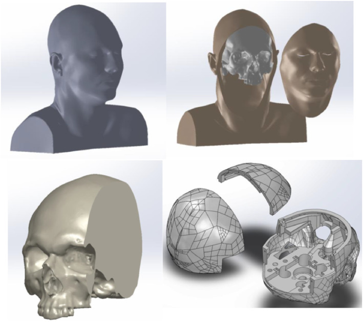
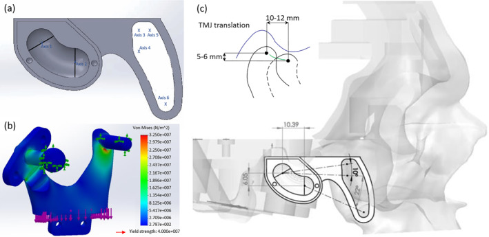
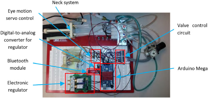
Methods: This paper presents a prototype medical simulator focusing on ocular and craniofacial trauma (OCF), for training in management of facial and upper airway injuries. It consists of a physical, electromechanical representation of head and neck structures, including the mandible, maxillary region, neck, orbit and peri-orbital regions to replicate different craniofacial traumas. Actuation and hydraulic systems are designed to control animatronic features and flow of simulated blood, tears, and cerebrospinal fluid.

Results: Experimentally validated, the OCF simulator achieves structural and functional characteristics as close as possible to those of a human body.

Conclusions: The OCF Simulator can be used as a stand-alone active simulator, it can be transported and used to train surgeons in simulated real-life scenarios.

Clinical trial registration: The authors declare that this statement is not applicable since no clinical tests have been performed.

Keywords: craniofacial trauma; medical simulators; physician training; robotic human head.


Conflict of interest statement

The authors declare no conflicts of interest.

Figures

FIGURE 1
FIGURE 1
Examples of medical simulators: (a) METI HPS mannequin [6]; (b) mannequin simulator in military scenario; (c) robotic chewing mechanisms developed by Takanishi Laboratories (Waseda University, Tokyo, Japan) [7]; (d) Laerdal SimMan mannequin [8].
FIGURE 2
FIGURE 2
OCF initial set‐up: unanimated mannequin and instrumentation.
FIGURE 3
FIGURE 3
A Computed Tomography (CT) of a real human head, with the skull and the resulted CAD file.
FIGURE 4
FIGURE 4
Jaw anatomy and main structure of the Temporomandibular Joint (TMJ). Adapted from ‘Gray's Anatomy’, by H. Gray, Crown Publishers, 1977 [12, 13].
FIGURE 5
FIGURE 5
Frontal and sagittal view of the Posselt's diagram with relevant movements and positions. Adapted from ‘Dynamics of the human masticatory system, Critical reviews in oral biology & medicine’, by J. H. Koolstra, 2002 [16]. Frontal view focal points: CO, complete occlusion of the mouth; a, maximum opening due only to condylar rotation; b, maximum opening reachable; c, maximum protrusion. Lateral view focal points: MI, coincident with the c point of the lateral; b, maximum opening; d, maximum right lateral movement; e, maximum left lateral movement. During opening, normally the mandible follows the border of the Posselt's Diagram from CO to a, and then to b; while during closing it normally follows a continuous curve profile linking directly b with CO.
FIGURE 6
FIGURE 6
Human neck anatomy and relevant components. Seven cervical vertebrae (C1 to C7) are stacked on top of each other and are separated by intervertebral discs. The cervical spine enables a wide range of motion, including flexion, extension, rotation, and lateral bending, while stabilising the head and maintaining balance. Key muscles include: Sternocleidomastoids and Scalene muscles for head rotation and flexion, and Longus Colli and Longus Capitis for cervical spine support and neck flexion. Lots of ligaments, which are fibrous connective tissues, connect bones, providing cervical spine stability. Trachea and Oesophagus allow air to the lungs and food to the stomach. Thyroid Gland regulates metabolism and energy levels in the body.
FIGURE 7
FIGURE 7
Human neck Range of Motion (RoM) and typical movements.
FIGURE 8
FIGURE 8
Facial blood circulatory system highlighting main arteries and veins. Adapted from ’Gray's Anatomy’, by H. Gray, Crown Publishers, 1977 [12].
FIGURE 9
FIGURE 9
CAD model and physical prototype of the EYE‐MECH (a); exploded view of one side of the EYE‐MECH cavity (b) [10].
FIGURE 10
FIGURE 10
CAD design of the jaw and preparation of its seat in the skull: (a) jaw socket to allow roto‐translational motions; (b) mandible mechanism design and preliminary FEM analysis; (c) skull preparation to accommodate the jaw.
FIGURE 11
FIGURE 11
Jaw assembly CAD design and relevant mechanical components (a); jaw prototype (b).
FIGURE 12
FIGURE 12
Jaw mechanism integrated in the skull (a); forces and torques applied to jaw mechanism (b).
FIGURE 13
FIGURE 13
Clutch schematisation with relevant loads and parameters.
FIGURE 14
FIGURE 14
Preliminary control scheme of the jaw mechanism [9].
FIGURE 15
FIGURE 15
Neck mechanism: (a) neck as a reproduction of cervical vertebrae; Inflating the Mckibben muscles, with a pressure of almost 38 psi, allows (b) a right lateral bending of up to 20°; (c) a left lateral bending of up to 20°; (d) a flexion of up to 24°; (e) an extension of up to 11°.
FIGURE 16
FIGURE 16
Neck rotational mechanism prototype and its integration into the skull and with the jaw mechanism.
FIGURE 17
FIGURE 17
Preliminary control scheme of the neck mechanism.
FIGURE 18
FIGURE 18
Robotic blood circuit system: (a) CAD design of the valve distribution system; (b) real prototype of the valve distribution system; (c) integration of the valve distribution system into the skull [9].
FIGURE 19
FIGURE 19
OCF prototype testing with integrated eye, jaw and neck systems.
FIGURE 20
FIGURE 20
CAD assembly of the OCF prototype with integrated eye, jaw, neck and cardiovascular circuit systems.
FIGURE 21
FIGURE 21
Preliminary control scheme of the OCF system with integrated eye, neck and cardiovascular circulatory systems [9].
FIGURE 22
FIGURE 22
From (a) to (c) are reported examples of maxillofacial injuries: (a) tripod or zygomaticomaxillary complex fracture; (b) Le Fort I complex fracture; (c) most common mandible fractures. From (d) to (f) are presented mandible and facial modules to replicate common fractures.

Similar articles

References

    1. Carey M. E., “Learning From Traditional Combat Mortality and Morbidity Data Used in the Evaluation of Combat Medical Care,” Military Medicine 152, no. 1 (1987): 6–13, 10.1093/milmed/152.1.6. - DOI - PubMed
    1. Montgomery S. P., Swiecki C. W., and Shriver C. D., “The Evaluation of Casualties From Operation Iraqi Freedom on Return to the Continental United States From March to June 2003,” Journal of the American College of Surgeons 201, no. 1 (2005): 7–12, 10.1016/j.jamcollsurg.2005.03.038. - DOI - PubMed
    1. Wade A. L., Dye J. L., Mohrle C. R., and Galarneau M. R., “Head, Face, and Neck Injuries During Operation Iraqi Freedom Ii: Results From the us Navy‐Marine Corps Combat Trauma Registry,” Journal of Trauma and Acute Care Surgery 63, no. 4 (2007): 836–840, 10.1097/01.ta.0000251453.54663.66. - DOI - PubMed
    1. Mehrotra D. and Markus A., “Emerging Simulation Technologies in Global Craniofacial Surgical Training,” Journal of Oral Biology and Craniofacial Research 11, no. 4 (2021): 486–499, 10.1016/j.jobcr.2021.06.002. - DOI - PMC - PubMed
    1. Ziv A., Ben‐David S., and Ziv M., “Simulation Based Medical Education: An Opportunity to Learn From Errors,” Medical Teacher 27, no. 3 (2005): 193–199, 10.1080/01421590500126718. - DOI - PubMed