. 2025 Dec;14(1):2449083.
doi: 10.1080/22221751.2024.2449083. Epub 2025 Jan 12.

A surrogate BSL2-compliant infection model recapitulating key aspects of human Marburg virus disease

Affiliations

A surrogate BSL2-compliant infection model recapitulating key aspects of human Marburg virus disease

Wanying Yang et al. Emerg Microbes Infect. 2025 Dec.

Abstract

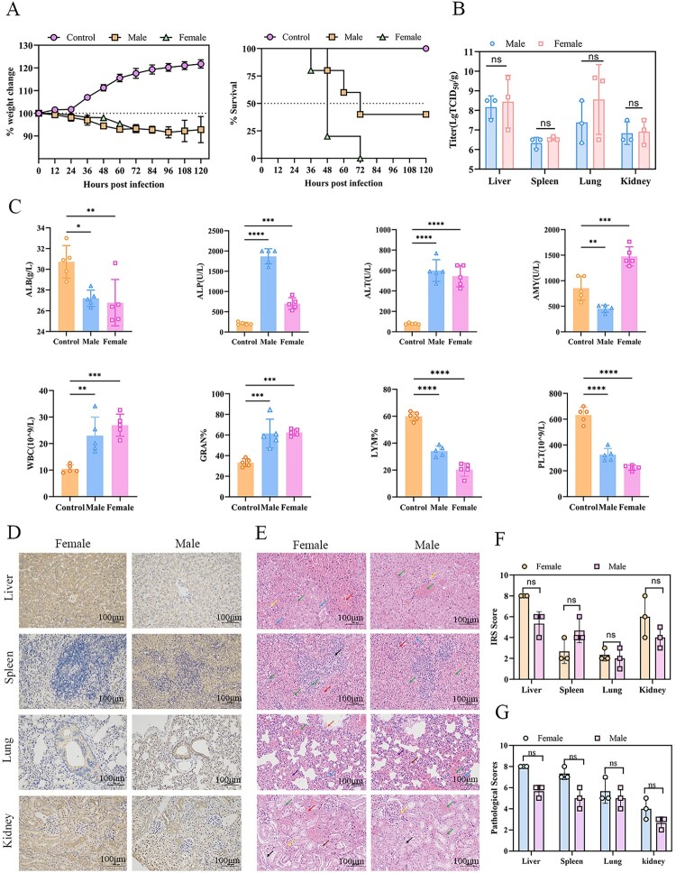
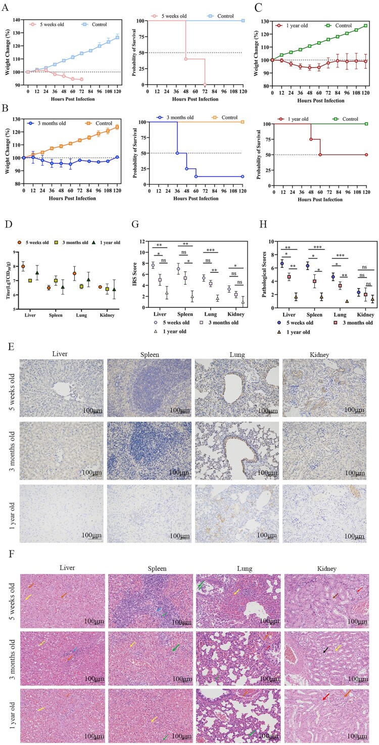
Marburg virus disease (MVD) is a severe infectious disease caused by the Marburg virus (MARV), posing a significant threat to humans. MARV needs to be operated under strict biosafety Level 4 (BSL-4) laboratory conditions. Therefore, accessible and practical animal models are urgently needed to advance prophylactic and therapeutic strategies for MARV. In this study, we constructed a recombinant vesicular stomatitis virus (VSV) expressing the Marburg virus glycoprotein (VSV-MARV/GP). Syrian hamsters infected with VSV-MARV/GP presented symptoms such as thrombocytopenia, lymphopenia, haemophilia, and multiorgan failure, developing a severe systemic disease akin to that observed in human MARV patients. Notably, the pathogenicity was found to be species-specific, age-related, sex-associated, and challenge route-dependent. Subsequently, the therapeutic efficacy of the MR191 monoclonal antibody was validated in this model. In summary, this alternative model is an effective tool for rapidly screening medical countermeasures against MARV GP in vivo under BSL-2 conditions.

Keywords: Marburg virus; Syrian hamster; recombinant vesicular stomatitis virus; recurrence of classic symptoms; surrogate model; vaccine evaluation and drug screening.


Conflict of interest statement

No potential conflict of interest was reported by the author(s).

Figures

Figure 1.
Figure 1.
The generation and the One-step growth curves of the recombinant vesicular stomatitis virus VSV-MARV/GP. (A) Schematic diagram of the generation of the recombinant vesicular stomatitis virus VSV-MARV/GP. The recombinant full-length plasmid (p3.1-VSVΔG-MARV GP) and four helper plasmids (pcDNA3.1-VSV-N, pcDNA3.1-VSV-P, pcDNA3.1-VSV-L, and pcDNA3.1-VSV-G) were co-transfected into BSR/T7 cells to obtain a recombinant virus bearing the glycoprotein of MARV. (B) One-step growth curves of WT VSV and VSV-MARV/GP inoculated into Vero E6 cells at an MOI of 0.1. Viruses were collected at 12 h intervals to measure titers.
Figure 2.
Figure 2.
Changes in weight and percent survival of rodents infected with VSV-MARV/GP. BALB/c mice (A), Sprague‒Dawley (SD) rats (B), Hartley guinea pigs (C), and Syrian hamsters (D) were inoculated with VSV-MARV/GP via the intraperitoneal (i.p.) route. Weight changes and percent survival were monitored.
Figure 3.
Figure 3.
Characterization of VSV-MARV/GP infection in Syrian hamsters. (A) Weight change and survival of Syrian hamsters infected with different doses of VSV-MARV/GP. (B) Blood biochemistry and blood cell counts were analyzed at 36 hpi. (C) Viral loads, including those in the heart, liver, spleen, lungs, kidneys, stomach, intestines, and brain, were determined at 36 hpi. The data are presented as the means ± SEMs. Statistical analyses were performed via one-way ANOVA. *, P < 0.05; *, P < 0.01; **, P < 0.001; ***, P < 0.0001; ****. (D) Histopathological and immunohistochemistry assays of the liver, spleen, lung and kidney at 36 hpi. Scale bar = 50 μm. Hepatic lesions, including hepatocellular necrosis, nuclear fragmentation (black arrows) and hemorrhage (yellow arrows), were observed. Splenic lesions, including infiltrating macrophages (blue arrows) and hemorrhages (green arrows), were observed. The lung tissue showed diffuse mild thickening of the alveolar wall with inflammatory cell infiltration (red arrows). Kidney lesions were observed as hemorrhages (yellow arrows).
Figure 4.
Figure 4.
Comparison of sex-associated Syrian hamsters infected with VSV-MARV/GP.(A) Weight change and survival of the animals in the male and female groups. (B) Viral loads in the liver, spleen, lung and kidney at 1.5 dpi. (C) Hematological changes at 1.5 dpi. (D) Immunohistochemistry of the liver, spleen, lungs and kidneys at 1.5 dpi. Scale bar = 100 μm. (E) Histopathological changes in the liver, spleen, lungs and kidneys at 1.5 dpi. Scale bar = 100 μm. Pathological changes in the liver include hepatocellular steatosis (yellow arrows), venous and peripheral hepatic sinusoidal stasis (green arrows), hemorrhage (red arrows), and hepatocellular punctate necrosis (blue arrows). Splenic pathological changes include a low number, small size, and irregular shape of white marrow (black arrow), red marrow hemorrhage (red arrow), cellular necrosis (green arrow), and granulocyte infiltration (blue arrow). Pulmonary pathology includes granulocytic infiltration (blue arrow), congestion (purple arrow), bronchiole lumen eosinophilia (orange arrow), congestion (green arrow), and brown pigmentation (brown arrow). Renal pathological changes include glomerular capillary bruising (yellow arrows), hydropic degeneration of a small number of tubular epithelial cells (black arrows), tubular atrophy (brown arrows), vascular stasis (green arrows), and focal hemorrhage (red arrows). (F) IRS scores of the liver, spleen, lungs and kidneys at 1.5 dpi. (G) Pathological scores of the liver, spleen, lungs and kidneys at 1.5 dpi. The data are presented as the means ± SEMs. Statistical analyses were performed via one-way ANOVA. *, P < 0.05; *, P < 0.01; **, P < 0.001; ***, P < 0.0001; ****.
Figure 5.
Figure 5.
Comparison of the age distributions of Syrian hamsters infected with VSV-MARV/GP. Five-week-old (A), 3-month-old (B) and 1-year-old (C) Syrian hamsters were infected with VSV-MARV/GP via the i.p. route. Weight changes and percent survival were monitored. Viral loads were evaluated by TCID50 in the liver, spleen, lung and kidney at 1.5 dpi (D). Pathological changes (E) and immunohistochemistry (F) of the liver, spleen, lungs and kidneys 1.5 dpi. Hepatic pathology included hepatocellular steatosis (yellow arrows), venous and hepatic sinusoidal stasis (orange arrows) and focal infiltration of lymphocytes around the central vein (blue arrows). The splenic pathology revealed more sparsely arranged lymphocytes (blue arrows), granulocytic infiltration (green arrows) and hemorrhage (yellow arrows). Lung pathology included granulocytic infiltration (green arrows), bruising (orange arrows), and hydropic degeneration of fine bronchial epithelial cells (red arrows). Renal pathology includes hydropic degeneration of renal tubular epithelial cells (red arrows), intratubular eosinophilia (brown arrows), cytosolic consolidation of renal tubular epithelial cells (black arrows), detachment of renal tubular epithelial cells (yellow arrows), and bruising (orange arrows). Scale bar = 100 μm. IRS score (G) and pathological score (H). The data are presented as the means ± SEMs. Statistical analyses were performed via one-way ANOVA. *, P < 0.05; *, P < 0.01; **, P < 0.001; ***, P < 0.0001; ****.
Figure 6.
Figure 6.
The effects of different challenge routes, including the intraperitoneal (i.p.), subcutaneous (s.c.), intramuscular (i.m.), and intranasal (i.n.) routes, on Syrian hamsters infected with VSV-MARV/GP were compared. (A) Weight change and survival of the animals in each group. (B) Viral load in the liver, spleen, lungs and kidneys at 1.5 dpi. (C) Hematological changes at 1.5 dpi. (D) Immunohistochemistry of the liver, spleen, lungs and kidneys at 1.5 dpi. Scale bar = 100 μm. (E) Histopathological changes in the liver, spleen, lung and kidney at 1.5 dpi. Scale bar = 100 μm. Hepatic pathological changes include dilated hepatic sinusoids (black arrows), hepatocellular steatosis (yellow arrows), hemorrhage (brown arrows), hepatocellular hydropic degeneration (red arrows), and venous and hepatic sinusoidal stasis (orange arrows). Splenic pathological changes include lymphocytic punctate necrosis (black arrows), granulocytic infiltration (green arrows), and white marrow adhesions (blue arrows). The pathological changes in the lungs included necrotic cell debris (black arrows), granulocytic infiltration (green arrows), lymphocytic infiltration (blue arrows), fine bronchial hemorrhage (yellow arrows) and perivascular edema (purple arrows). Renal pathological changes include hydropic degeneration of tubular epithelial cells (red arrows), glomerular capillary stasis (yellow arrows) and bruising (orange arrows). (F) Immunohistochemical scores of the liver, spleen, lungs and kidneys at 1.5 dpi. (G) Pathological scores of the liver, spleen, lungs and kidneys at 1.5 dpi.
Figure 7.
Figure 7.
Assessment of the therapeutic efficacy of MR191 in a lethal Syrian hamster model. (A) Schematic diagram for assessing the therapeutic effect of MR191 in the Syrian hamster model. (B) Weight change and percent survival after VSV-MARV/GP infection. (C) Viral loads were evaluated by TCID50 in the liver, spleen, lung and kidney at 36 hpi and 5 dpi. (D) Hematological changes at 36 hpi and 5 dpi. (E) Immunohistochemistry of the liver, spleen, lungs and kidneys. Scale bar = 100 μm or 50 μm. (F) Pathological changes in the liver, spleen, lungs and kidneys. Scale bar = 100 μm. The hepatic lesions included hepatocellular ballooning (red arrows), hepatocellular edema (black arrows), bruising (yellow arrows), and lymphocytic infiltration (green arrows). Splenic lesions include cellular punctate necrosis (black arrows) and bruising (yellow arrows). Lung pathological changes include granulocyte infiltration (black arrows), edema of fine bronchial epithelial cells (red arrows), siltation of pulmonary veins and capillaries of alveolar walls (blue arrows), and perivascular edema (green arrows). Renal pathological changes include hydropic degeneration of tubular epithelial cells (red arrows), bruising (yellow arrows), and necrosis of tubular epithelial cells (black arrows). (G) IRS scores of the liver, spleen, lung and kidney. (H) Pathological scores of the liver, spleen, lung and kidney.

Similar articles

References

    1. Abir MH, Rahman T, Das A, et al. . Pathogenicity and virulence of Marburg virus. Virulence. 2022;13(1):609–633. doi:10.1080/21505594.2022.2054760 - DOI - PMC - PubMed
    1. Brauburger K, Hume AJ, Mühlberger E, et al. . Forty-five years of Marburg virus research. Viruses. 2012;4(10):1878–1927. doi:10.3390/v4101878 - DOI - PMC - PubMed
    1. Geisbert TW, Daddario-DiCaprio KM, Geisbert JB, et al. . Marburg virus Angola infection of rhesus macaques: pathogenesis and treatment with recombinant nematode anticoagulant protein c2. J Infect Dis. 2007;196(Suppl 2):S372–S381. doi:10.1086/520608 - DOI - PMC - PubMed
    1. Daddario-DiCaprio KM, Geisbert TW, Geisbert JB, et al. . Cross-protection against Marburg virus strains by using a live, attenuated recombinant vaccine. J Virol. 2006;80(19):9659–9666. doi:10.1128/JVI.00959-06 - DOI - PMC - PubMed
    1. Alves DA, Glynn AR, Steele KE, et al. . Aerosol exposure to the Angola strain of marburg virus causes lethal viral hemorrhagic fever in cynomolgus macaques. Vet Pathol. 2010;47(5):831–851. doi:10.1177/0300985810378597 - DOI - PubMed

LinkOut - more resources