. 2025 Jan 28;10(1):e0077524.
doi: 10.1128/msphere.00775-24. Epub 2024 Dec 31.

mRNA-LNP vaccines combined with tPA signal sequence elicit strong protective immunity against Klebsiella pneumoniae

Affiliations

mRNA-LNP vaccines combined with tPA signal sequence elicit strong protective immunity against Klebsiella pneumoniae

Ting Huang et al. mSphere. .

Abstract

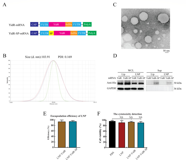
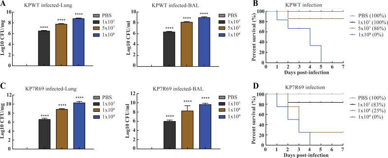
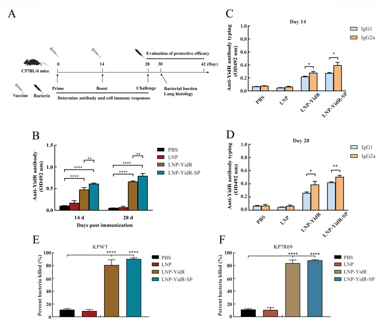
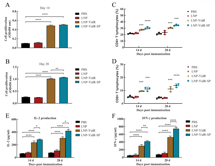
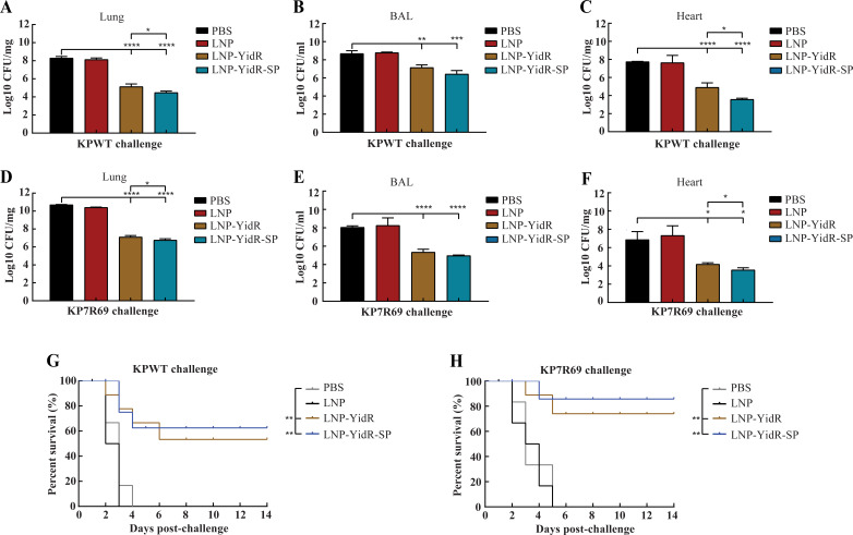
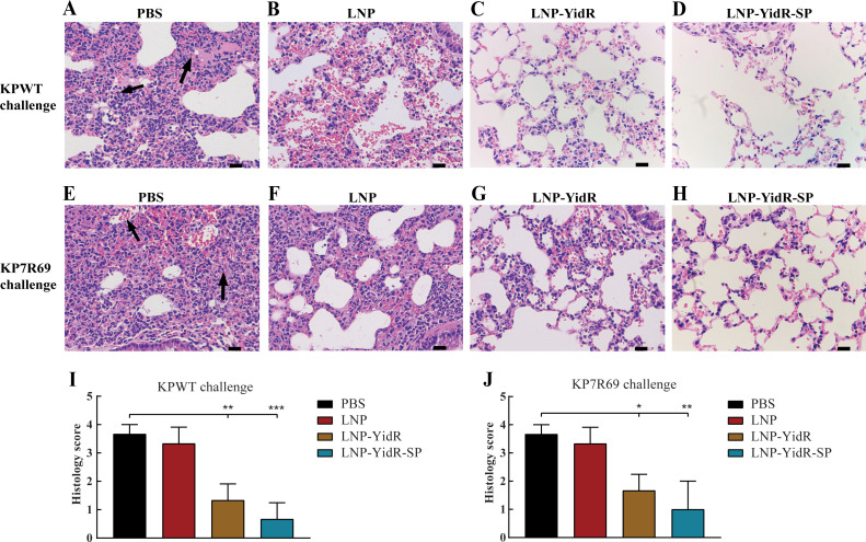
Klebsiella pneumoniae is a prominent Gram-negative and encapsulated opportunistic pathogen that causes a multitude of infections such as severe respiratory and healthcare-associated infections. Despite the widespread anti-microbial resistance and the high mortality rate, currently, no clinically vaccine is approved for battling K. pneumoniae. To date, messenger RNA (mRNA) vaccine is one of the most advancing technologies and are extensively investigated for viral infection, while infrequently applied for prevention of bacterial infections. In the present study, we aim to construct a new mRNA vaccine encoding YidR or combining with a tissue plasminogen activator signal sequence for preventing K. pneumoniae infection. Adaptive immunity was determined in mRNA vaccines-immunized mice and the protective effects of mRNA vaccines were evaluated in K. pneumoniae infected models. The results showed that lipid nanoparticle (LNP)-YidR-mRNA vaccine was produced with good morphology, high the encapsulation efficiency, and the specific antigen was highly expressed in cells in vitro. In addition, immunization with either LNP-YidR or LNP-YidR-SP elicited a Th1-biased immune response, reduced bacterial load, and provided broad protection in the lung infection models. Importantly, the LNP-YidR-SP mRNA vaccine induced strong adaptive humoral and cellular immunity and increased the survivability of mice compared to the other groups. Our findings serve as a focal point for developing a potential mRNA vaccine against K. pneumoniae, indicating the potential of mRNA vaccines for improving next-generation bacterial vaccine.IMPORTANCEK. pneumoniae is a notorious and clinical bacterium that is evolving in community-acquired and nosocomial settings. This opportunistic pathogen causes severe infectious diseases, including urinary tract infection and pneumonia, and causes a concerning global public burden. Despite efforts having been created to develop different types of K. pneumoniae vaccines, there is no licensed vaccine for preventing K. pneumoniae infection. Therefore, to develop an effective tactic is essential to combat K. pneumoniae-caused diseases. This study provides a novel vaccine strategy against K. pneumoniae and a potent platform to elicit high levels of humoral and cell-meditated immunity.

Keywords: Klebsiella pneumoniae; antibacterial immunity; mRNA vaccine; virulence.


Conflict of interest statement

The authors declare no conflict of interest.

Figures

Fig 1
Fig 1
In vitro characterization of YidR-mRNA vaccine. (A) The schematic illustration of YidR-mRNA constructs. The mRNA constructs consist of 5′ cap followed by 5′-untranslated regions, with or without signal peptide (SP), YidR, 6xHis tag, 3′ untranslated region (UTR), and polyadenylation (polyA) tail. (B) The size distribution of LNPs was measured by a Malvern instrument. (C) The morphology of LNPs was detected by transmission electron microscopy. Scar bar, 50 nm. (D) The expression and secretion of YidR-mRNA vaccines was transfected into HEK-293T cells and detected by Western blotting. (E) The encapsulation efficiency of LNPs was determined by a Ribogreen assay. (F) The cytotoxicity of blank LNP and mRNA vaccines in HEK293 cells. Lip, lipofectamine 2000; LNP, lipid nanoparticle; NS, not significant; PDI, polydispersity index; PBS, phosphate-buffered saline; Sup, supernatant; WCL, whole-cell lysate.
Fig 2
Fig 2
Determination of lethal dose using different loads of K. pneumoniae strains. (A and C) Bacterial burdens of tissues from different groups of infected mice were detected by a CFU assay. (B and D) Survival curves of infected mice were monitored for the subsequent 7 days. Data represent mean ± SEM. ****P < 0.0001.
Fig 3
Fig 3
Antibody responses and opsonophagocytic killing activity of mice induced by the mRNA vaccines. (A) The experimental timeline was presented by a diagram; C57BL/6 mice were vaccinated or treated with PBS, followed by infection with K. pneumoniae. Vaccinations are indicated by the empty syringes; bacterial challenge is indicated by the black syringe. Mice were immunized with two mRNA vaccines and were boosted at day 14. (B–D) The anti-YidR IgG and IgG subtype (IgG1 and IgG2a) were detected at different time points by enzyme-linked immunosorbent assay. The OPK activity was detected in the serum of mice immunized with LNP-YidR and LNP-YidR-SP. (E) OPK activity against KPWT strain. (F) OPK activity against KP7R69 strain. Data represent mean ± SEM. *P < 0.05, **P < 0.01, ****P < 0.0001.
Fig 4
Fig 4
Cellular immune responses of mice activated by immunization with mRNA vaccines. (A and B) The spleen lymphocytes of immunized mice were separated as described in Materials and Methods, and the proliferation of spleen lymphocytes was detected by CCK-8 assay. (C and D) The CD4+ and CD8+ T lymphocytes of immunized mice were analyzed by flow cytometry at 14 and 28 days post-immunization. (E and F) The production levels of IL-2 (E) and IFN-γ (F) from the suspension of the spleen lymphocytes were assessed via an enzyme-linked immunosorbent assay kit. Data represent mean ± SEM. *P < 0.05, **P < 0.01, ***P < 0.001, ****P < 0.0001.
Fig 5
Fig 5
Bacterial burden and survivability of mRNA-immunized mice challenged with K. pneumoniae. (A–H) The immunized mice were infected intranasally at day 28 post-immunization with KPWT or KP7R69 strains. The bacterial loads of lungs (A and D), BAL (B and E), and hearts (C and F) were detected by CFU assay. Survivability of mRNA-immunized mice was monitored for 2 weeks (G and H). Survival curves of the immunized mice were assessed by log-rank (Mantel-Cox) test. Data represent mean ± SEM. *P < 0.05, **P < 0.01, ***P < 0.001, ****P < 0.0001.
Fig 6
Fig 6
Histopathologic analysis of lungs in the mRNA-immunized mice challenged with K. pneumoniae. (A–H) The mice were infected with KPWT strain (A–D) or KP7R69 strain (E–H) at day 28. The lungs of mRNA-immunized mice were obtained for histopathologic analysis. (A and E) The cellular debris and abnormal cells of lungs are presented by black arrows. (I and J) Histology score in the infected lungs. Scale bar, 20 µm. *P < 0.05, **P < 0.01, ***P < 0.001.

Similar articles

Cited by

References

    1. Hussein KE, Bahey-El-Din M, Sheweita SA. 2018. Immunization with the outer membrane proteins OmpK17 and OmpK36 elicits protection against Klebsiella pneumoniae in the murine infection model. Microb Pathog 119:12–18. doi:10.1016/j.micpath.2018.04.004 - DOI - PubMed
    1. Paczosa MK, Mecsas J. 2016. Klebsiella pneumoniae: going on the offense with a strong defense. Microbiol Mol Biol Rev 80:629–661. doi:10.1128/MMBR.00078-15 - DOI - PMC - PubMed
    1. Choby JE, Howard-Anderson J, Weiss DS. 2020. Hypervirulent Klebsiella pneumoniae - clinical and molecular perspectives. J Intern Med 287:283–300. doi:10.1111/joim.13007 - DOI - PMC - PubMed
    1. Martin RM, Bachman MA. 2018. Colonization, infection, and the accessory genome of Klebsiella pneumoniae Front Cell Infect Microbiol 8:4. doi:10.3389/fcimb.2018.00004 - DOI - PMC - PubMed
    1. Bassetti M, Righi E, Carnelutti A, Graziano E, Russo A. 2018. Multidrug-resistant Klebsiella pneumoniae: challenges for treatment, prevention and infection control. Expert Rev Anti Infect Ther 16:749–761. doi:10.1080/14787210.2018.1522249 - DOI - PubMed

MeSH terms

LinkOut - more resources