

Reactivation of hidden-latent Brucella infection after doxycycline and streptomycin treatment in mice
- PMID: 39745377
- PMCID: PMC11823614
- DOI: 10.1128/aac.01302-24
Reactivation of hidden-latent Brucella infection after doxycycline and streptomycin treatment in mice
Abstract
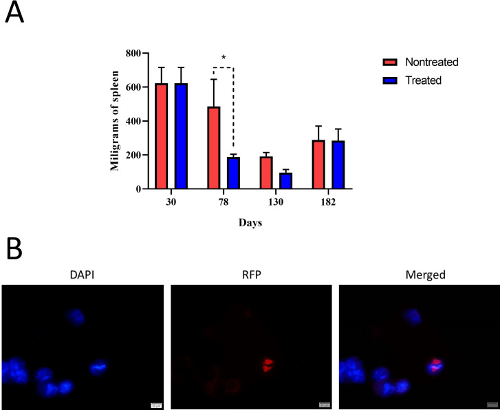
Brucellosis has therapeutic challenges due to 3%-15% relapses/therapeutic failures (R/TF) after antibiotic treatment. Therefore, determining the antibiotic concentration in tissues, the physiopathological parameters, and the R/TF after treatment is relevant. After exploring different antibiotic quantities, we found that a combined dose of 100 µg/g of doxycycline (for 45 days) and 7.5 µg/g of streptomycin (for 14 days), respectively, achieved therapeutic levels of more than fourfold minimum inhibitory concentrations (MICs) against Brucella abortus in the spleen, liver, bone marrow, and plasma of mice, causing minimal pathophysiological effects. After 30 days of infection, mice received antibiotics, and hematological, histopathological, biochemical, and immunological analyses were performed. After antibiotic therapy, the pathological, hematological, immunological, and physiological profiles paralleled those described in human brucellosis. Treatment lowered antibody titers, reduced proinflammatory cytokines, and reduced inflammation in the target organs for a protracted period. No bacteria were detected in tissues 8 weeks after treatment, suggesting complete recovery. However, despite high doxycycline and streptomycin concentrations in tissues, relapses appeared in 100% of the animals after 182 days post-infection, estimated by the bacterial counts and PCR from organs. This proportion contrasts with the 15% R/TF observed in humans after antibiotic treatments. None of the B. abortus isolated from relapses showed augmented MICs or mutations coding for antibiotic resistance in chromosomal-relevant regions. We discuss whether our findings constitute a general phenomenon or differences in the exhaustive screening method for bacteria detection related to the murine model. Along these lines, we envision likely mechanisms of bacterial persistence in tissues after antibiotic treatment.
Keywords: INF-γ; antibiotic resistance; antibiotic treatment; brucellosis; cytokines; doxycycline; relapses; streptomycin.
Conflict of interest statement
The authors declare no conflict of interest.
Figures


Similar articles
-
Antibiotics for treating human brucellosis.Cochrane Database Syst Rev. 2012 Oct 17;10(10):CD007179. doi: 10.1002/14651858.CD007179.pub2. Cochrane Database Syst Rev. 2012. PMID: 23076931 Free PMC article.
-
Prevalence of Brucella melitensis and Brucella abortus tetracyclines resistance: A systematic review and meta-analysis.Microb Pathog. 2023 Oct;183:106321. doi: 10.1016/j.micpath.2023.106321. Epub 2023 Sep 7. Microb Pathog. 2023. PMID: 37673354
-
Antibiotic prophylaxis for leptospirosis.Cochrane Database Syst Rev. 2024 Mar 14;3(3):CD014959. doi: 10.1002/14651858.CD014959.pub2. Cochrane Database Syst Rev. 2024. PMID: 38483067 Free PMC article.
-
Antibiotics for treatment of leptospirosis.Cochrane Database Syst Rev. 2024 Mar 14;3(3):CD014960. doi: 10.1002/14651858.CD014960.pub2. Cochrane Database Syst Rev. 2024. PMID: 38483092 Free PMC article.
-
Optimized solid lipid nanoparticles for co-delivery of gentamicin and doxycycline: a novel approach to combat intracellular brucella abortus infections.World J Microbiol Biotechnol. 2025 Jun 27;41(7):232. doi: 10.1007/s11274-025-04427-2. World J Microbiol Biotechnol. 2025. PMID: 40571847
References
-
- Ariza J, Bosilkovski M, Cascio A, Colmenero JD, Corbel MJ, Falagas ME, Memish ZA, Roushan MRH, Rubinstein E, Sipsas NV, Solera J, Young EJ, Pappas G, International Society of Chemotherapy, Institute of Continuing Medical Education of Ioannina . 2007. Perspectives for the treatment of brucellosis in the 21st century: the Ioannina recommendations. PLoS Med 4:e317. doi:10.1371/journal.pmed.0040317 - DOI - PMC - PubMed
-
- Ruiz-Castañeda M. 1986. Brucelosis. Tercera, La Prensa Médica Mexicana, S.A. - PubMed
-
- Spink WW. 1956. The nature of brucellosis. University of Minnesota Press.
Publication types
MeSH terms
Substances
Grants and funding
LinkOut - more resources
Full Text Sources
Medical
Miscellaneous
