Observational Study
. 2025 Feb 5;16(2):e0372124.
doi: 10.1128/mbio.03721-24. Epub 2024 Dec 31.

Are we serologically prepared against an avian influenza pandemic and could seasonal flu vaccines help us?

Affiliations
Observational Study

Are we serologically prepared against an avian influenza pandemic and could seasonal flu vaccines help us?

Iván Sanz-Muñoz et al. mBio. .

Abstract

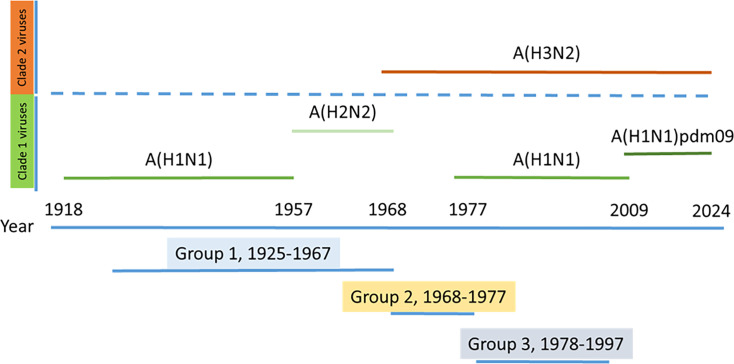
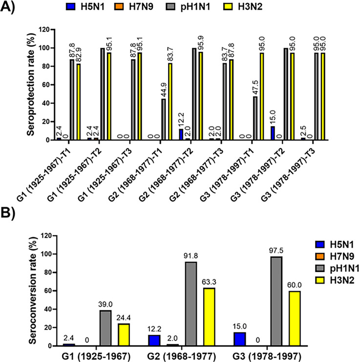
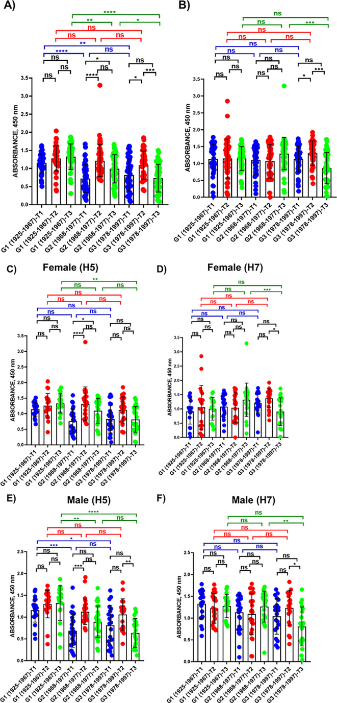
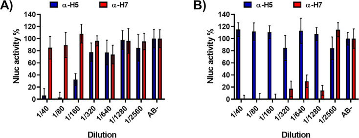
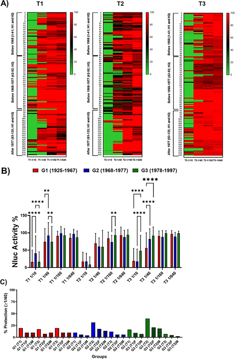
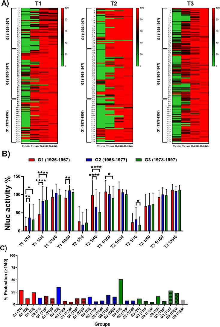
The current situation with H5N1 highly pathogenic avian influenza virus (HPAI) is causing a worldwide concern due to multiple outbreaks in wild birds, poultry, and mammals. Moreover, multiple zoonotic infections in humans have been reported. Importantly, HPAI H5N1 viruses with genetic markers of adaptation to mammals have been detected. Together with HPAI H5N1, avian influenza viruses H7N9 (high and low pathogenic) stand out due to their high mortality rates in humans. This raises the question of how prepared we are serologically and whether seasonal vaccines are capable of inducing protective immunity against these influenza subtypes. An observational study was conducted in which sera from people born between years 1925-1967, 1968-1977, and 1978-1997 were collected before or after 28 days or 6 months post-vaccination with an inactivated seasonal influenza vaccine. Then, hemagglutination inhibition, viral neutralization, and immunoassays were performed to assess the basal protective immunity of the population as well as the ability of seasonal influenza vaccines to induce protective responses. Our results indicate that subtype-specific serological protection against H5N1 and H7N9 in the representative Spanish population evaluated was limited or nonexistent. However, seasonal vaccination was able to increase the antibody titers to protective levels in a moderate percentage of people, probably due to cross-reactive responses. These findings demonstrate the importance of vaccination and suggest that seasonal influenza vaccines could be used as a first line of defense against an eventual pandemic caused by avian influenza viruses, to be followed immediately by the use of more specific pandemic vaccines.IMPORTANCEInfluenza A viruses (IAV) can infect and replicate in multiple mammalian and avian species. Avian influenza virus (AIV) is a highly contagious viral disease that occurs primarily in poultry and wild water birds. Due to the lack of population immunity in humans and ongoing evolution of AIV, there is a continuing risk that new IAV could emerge and rapidly spread worldwide, causing a pandemic, if the ability to transmit efficiently among humans was gained. The aim of this study is to analyze the basal protection and presence of antibodies against IAV H5N1 and H7N9 subtypes in the population from different ages. Moreover, we have evaluated the humoral response after immunization with a seasonal influenza vaccine. This study is strategically important to evaluate the level of population immunity that is a major factor when assessing the impact that an emerging IAV strain would have, and the role of seasonal vaccines to mitigate the effects of a pandemic.

Keywords: avian influenza; influenza A virus; influenza vaccine; nanoluciferase; neutralizing antibodies; pandemic; seroconversion; seroprotection.


Conflict of interest statement

The authors declare no conflict of interest.

Figures

Fig 1
Fig 1
Distribution of age groups regarding the most likely imprinting by both clade 1 and 2 IAVs. The participants in the study were divided into three age groups depending on the most likely first encounter with influenza A viruses. Group 1 included people born between 1925 and 1967 who were likely primed by both the A(H1N1) and A(H2N2) clade 1 viruses. Group 2 included people born between 1968 and 1977 who were likely primed by the A(H3N2) subtype clade 2 virus. Group 3 included people born between 1978 and 1997 who were likely primed by both A(H3N2) and A(H1N1) re-emergent viruses since 1977; these groups included both clade 2 and clade 1, respectively.
Fig 2
Fig 2
In vitro characterization of the recombinant viruses generated in this study. (A) Analysis of protein expression by immunofluorescence. MDCK cells (24-well plates, 2 × 10⁵ cells/well) were infected with the indicated viruses (MOI, 0.01) or mock infected. Infected cells were fixed and permeabilized at 24 h p.i. The cells were stained using specific pAbs against H5 (NR-2705), N1 (NR-9598), H7 (NR-48597), or N9 (NR-49276). A MAb against viral NP was used as control. Representative images are shown. Bars, 100 µm. (B and C) Plaque phenotype. MDCK cells (six-well plate format, 1 × 10⁶ cells/well) were infected with ~50 FFU of PR8(H5N1) or PR8(H5N1)-NLuc in the presence or absence of TPCK-trypsin (B) or infected with PR8(H7N9) or PR8(H7N9)-NLuc (C) and incubated at 37°C for 3 days. Plaques were evaluated by immunostaining using a MAb against IAV NP (MAb HB-65). (D and E) Analysis of protein expression by Western blot. MDCK cells (six-well plates, 106 cells/well) were infected with the indicated viruses or mock infected. Protein expression was examined by Western blotting using specific antibodies against HA (NR-2705 for H5 and NR-48597 for H7), NP, and NLuc. Actin was used as a loading control. The numbers on the left indicate the molecular size of the protein markers (in kilodaltons). Growth kinetics of the generated recombinant viruses. (F and H) Multicycle growth kinetics. Viral titers (in FFU per milliliter) in culture supernatants from MDCK cells (12-well plates, 5 × 10⁵ cells/well, triplicates) infected with PR8(H5N1) or PR8(H5N1)-NLuc (A) or infected with PR8(H7N9) or PR8(H7N9)-NLuc (C) (MOI, 0.001) were determined by immunofocus assay at the indicated times post-infection. The data represent the means ± SD of triplicate samples. *, P < 0.05, using two-way ANOVA. (G and I) NLuc expression. NLuc was evaluated in the same culture supernatants obtained from the experiment, and the results are presented in panel A. RLU, relative light units.
Fig 3
Fig 3
Seroprotection and seroconversion rates for IAV subtypes H1, H3, H5, and H7 in human serum samples. The seroprotection rate (A) and seroconversion rate (B) for the indicated IAV subtypes were determined in human serum samples from individuals born in 1925–1967 (G1), 1968–1977 (G2), and 1978–1997 (G3) and collected immediately before vaccine administration (T1), and post-vaccination at 28 days after vaccination (T2) or 6 months after vaccination (T3).
Fig 4
Fig 4
Violin plots (with all points) showing GMTs of HAI antibodies against IAV subtypes H1 (C), H3 (D), H5 (A), and H7 (B) in human serum samples. HAI titers against the indicated IAV subtypes were determined in human serum samples corresponding to individuals born in 1925–1967 (G1), 1968–1977 (G2), and 1978–1997 (G3) and collected immediately before vaccine administration (T1), and post-vaccination at 28 days after vaccination (T2) or 6 months after vaccination (T3). Statistical analysis was performed using one-way ANOVA with Bonferroni’s multiple comparison test. *P < 0.05; **P > 0.01; ***P < 0.001; ****P < 0.0001.
Fig 5
Fig 5
ELISA. The presence of IgG antibodies recognizing H5 (A) or H7 (B) was evaluated by ELISA using purified recombinant H5 (NR-59424, A/bald Eagle/Florida/125/2017) or H7 (NR-44365, A/Anhui/1/2013) proteins. Human serum samples corresponding to individuals born between 1925 and 1967 (G1), 1968 and 1977 (G2), and 1978 and 1997 (G3) were collected immediately before vaccine administration (T1), and post-vaccination at 28 days after vaccination (T2) or 6 months after vaccination (T3). *, P < 0.05, using one-way ANOVA. Data were also evaluated for the serum samples obtained from female (C and D) or male (E and F) subjects.
Fig 6
Fig 6
NLuc-based microneutralization assay for evaluating IAV NAbs. One to 200 FFU of PR8(H5N1)-NLuc (A) or PR8(H7N9)-NLuc (B) viruses were preincubated with twofold serial dilutions (starting dilution 1/40) of the indicated sera for PR8 H5N1 (NR-665) or PR8 H7N9 NR-48597) for 1 h. Subsequently, MDCK cells (96-well plates, 2 × 10⁴ cells/well, and triplicates) were infected with the antibody-virus mixture. Virus neutralization was determined by quantitating NLuc reporter expression at 48 h p.i. Mock-infected cells were used as internal controls for basal levels of luciferase (NLuc) expression. Infected cells in the absence of serum were used to determine the maximum reporter expression. The data are presented as the means of triplicate analyses.
Fig 7
Fig 7
Identification of cross-neutralizing antibodies against H5N1. One to 200 FFU of PR8(H5N1)-NLuc was preincubated for 1 h with fourfold serial dilutions (starting dilution 1/10) of the human serum samples corresponding to individuals born in 1925–1967 (G1), 1968–1977 (G2), and 1978–1997 (G3) and collected immediately before vaccine administration (T1), and post-vaccination at 28 days after vaccination (T2) or 6 months after vaccination (T3). Subsequently, MDCK cells (96-well plates, 2 × 10⁴ cells/well, and triplicates) were infected with the antibody-virus mixture. Virus neutralization was determined by quantitating NLuc reporter expression at 48 h p.i. Mock-infected cells were used as internal controls for basal levels of luciferase (NLuc) expression. Infected cells in the absence of serum were used to determine maximum (100%) reporter expression. NLuc activity or neutralization for the individual serum samples is displayed using a heatmap visualization method (A). The average for the different groups is also represented (B). *P < 0.05; **P > 0.01; ***P < 0.001; ****P < 0.0001, using two-way ANOVA. Red indicates more NLuc activity or less neutralization, and green indicates less NLuc activity or more neutralization. (C) The percentage of individuals (total, female or male) showing protection (NAb titers ≥1/40) is also presented.
Fig 8
Fig 8
Identification of cross-neutralizing antibodies against H7N9. One to 200 FFU of PR8(H7N9)-NLuc was preincubated for 1 h with fourfold serial dilutions (starting dilution 1/10) of the human serum samples corresponding to individuals born in 1925–1967 (G1), 1968–1977 (G2), and 1978–1997 (G3) and collected immediately before vaccine administration (T1), and post-vaccination at 28 days after vaccination (T2) or 6 months after vaccination (T3). Subsequently, MDCK cells (96-well plates, 2 × 10⁴ cells/well, and triplicates) were infected with the antibody-virus mixture. Virus neutralization was determined by quantitating NLuc reporter expression at 48 h p.i. Mock-infected cells were used as internal controls for basal levels of luciferase (NLuc) expression. Infected cells in the absence of serum were used to determine maximum (100%) reporter expression. NLuc activity or neutralization for the individual sera samples was displayed using a heatmap visualization method (A). The average for the different groups is also represented (B). *P < 0.05; **P > 0.01; ***P < 0.001; ****P < 0.0001, using two-way ANOVA. Red indicates more NLuc activity or less neutralization, and green indicates less NLuc activity or more neutralization. (C) The percentage of individuals (total, female or male) showing protection (NAb titers ≥1/40) is also indicated.

Similar articles

Cited by

References

    1. Dou D, Revol R, Östbye H, Wang H, Daniels R. 2018. Influenza A virus cell entry, replication, virion assembly and movement. Front Immunol 9:1581. doi:10.3389/fimmu.2018.01581 - DOI - PMC - PubMed
    1. Martínez-Sobrido L, Peersen O, Nogales A. 2018. Temperature sensitive mutations in influenza A viral ribonucleoprotein complex responsible for the attenuation of the live attenuated influenza vaccine. Viruses 10:560. doi:10.3390/v10100560 - DOI - PMC - PubMed
    1. Short KR, Richard M, Verhagen JH, van Riel D, Schrauwen EJA, van den Brand JMA, Mänz B, Bodewes R, Herfst S. 2015. One health, multiple challenges: the inter-species transmission of influenza A virus. One Health 1:1–13. doi:10.1016/j.onehlt.2015.03.001 - DOI - PMC - PubMed
    1. Sutton TC. 2018. The pandemic threat of emerging H5 and H7 avian influenza viruses. Viruses 10:461. doi:10.3390/v10090461 - DOI - PMC - PubMed
    1. Wille M, Holmes EC. 2020. The ecology and evolution of influenza viruses. Cold Spring Harb Perspect Med 10:a038489. doi:10.1101/cshperspect.a038489 - DOI - PMC - PubMed

Publication types

MeSH terms

LinkOut - more resources