Review
. 2025 Jan;329(1):e13432.
doi: 10.1111/imr.13432. Epub 2024 Dec 29.

Biophysical and Structural Features of αβT-Cell Receptor Mechanosensing: A Paradigmatic Shift in Understanding T-Cell Activation

Affiliations
Review

Biophysical and Structural Features of αβT-Cell Receptor Mechanosensing: A Paradigmatic Shift in Understanding T-Cell Activation

Robert J Mallis et al. Immunol Rev. 2025 Jan.

Abstract

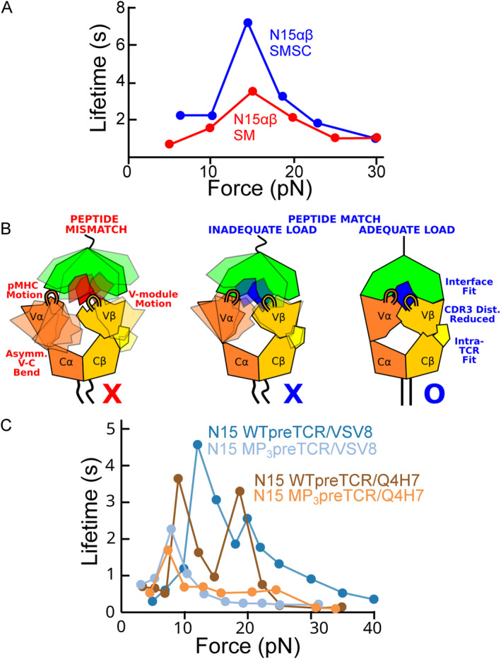
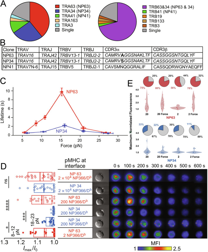
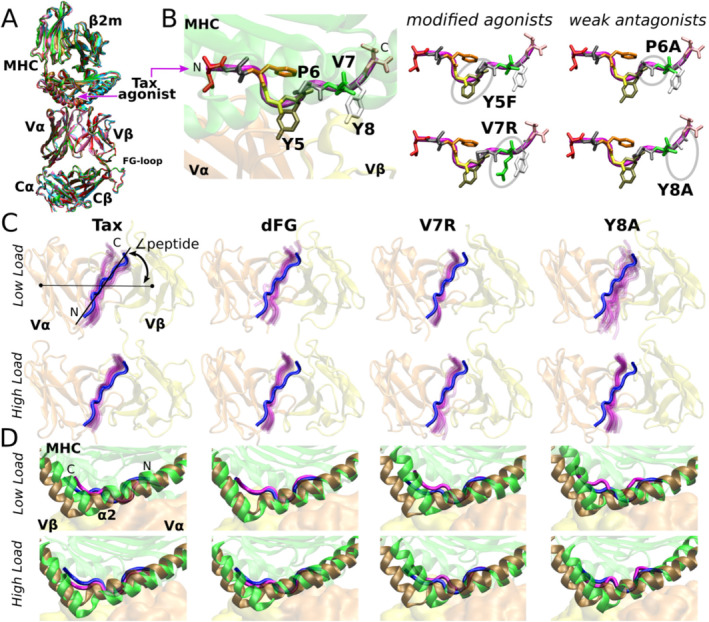
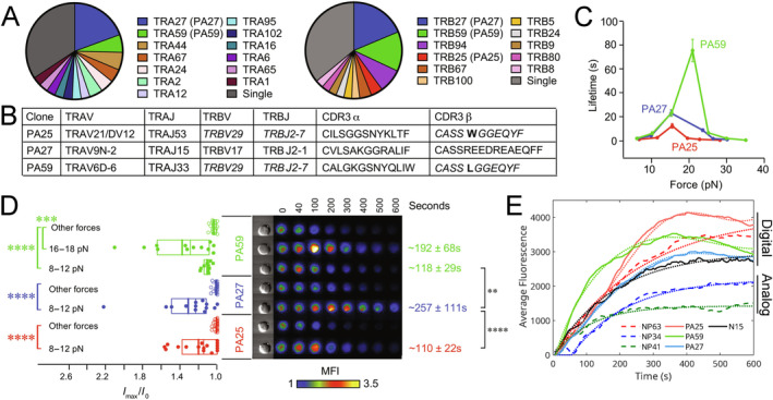
αβT cells protect vertebrates against many diseases, optimizing surveillance using mechanical force to distinguish between pathophysiologic cellular alterations and normal self-constituents. The multi-subunit αβT-cell receptor (TCR) operates outside of thermal equilibrium, harvesting energy via physical forces generated by T-cell motility and actin-myosin machinery. When a peptide-bound major histocompatibility complex molecule (pMHC) on an antigen presenting cell is ligated, the αβTCR on the T cell leverages force to form a catch bond, prolonging bond lifetime, and enhancing antigen discrimination. Under load, the αβTCR undergoes reversible structural transitions involving partial unfolding of its clonotypic immunoglobulin-like (Ig) domains and coupled rearrangements of associated CD3 subunits and structural elements. We postulate that transitions provide critical energy to initiate the signaling cascade via induction of αβTCR quaternary structural rearrangements, associated membrane perturbations, exposure of CD3 ITAMs to phosphorylation by non-receptor tyrosine kinases, and phase separation of signaling molecules. Understanding force-mediated signaling by the αβTCR clarifies long-standing questions regarding αβTCR antigen recognition, specificity and affinity, providing a basis for continued investigation. Future directions include examining atomistic mechanisms of αβTCR signal initiation, performance quality, tissue compliance adaptability, and T-cell memory fate. The mechanotransduction paradigm will foster improved rational design of T-cell based vaccines, CAR-Ts, and adoptive therapies.

Keywords: T cell; T‐cell receptor (TCR); cell signaling; mechanosensing; molecular dynamics (MD); optical tweezers (OT); preTCR; single molecule (SM).


Conflict of interest statement

The authors declare no conflicts of interest.

Figures

FIGURE 1
FIGURE 1
Organization, development and canonical ligand recognition of the αβTCR. (A) The αβTCR is a cell surface transmembrane receptor composed of eight subunits including the ligand binding TCRαβ heterodimer, signaling CD3 heterodimers εγ and εδ and CD3 homodimer ζζ. ITAM motifs including dual Tyr residues (denoted by a circled Y) are indicated in proper stoichiometry. The Cβ FG loop is labeled. Terminology for the αβTCR components from N to C terminus (top to bottom): Variable (V) domains; Constant (C) domains or CD3 ectodomains (CD3); Connecting Peptide region (CP); Transmembrane region (TM); Cytoplasmic Tail including ITAMs (Cyto). (B) Thymic selection occurs as hematopoietic progenitors migrate to the thymus, are committed to the T‐cell lineage (Thymus seeding progenitors; TSP) and undergo β‐rearrangement, β‐selection, α‐rearrangement and then positive and negative selection to mature into CD4 or CD8 single positive (SP) thymocytes prior to emigrating from the thymus to the peripheral tissues as naïve T cells. Immature SPs are omitted for clarity. Panel adapted from [37]. (C) The preTCR shares most components with the αβTCR except for substitution of pTα for TCRα. Annotation as in panel A. (D) Schematic footprint of TCRαβ interacting with pMHC ligand. View is from the TCR side where pMHC is behind the TCR. MHCI α‐helices from α1 and α2 subunits are shown. The CDR1‐3 ligand recognition loops' approximate interaction positions are shown with numbers within the red or blue ellipse for positioning of TCR Vα or Vβ domains. The peptide N and C termini are indicated. Positions are derived from the crystal structure of the N15αβ‐VSV8/Kb complex [38].
FIGURE 2
FIGURE 2
The preTCR recognizes pMHC ligand via a combined recognition surface utilizing CDR3 and the Vβ patch region with similarities and differences from TCR‐pMHC ligation. (A) Chemical shift perturbation data showing changes in 1H‐15N TROSY‐HSQC spectra with addition of VSV8/Kb to N15β used as a model for the preTCR ligand binding. Peaks are from N15β alone (red), with 200 (dark blue) and 500 μM (light blue) pMHC ligand added. (B) x‐ray crystallographic models of N15β with residues interacting with ligand highlighted. Left is a cartoon structure with a sphere indicating each interacting residue; Right is the corresponding surface model. Panels A‐B adapted from [51]. (C) Crystal structure of preTCR‐pMHC model N15β‐VSV8/Kb highlighting the interaction of CDR3 (cyan) with the C‐terminal residues of the peptide (yellow) and the Vβ framework residues including the Vβ patch (C″‐C′‐C‐F face; tan) interacting with MHC residues, particularly with the MHC α2 helix. (PDB code 6WL2). (D) Models of preTCR‐pMHC (left) and αβTCR‐pMHC (right) using crystal structures of N15β‐VSV8/Kb [53], N15αβ‐VSV8/Kb [38], and preTCR [55] overlaid on the cryoEM structure of the αβTCR [15]. Subunit coloring is as in Figure 1A,C. Heavy chain of Kb is purple, β2m is cyan and peptide is yellow. Despite the local differences in interaction angles, the longitudinal distance remains essentially the same, as does the incident angle with the antigen presenting cell (APC) plasma membrane assuming an upright TCR/preTCR. A dotted line depicts the C‐terminus of the MHC heavy chain connecting to the plasma membrane. The single‐span TM of MHC class I structure has not been determined to date. All structures rendered with PyMol [56].
FIGURE 3
FIGURE 3
T‐cell motility engenders mechanical forces to modulate TCR ligand binding strength and reversibly deform the TCR to activate T‐cell signaling. (A) Serial images of a T cell scanning a stationary layer of epithelia as APCs in search of cognate antigen [57]. A T cell is highlighted in yellow to show approximate position and shape changes over time (frames 1 through 6 span approximately 1 s). This and another movie are available as Video S1 and S2. (B) Schematic bond‐lifetime vs. force curves showing two catch bond profiles of two different TCRs interacting with the same cognate ligand (red and black) and the slip bond profiles of an irrelevant ligand. Relevant force regimes are highlighted. (C) TCR reversible transitioning model. Cycles of extension and contraction under load as observed in OT experiments are shown for ectodomains of T‐cell surface αβTCR and pMHC. CD3 subunits, TM regions and cytoplasmic tails are not shown for simplicity. (D) Schematic SM traces contrasting low‐ and high‐frequency transitions. (B–D) adapted from [58]. (E) Variability in the conformation of the αβTCR with addition of pMHC imaged with cryoEM. Left: Unliganded, compact state obtained in nanodisc. Middle: Unliganded, extended state obtained in detergent micelle. Right: pMHC liganded, extended state obtained in nanodisc. Approximate boundaries are indicated: V, TCRαβ Variable region; C, TCRαβ Constant region and CD3 ectodomains; TM, TCRαβ and CD3 TM regions. On the right panel, αβTCR and pMHC are colored for clarity. Structural image adapted from [17].
FIGURE 4
FIGURE 4
Biophysical investigation of TCR mechanosensor function. Each panel illustrates the experimental setup of the indicated assay. (A) Single molecule (SM). (B) Single molecule‐Single Cell (SMSC) with inset showing a bead approaching the T cell. Panels A‐B adapted from [31]. (C) Single Cell Activation Requirement (SCAR). Adapted from [32]. (D) Dual Trap Single Molecule (DTSM). Adapted from [60]. (E) Molecular dynamics (MD) simulation. Loads are applied via the harmonic restraining potential imposed on the Cα atoms of the terminal residues. Added strands allow transverse fluctuation of the complex, so that the applied load has both tensional and transverse components. Adapted from [76].
FIGURE 5
FIGURE 5
Measuring the relationship between force and the stability of the TCRαβ‐pMHC and preTCR‐pMHC interactions. (A) Force‐bond lifetime relationship for isolated N15 TCRαβ ectodomains (red trace) or αβTCR holoreceptor (blue trace) in binding VSV8/Kb using SM or SMSC, respectively. SMSC data is derived from experiments using an α3 mutant Kb molecule which does not bind CD8 to directly compare only TCRαβ‐pMHC interaction between experiments. Plots have been simplified for clarity. Panel adapted from [31]. (B) Atomistic basis for load sensitivity of the TCR. Due to the uneven compliance of the interfaces between the 4 subdomains, Vα moves more than Vβ does relative to the C‐module. Without an adequate load or the matching peptide, the asymmetric motion of the TCR chassis destabilizes the interface with pMHC (left). When a proper amount of load is applied while a cognate pMHC is bound, the subdomain motion can be suppressed and interfaces can be stabilized (right), which is the basis for ligand‐dependent catch bond formation. Panel adapted from [61]. (C) Force‐bond lifetime relationship for wild‐type (WT, dark shaded traces) or Vβ patch mutant (MP3, light shaded traces) N15 preTCR in binding VSV8/Kb (blue traces) or Q4H7/Kb (brown traces) using SM. Plots have been simplified for clarity. Panel adapted from [35].
FIGURE 6
FIGURE 6
TCRα transmembrane and connecting peptide structure directly impacts parameters of mechanosensing. (A) NMR structures of two conformers of TCRα TM region showing “L” state (red) and “E” state (raspberry). Residues important for TCR function are shown in sticks. (B) Bond‐lifetime vs. force curves of SMSC measurement of TCRα TM and TCRα and CD3δ CP mutants as compared to the WT N15αβTCR. Plots have been simplified from the original for clarity. (C) SCAR assay of mutants assayed in SMSC in panel B. Left: Plots of relative maximum fluorescence intensity with triggering (Imax/I0) with each symbol representing a single cell measured. Right: Representative triggering trace and the fluorescence image at maximum fluorescence (Imax) and the time for which it occurred. Each mutant is shown at high pMHC copy number (2 × 104 interfacial pMHC) and at lowest pMHC copy number in which a response was seen and at optimal force needed to trigger. All are compared in relation to the WT N15αβ TCR. Figure adapted from [63].
FIGURE 7
FIGURE 7
Differential sensitivity of highly related TCRs specific for high‐copy ligand NP366‐374/Db display in mice challenged with IAV infection. (A) Proportions of V gene usage by NP366‐374/Db specific T cells in recall responses to IAV. The top two TCRs (NP63 and NP34) account for the majority of NP366‐374/Db‐specific clonotypes. (B) Gene usage for top three most common clonotypic TCRs as well as translation of CDR3 regions for TCRα and TCRβ. Note the near identity of NP34 and NP63. (C) SMSC Force‐lifetime plots of NP34 and NP63. (D) SCAR profiles of NP34 and NP63. Left: Plots of relative maximum fluorescence intensity with triggering of individual cells given as symbols (Imax/I0). Right: Representative triggered trace for each. Each TCR is shown at high pMHC copy number (2 × 104 interfacial pMHC) and at minimum number in which a response was seen and at optimal force needed to trigger. (E) Violin plots depicting the maximum normalized fluorescence of each tested cell for NP63 and NP34 after pMHC stimulation. Interfacial numbers of pMHC are shown below the plots. Pie charts show triggering percentage. Solid color represents percentage triggered, gray color represents non‐triggered. A recovery of triggering percentage and maximum normalized fluorescence is observed in NP63 for two interfacial pMHC when force is applied to the TCR‐pMHC bond, confirming its digital capability. However, NP34 shows no recovery with force application, indicating it is analog. Figure adapted from [60].
FIGURE 8
FIGURE 8
MD simulations show point mutations of the Tax agonist peptide bound to A6 TCR alter the TCR‐pMHC interface in a load‐dependent manner. (A) Overlay of five A6 TCR‐pMHC crystal structures with different bound peptides: Wild‐type Tax agonist [brown, PDB 1AO7, [12]], Y5F [red, PDB 3QFJ, [122]], V7R [pink, PDB 1QSE, [123]], P6A [cyan, PDB 1QRN, [123]], and Y8A [green, PDB 1QSF, [123]]. Note all 5 structures align very closely. (B) Tax peptide residues from crystal structures are shown in licorice representation. Point mutations included in MD study are labeled. The peptide conformations are essentially identical even though point mutations lead to different functional outcomes, especially for weak antagonists. (C) Peptide motion relative to the V‐module during simulation (magenta). Viewing direction is from pMHC into the V‐module interface. The peptide from crystal structure is colored blue. In each panel, more than 10 snapshots from over 1‐μs long MD simulation are shown in semi‐transparent magenta, revealing degrees of motion. dFG: Cβ FG loop deletion mutant. The peptide angle is one of many measures that were used to quantify interfacial stability [61, 76, 86]. WT under high load (18.2 pN) best maintains the peptide position relative to the V‐module, as the peptide orientation stays closest to that of the crystal structure. (D) Positional shift of the MHC relative to the V‐module highlighted by the MHC α2 helix. For WT (“Tax”) under high load, the last frame from panel C (green and magenta for MHC α2 and peptide, respectively) is close to the crystal structure (brown and blue). Figure adapted from [61].
FIGURE 9
FIGURE 9
High sensitivity of TCRs specific for low‐copy ligand PA224‐233/Db in mice challenged with IAV infection. (A) Proportions of V gene usage by PA224‐233/Db specific T cells in recall responses to IAV. Note the top two TCRs (PA27 and PA59) account for a significant, but not as large, percentage of the repertoire as seen in the NP366‐374/Db experiment (Figure 7A). (B) Gene usage for top three most common clonotypic TCRs as well as translation of CDR3 regions for TCRα and TCRβ. (C) SMSC Force‐lifetime plots of PA25, PA27 and PA59. (D) SCAR profiles of PA25, PA27 and PA59. Left: Relative maximum fluorescence intensity with triggering (Imax/I0). Right: Representative triggered trace for each. Far Right: Average time to reach maximum fluorescence. Each TCR is shown at a copy number of 2 pMHC and at various forces and at optimal force needed to trigger. PA59 does not trigger at 8–12 pN but triggers well at 16–18 pN. (E) Metric for discrimination of digital versus analog TCRs by plotting average fluorescence versus time. Figure adapted from [60].
FIGURE 10
FIGURE 10
The γδTCR is not inherently force‐sensitive but can be engineered to be so by replacement of TCRγδ constant domains with those of TCRαβ. (A) Comparison between TCRαβ (top) and TCRγδ (bottom) ectodomains. Cβ FG loop is highlighted in light blue and Cγ FG loop is in yellow. Right column shows magnified views of the squared region. The FG loop has a broader area of interaction with the V domain in the TCRαβ compared to that of TCRγδ. (B) Construct design for SM experiments delineating allosteric mechanotransduction of the C domains of TCRαβ compared to that of DP10.7 TCRγδ. The leucine zipper (LZ) pairs allow interrogation of TCRγδ with CD1d‐sulfatide or CD1d (unloaded) in SM experiments. (C) SM bond lifetime vs. force curve of each construct with indicated ligand as compared to the N15αβ‐VSV8/Kb interaction. (D–G) SM traces of TCRγδ or γδ‐αβ chimera binding to CD1d or CD1d‐sulfatide. Each panel shows one or more single pulls with ligand to a specified distance (black trace), a pre‐transition dwell (green), if a transition is present, a post‐transition dwell (blue), and a rupture (red). Applied force is shown in each trace. Note that transitions are only common in experiments with chimeras and with CD1d‐sulfatide at most forces. Figure adapted from [36].
FIGURE 11
FIGURE 11
Models of TCR triggering and relationship with cytoskeletal elements. (A) Digital T cell triggering via sparse pMHC ligand results in highly energized TCR that transduces a strong signal despite low copy number. Signal initiation may be highly localized. (B) Analog T cell produces a weaker, more diffuse signal that relies on high copy number. (C) Linkage of TCR and pMHC to respective cell actomyosin machinery results in multiple cycles of opening and closing transitions generating local energy from cellular pools. Also see Figure 3C. (D) Dynamic oscillation of the free energy landscape. Transition to the extended state occurs when the applied load is high, causing rightward tilting of the free energy landscape. Once the extended state is reached, the larger span of the complex reduces force, resulting in leftward tilting of the free energy landscape. This leads to transition back to the compact state. With dissipative elements coupled to the TCR‐pMHC complex (e.g., membrane and the cytoskeleton; Figure 12), energy input is needed to maintain repeated cycling. Panels A‐D adapted from [58]. (E) Schematic of dynamic oscillation of the full αβTCR‐pMHC complex through repeated rounds of extension and contraction under load. This oscillation can agitate the membrane, re‐organize TM domains (e.g., dissociation of CD3ζζ), release ITAMs from the inner membrane leaflet, followed by their phosphorylation by Lck located on the cytoplasmic tail of CD8 (or CD4 on CD4+ T cells). Within the confined space between cells, pull on the membrane will be more during the compact (middle panel) and less during the extended phases (right panel) of the αβTCR‐pMHC complex. Transitions therefore bend the membrane as shown and agitate associations within the CD3 complex dimers. Similarly, since the conformational transition is thought to reside in Cαβ, local congestion due to the transient presence and absence of folded Cα and Cβ ectodomains will also agitate CD3 associations. Given that the CD8αβ coreceptor is positioned such that it extends from the membrane and interacts with the α3 domain of MHCI spanning the location of the conformational change, the extended state will yank on the CD8 tail driving Lck closer to ITAMs on the inside of the cell. Such positioning of the CD8 coreceptor can enhance signaling. The pMHC unbound state is in the left panel for completeness. Coloring of αβTCR is as in Figure 1A with CD8 in brown.
FIGURE 12
FIGURE 12
Mechanical properties of tissues have potential to impact mechanosensing. (A) Model of TCR mechanoregulation in tissues of varying stiffness showing forms of coupling, shear forces, conformational transitions and motor activation. Adapted from [58]. (B, C) Fluorescence microscopy of lungs isolated from IAV‐infected mice 7 days following recall response. (B) E‐cadherin stained cells (red) highlight the airway epithelial layer. CD103‐stained cells (green) show T cells and APCs. DAPI reagent (blue) stains nuclei. Insets at bottom show infiltration of epithelial cells with CD103+ cells. (C) CD8+ T cells (red) with Vβ13.1 [Vβ8.3+ = Vβ13.1 in modern nomenclature; [140]] cells (blue) and Vβ13.1+CD8+ cells (magenta), Vβ29 (TCRVβ7+ = Vβ29 in modern nomenclature) (green) and TCRVβ29+CD8+ cells (yellow). TRBV29 is the most common TRBV in recall responses to PA224‐233/Db (Figure 9A,B) and TRBV13.1 is the most common TRBV in recall response to NP366‐374/Db (Figure 7A,B). (D) Fluorescence microscopy of lungs isolated from IAV‐infected mice 14 days following primary infection. The lung was stained with anti‐TGFβ (red) and CD11b (green), the latter staining only macrophages (mΦ). Br = bronchioles. (B–D) adapted from [141].
FIGURE 13
FIGURE 13
Design of therapeutics receptors (A). Commonly used BBζ CAR‐T receptor utilizing cancer‐specific scFV fragment fused to CD8α TM region, 4‐1BB cytoplasmic region, and CD3ζ ITAM region (B). Newer design of HITs or STARs, fusing light and heavy chain V regions individually with TCR C domains to form a TCR‐like CAR with potential for TCR‐like properties. Compare with domain organization of WT αβTCR (Figure 1A).

Similar articles

Cited by

References

    1. Reinherz E. L., Hussey R. E., and Schlossman S. F., “A Monoclonal Antibody Blocking Human T Cell Function,” European Journal of Immunology 10 (1980): 758–762. - PubMed
    1. Allison J. P., McIntyre B. W., and Bloch D., “Tumor‐Specific Antigen of Murine T‐Lymphoma Defined With Monoclonal Antibody,” Journal of Immunology 129 (1982): 2293–2300. - PubMed
    1. Marrack P., Shimonkevitz R., Hannum C., Haskins K., and Kappler J., “The Major Histocompatibility Complex‐Restricted Antigen Receptor on T Cells. IV. An Antiidiotypic Antibody Predicts Both Antigen and I‐Specificity,” Journal of Experimental Medicine 158 (1983): 1635–1646. - PMC - PubMed
    1. Meuer S. C., Acuto O., Hussey R. E., et al., “Evidence for the T3‐Associated 90K Heterodimer as the T‐Cell Antigen Receptor,” Nature 303 (1983): 808–810. - PubMed
    1. Hedrick S. M., Cohen D. I., Nielsen E. A., and Davis M. M., “Isolation of cDNA Clones Encoding T Cell‐Specific Membrane‐Associated Proteins,” Nature 308 (1984): 149–153. - PubMed

Publication types

MeSH terms

Substances

LinkOut - more resources