. 2025 Feb 25;99(2):e0144324.
doi: 10.1128/jvi.01443-24. Epub 2024 Dec 31.

Dengue and Zika virus NS4B proteins differ in topology and in determinants of ER membrane protein complex dependency

Affiliations

Dengue and Zika virus NS4B proteins differ in topology and in determinants of ER membrane protein complex dependency

Samuel S Porter et al. J Virol. .

Abstract

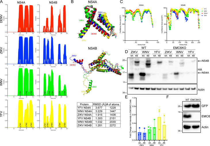
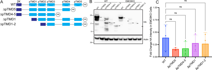
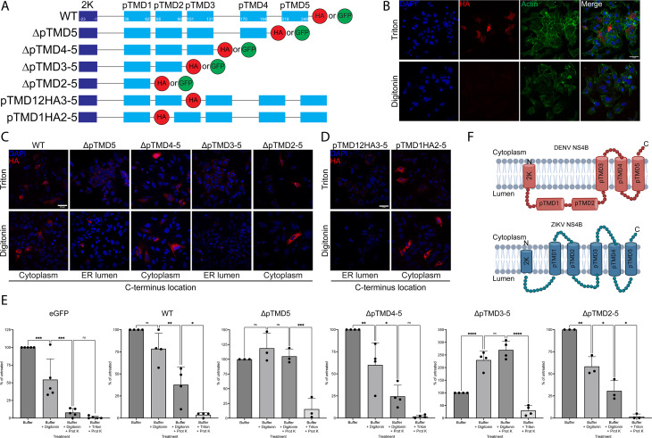
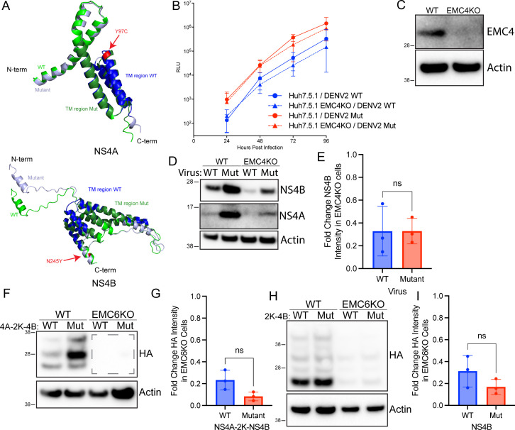
Flaviviruses utilize the cellular endoplasmic reticulum (ER) for all aspects of their lifecycle. Genome replication and other viral activities take place in structures called replication organelles (ROs), which are invaginations induced in the ER membrane. Among the required elements for RO formation is the biogenesis of viral nonstructural proteins NS4A and NS4B. We have previously shown that NS4A and NS4B from Dengue virus (DENV) and Zika virus (ZIKV) depend on the cellular ER membrane protein complex (EMC) for biogenesis. Here, we find that this dependency extends to the NS4A and NS4B proteins of Yellow Fever virus (YFV) and West Nile virus (WNV), which share similar computationally predicted membrane topologies. However, we demonstrate that ZIKV NS4B has different determinants of its dependency on the EMC than those for DENV NS4B, as well as a different membrane topology. Furthermore, we characterize mutant isolates of DENV and ZIKV that were serially passaged in EMC knockout cells and find that none are completely independent of the EMC for infection, and that mutant NS4B proteins remain sensitive to EMC depletion, suggesting a high genetic barrier to EMC depletion. Collectively, our findings are consistent with a model in which the EMC recognizes multiple determinants in the NS4B protein to support infection in several flaviviruses of critical public health importance.IMPORTANCEThe NS4A and NS4B proteins of flaviviruses are critically important to replication, but little is known about their function. It has been previously reported that the cellular EMC supports the biogenesis of NS4A and NS4B from Dengue and Zika virus. In this work, we demonstrate that this dependency on the EMC for NS4A and NS4B biogenesis extends to the West Nile and Yellow Fever viruses. Furthermore, we examine the features of ZIKV NS4B and find that its membrane topology of ZIKV NS4B and its determinants of dependency on the EMC are different from those previously described in DENV NS4B. Finally, we present evidence that there is a high genetic barrier for Dengue and Zika viruses to overcome EMC depletion.

Keywords: Dengue fever; Zika; flavivirus; virology.


Conflict of interest statement

The authors declare no conflict of interest.

Figures

Fig 1
Fig 1
Predicted topologies, structure, and EMC dependence of flavivirus NS4A and NS4B proteins. (A) Probability of indicated amino acid being within a transmembrane domain for the indicated flavivirus NS4A and NS4B proteins, based on TMHMM 2.0 hydrophobicity predictions. (B) Alignments of AlphaFold2-predicted protein structures of flavivirus NS4A (top) and NS4B (bottom). Table indicates root-mean-square deviation of indicated protein from DENV NS4A- or NS4B-predicted structures. (C) Confidence scores for AlphaFold2 predictions. (D) Wild-type 293T cells or pooled EMC6 knockout cells were transiently transfected with plasmids encoding NS4A-HA or 2K-NS4B-HA from indicated flavivirus species. Proteins were resolved using SDS-PAGE followed by immunoblotting for the indicated proteins. Blots are representative of three independent experiments. (E) Quantitation of D. Bars represent means ± SD. (F) Wild-type 293T cells or pooled EMC6 knockout cells were transiently transfected with a plasmid encoding GFP. Proteins were resolved using SDS-PAGE followed by immunoblotting for the indicated proteins. Blots are representative of three independent experiments.
Fig 2
Fig 2
ZIKV NS4B has multiple determinants of EMC dependency. (A) Schematic of truncation mutations of ZIKV NS4B. (B) Wild-type 293T cells or EMC6 KO cells were transfected to express NS4B-HA truncation mutants. Proteins were resolved using SDS-PAGE followed by Western blotting for the indicated proteins. Arrows indicated NS4B bands of interest. Blots are representative of three independent experiments. (C) Quantitation of B. Bars represent means ± SD.
Fig 3
Fig 3
Topology of ZIKV NS4B. (A) Schematic of truncation and insertion mutants of ZIKV 2K-NS4B. B) Huh7.5.1 cells on coverslips were transfected with a plasmid expressing NHK-HA. Cells were fixed with PFA, permeabilized with Triton X-100 or digitonin, stained with anti-HA and anti-actin antibodies and DAPI, and imaged with a confocal microscope. Micrographs are representative of three independent experiments. Scale bar represents 50 µm. (C) Huh7.5.1 cells on coverslips were transfected with plasmids expressing WT or truncated deletion mutations of ZIKV NS4B. Cells were fixed with PFA, permeabilized with Triton X-100 or digitonin, stained with an anti-HA antibody and DAPI, and imaged with a confocal microscope. Micrographs are representative of three independent experiments. Scale bar represents 50 µm. (D) Huh7.5.1 cells on coverslips were transfected with plasmids expressing ZIKV NS4B with HA inserted after either pTMD1 or pTMD2. Cells were fixed with PFA, permeabilized with Triton X-100 or digitonin, stained with anti-HA antibody and DAPI, and imaged with a confocal microscope. Micrographs are representative of three independent experiments. Scale bar represents 50 µm. (E) 293T cells were transfected with plasmids expressing GFP-tagged WT or truncated deletion mutations of ZIKV NS4B. Live cells were washed with buffer and imaged on a confocal microscope. Cells were then permeabilized with digitonin and imaged. Permeabilized cells were then exposed to proteinase K and imaged. Then, cells were incubated with proteinase K and Triton X-100 and imaged. Total integrated GFP signal from each step was measured and plotted as percentage of buffer-only treatment. Micrographs are representative of three independent experiments. Scale bar represents 50 µm. (F) Diagram of experimentally determined DENV (top) (10) and ZIKV (bottom) NS4B topology. Created in BioRender.
Fig 4
Fig 4
Characterization of potential EMC-independent DENV mutants. (A) Alignments of AlphaFold3-predicted structures of WT and putative EMC escape mutants of DENV NS4A and NS4B. Point mutations are indicated in red. (B) Wild-type Huh7.5.1 cells or EMC4 KO cells were infected with WT or NS4A Y97C/NS4B N245Y mutant DENV2-Luc reporter viruses at 0.1 MOI. Luciferase activity was measured at the indicated timepoints as a readout of viral infection. Points represent means ± SD of four independent experiments with three technical replicates in each experiment. (C) Wild-type Huh7.5.1 cells or EMC4 stable KO cell pools were lysed, and proteins were resolved using SDS-PAGE followed by immunoblotting with the indicated antibodies. (D) Wild-type Huh7.5.1 cells or EMC4 stable KO cell pools were infected with WT or NS4A Y97C/NS4B N245Y mutant DENV2-Luc viruses at 0.1 MOI. At 48 hpi, cells were lysed, and proteins were resolved using SDS-PAGE followed by immunoblotting with the indicated antibodies. Blots are representative of three independent experiments. (E) Quantitation of D. Bars represent means ± SD. (F) Wild-type 293T cells or EMC6 KO cells were transfected to express either WT or mutant (Y97C/N245Y) DENV NS4A-2K–NS4B-HA. Proteins were resolved using SDS-PAGE followed by immunoblotting for the indicated proteins. Blots are representative of three independent experiments. (G) Quantitation of F. Dashed lines on F represent quantitation area for KO cells. Bars represent means ± SD. (H) Wild-type 293T or EMC6 KO cells were transfected to express either WT or mutant (N245Y) DENV 2K-NS4B-HA. Proteins were resolved using SDS-PAGE followed by immunoblotting for the indicated proteins. Blots are representative of three independent experiments. (I) Quantitation of F. Bars represent means ± SD.
Fig 5
Fig 5
Characterization of potential EMC-independent ZIKV mutants. (A) Huh7.5.1 EM6 KO cells were infected with ZIKV PRVABC59 at an initial MOI of 2.2 and serially passaged every 3–4 days. After 12–24 passages, supernatant was collected, RNA was extracted and subjected to RT-PCR to amplify the NS4A-2K–NS4B region. PCR products were sequenced, and SNPs were compared with wild-type genome. (B) Wild-type or EMC6 KO Huh7.5.1 cells were infected with WT or NS4A E19G/L57F mutant ZIKV at 0.2 MOI or mock infected. At 72 hpi, cells were fixed with 4% PFA and stained with methylene blue. (C) Wild-type or EMC6 KO Huh7.5.1 cells were infected with WT or NS4A E19G/L57F mutant ZIKV at 0.1 MOI. At 48 hpi, cells were lysed, and proteins were resolved using SDS-PAGE followed by immunoblotting with the indicated antibodies. Blots are representative of three independent experiments. (D) Quantitation of C. Bars represent means ± SD. (E) Wild-type or EMC6 KO 293T cells were transfected with plasmids expressing either WT ZIKV NS4A or the indicated mutant. Proteins were resolved using SDS-PAGE followed by Western blotting for the indicated proteins. Blots are representative of three independent experiments. (F) Quantitation of E. Bars represent means ± SD.

Similar articles

Cited by

References

    1. Cortese M, Goellner S, Acosta EG, Neufeldt CJ, Oleksiuk O, Lampe M, Haselmann U, Funaya C, Schieber N, Ronchi P, Schorb M, Pruunsild P, Schwab Y, Chatel-Chaix L, Ruggieri A, Bartenschlager R. 2017. Ultrastructural characterization of Zika Virus replication factories. Cell Rep 18:2113–2123. doi:10.1016/j.celrep.2017.02.014 - DOI - PMC - PubMed
    1. Welsch S, Miller S, Romero-Brey I, Merz A, Bleck CKE, Walther P, Fuller SD, Antony C, Krijnse-Locker J, Bartenschlager R. 2009. Composition and three-dimensional architecture of the dengue virus replication and assembly sites. Cell Host Microbe 5:365–375. doi:10.1016/j.chom.2009.03.007 - DOI - PMC - PubMed
    1. Lin DL, Inoue T, Chen Y-J, Chang A, Tsai B, Tai AW. 2019. The ER membrane protein complex promotes biogenesis of Dengue and Zika virus non-structural multi-pass transmembrane proteins to support infection. Cell Rep 27:1666–1674. doi:10.1016/j.celrep.2019.04.051 - DOI - PMC - PubMed
    1. Ngo AM, Shurtleff MJ, Popova KD, Kulsuptrakul J, Weissman JS, Puschnik AS. 2019. The ER membrane protein complex is required to ensure correct topology and stable expression of flavivirus polyproteins. Elife 8:e48469. doi:10.7554/eLife.48469 - DOI - PMC - PubMed
    1. Volkmar N, Christianson JC. 2020. Squaring the EMC - how promoting membrane protein biogenesis impacts cellular functions and organismal homeostasis. J Cell Sci 133:jcs243519. doi:10.1242/jcs.243519 - DOI - PMC - PubMed

Substances

LinkOut - more resources