. 2025 Feb 25;99(2):e0180924.
doi: 10.1128/jvi.01809-24. Epub 2024 Dec 31.

Octahedral small virus-like particles of dengue virus type 2

Affiliations

Octahedral small virus-like particles of dengue virus type 2

Adam Johnson et al. J Virol. .

Abstract

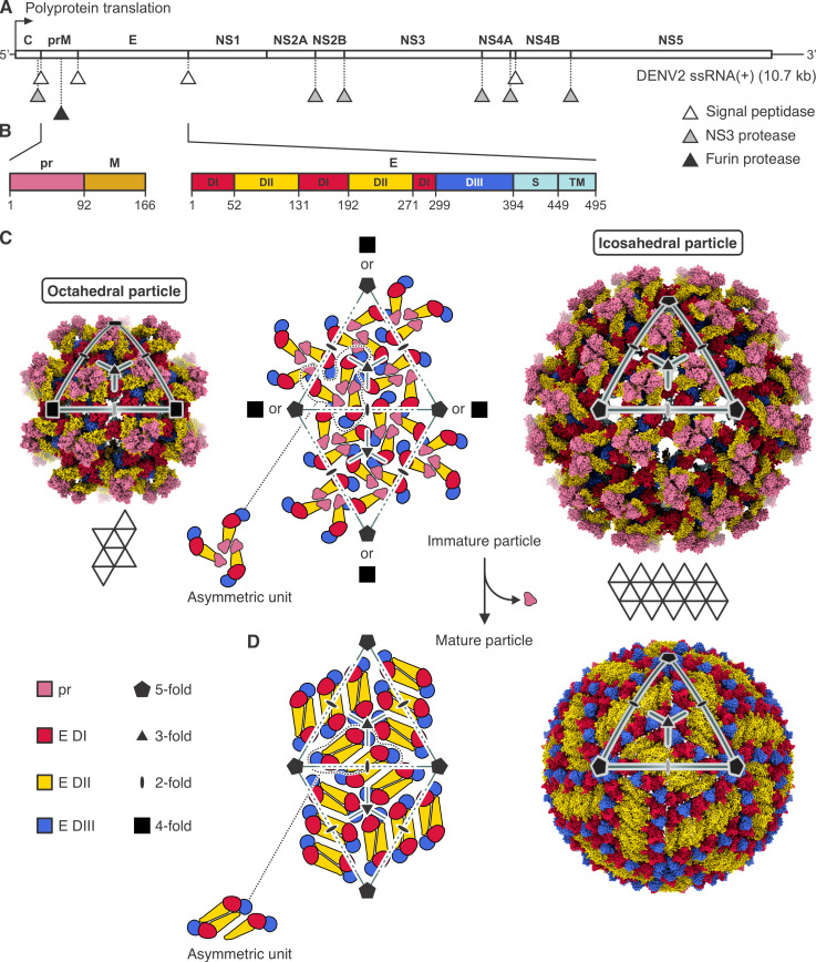
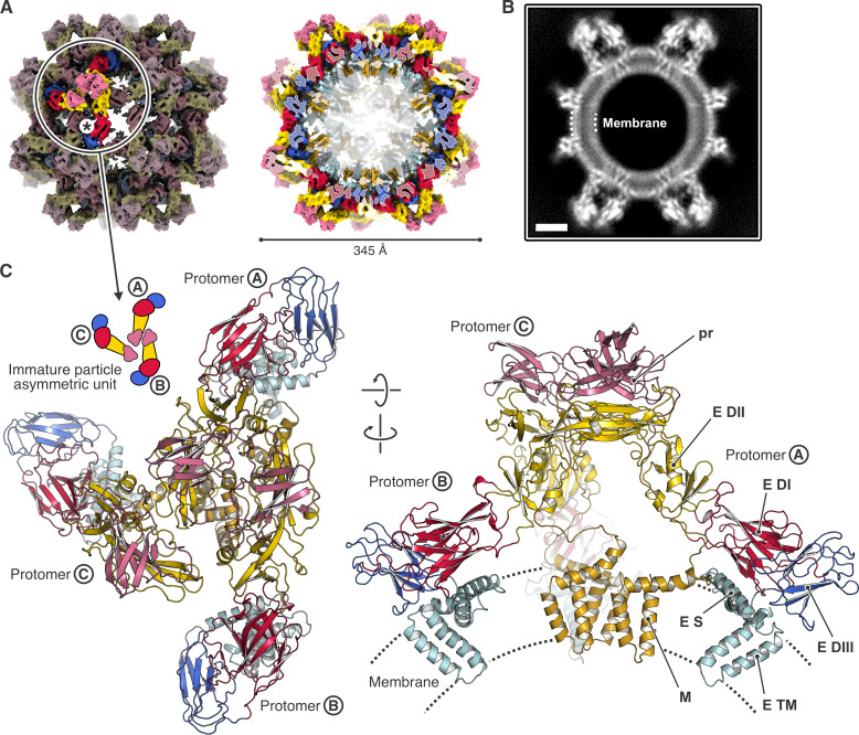
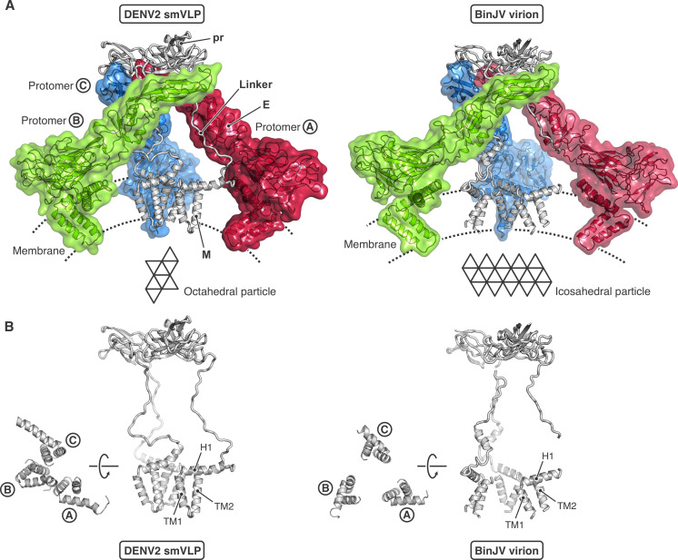
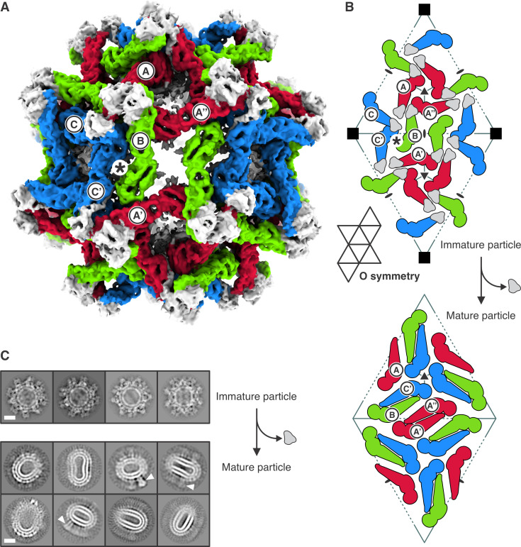
Flavivirus envelope (E) and precursor M (prM) proteins, when ectopically expressed, assemble into empty, virus-like particles (VLPs). Cleavage of prM to M and loss of the pr fragment converts the VLPs from immature to mature particles, mimicking a similar maturation of authentic virions. Most of the VLPs obtained by prM-E expression are smaller than virions; early, low-resolution cryo-EM studies suggested a simple, 60-subunit, icosahedral organization. We describe here the cryo-EM structure of immature, small VLPs (smVLPs) from dengue virus type 2 and show that they have octahedral rather than icosahedral symmetry. The asymmetric unit of the octahedral particle is an asymmetric trimer of prM-E heterodimers, just as it is on icosahedral immature virions; the full, octahedrally symmetric particle thus has 24 such asymmetric trimers or 72 prM-E heterodimers in all. Cleavage of prM and release of pr generates ovoid, somewhat irregular, mature particles. Previous work has shown that mature smVLPs have fusion properties identical to those of virions, consistent with local, virion-like clustering of 36 E dimers on their surface. The cryo-EM structure and the properties of the smVLPs described here relate directly to ongoing efforts to use them as vaccine immunogens.

Importance: Ectopic expression of flavivirus envelope (E) and precursor M (prM) proteins leads to the formation and secretion of empty, virus-like particles (VLPs). We show that a major class of VLPs, of smaller diameter than those of virion size ("small VLPs": smVLPs), are octahedrally symmetric particles. The known characteristics of immature virions (asymmetric trimers of prM-E heterodimers) allow us to understand the assembly of an octahedral (rather than icosahedral) surface lattice. Cleavage of prM and formation of mature, fusogenic smVLPs yield somewhat irregular, ovoid particles. These observations are directly relevant to proposals for using immunogenic but non-infectious VLPs as components of specific flavivirus vaccines.

Keywords: cryo-EM; flavivirus; structure; vaccine.


Conflict of interest statement

The authors declare no conflict of interest.

Figures

Fig 1
Fig 1
Flavivirus structural organization. (A) Flavivirus proteins are produced in the host cell from an approximately 10.7 kb long single-stranded positive sense genome/messenger RNA, ssRNA(+). A polyprotein is translated, which is co-translationally and post-translationally processed by different host and viral proteases (signal peptidase, NS3 protease, furin protease) at positions indicated by triangles. C, (anchored) capsid protein; prM, membrane glycoprotein precursor; E, envelope protein; NS1, nonstructural protein 1; NS2A, nonstructural protein 2A; NS2B, nonstructural protein 2B; NS3, nonstructural protein 3 (protease); NS4A, nonstructural protein 4A; NS4B, nonstructural protein 4B; NS5, nonstructural protein 5 (methyltransferase and RNA-dependent RNA polymerase). (B) Primary sequence and domain organization of the prM and E proteins. Residues at domain boundaries are numbered. pr sequence is colored pink, M is brown. E protein domains are labeled I (red), II (yellow), III (blue), S (stem, cyan), and TM (transmembrane region, cyan). prM is cleaved by furin protease after residue 91 (dengue virus type 2 [DENV2] sequence numbering). (C) Structural organization of immature flavivirus particles. The middle panel shows schematically the arrangement of prM-E protomers on a pseudohexagonal lattice. Folding of 20 triangular faces around fivefold axes results in icosahedral particles, such as the one shown in the right panel, which is the structure of an immature virion (immature Spondweni virus [SPOV] particle, PDB-ID 6ZQW). Folding of eight triangular faces around fourfold axes results in octahedral particles, such as the one shown in the left panel, which is the structure of the DENV2 small VLP (smVLP) reported here. In both the octahedral and the icosahedral particles, the triangulation number is T = 1 with three prM-E protomers per asymmetric unit. Protein domains are colored as in (B). (D) Structural organization of mature flavivirus particles. Proteolytic cleavage by furin in the TGN, exposure to mildly acidic pH, and release of virions from the host cell with dissociation of the pr protein are accompanied by substantial structural rearrangements of the M and E proteins to generate the mature particle, in which M and E form dimers. The structure of the mature particle has the same icosahedral symmetry as the immature particle (mature SPOV particle, PDB-ID 6ZQV).
Fig 2
Fig 2
Structure of the DENV2 octahedral small virus-like particle (smVLP). (A) Cryo-EM map of the octahedral smVLP at 6.5 Å resolution, viewed along a twofold symmetry axis. prM-E protein domains are colored according to the following scheme: pr, pink; M, brown; E DI, red; E DII, yellow; E DIII, blue; E stem and C-terminal domains, cyan. Left, one asymmetric unit, consisting of an asymmetric trimer of three prM-E protomers (labeled Ⓐ, Ⓑ, and Ⓒ), is highlighted. Asterisk shows position of local twofold axis. Right, the particle is cut to allow inside view of the M and E transmembrane α helices. (B) Projection of a 12 Å-thick central slice of the octahedral smVLP cryo-EM map showing density arising from the lipid bilayer. The scale bar corresponds to 50 Å. (C) Ribbon representation of the asymmetric trimer structure, viewed from the outside of the particle (left), and viewed from the side (right). The membrane is indicated schematically. Domains are colored as in (A).
Fig 3
Fig 3
Comparison of the prM-E asymmetric trimer from the octahedral DENV2 smVLP and the icosahedral BinJV immature flavivirus virion. (A) DENV2 smVLP (left) and BinJV virion (right, PDB-ID 7L30). The E subunits are shown in transparent surface representation and colored differently: protomer Ⓐ, red; protomer Ⓑ, green; protomer Ⓒ, blue. prM is shown in gray ribbon representation for all three protomers. The octahedral DENV2 smVLP (left) contains 72 prM-E protomers, with three asymmetric trimers within each of the eight triangular faces (3 × 3 × 8 = 72). The icosahedral immature BinJV virion (right) contains 180 prM-E protomers, with three asymmetric trimers within each of the 20 triangular faces (3 × 3 × 20 = 180). (B) View of the prM within one asymmetric trimer for the octahedral DENV2 smVLP (left) and the icosahedral immature BinJV virion (right).
Fig 4
Fig 4
Immature and mature conformations of DENV2 smVLPs. (A) Structure of the octahedral particle with the asymmetric trimers in immature conformation. Protomers are colored red, green, and blue. prM of all protomers is shown in gray. Protomers of the reference asymmetric unit are labeled Ⓐ, Ⓑ, and Ⓒ. Protomers of neighboring asymmetric units are labeled with a prime or double prime, e.g., A′, and A″. Asterisk shows the position of a local twofold axis relating protomers B and C′. (B) Arrangement of prM-E protomers in the immature particle (top). Subunit labels and colors as in (A). Note that the stem-TM of E (not shown explicitly) will enter the membrane beneath the center of an adjacent asymmetric ABC trimer. Thus, the stem-TM of the E subunit labeled C′ will contact the stem-TM of prM on subunit B. In the transition to a mature particle, E subunits C′ and B dimerize, and their associated M fragments (stem-TM of prM after the pr fragment dissociates) remain together. Retention of octahedral symmetry during maturation (i.e., cleavage of pr-M and loss of pr) would yield the packing of M-E protomers shown in the bottom panel. The elongated, mature smVLPs show, however, that octahedral symmetry is not retained. We describe in the text the reasons to propose that the dimer pairing illustrated is present. (C) 2D class averages of immature (top) and mature (bottom) DENV2 smVLPs. White arrowheads indicate what appear to be E trimers in post-fusion conformation. The scale bar corresponds to 100 Å.

References

    1. Kostyuchenko VA, Zhang Q, Tan JL, Ng TS, Lok SM. 2013. Immature and mature dengue serotype 1 virus structures provide insight into the maturation process. J Virol 87:7700–7707. doi:10.1128/JVI.00197-13 - DOI - PMC - PubMed
    1. Prasad VM, Miller AS, Klose T, Sirohi D, Buda G, Jiang W, Kuhn RJ, Rossmann MG. 2017. Structure of the immature Zika virus at 9 Å resolution. Nat Struct Mol Biol 24:184–186. doi:10.1038/nsmb.3352 - DOI - PMC - PubMed
    1. Yu IM, Holdaway HA, Chipman PR, Kuhn RJ, Rossmann MG, Chen J. 2009. Association of the pr peptides with dengue virus at acidic pH blocks membrane fusion. J Virol 83:12101–12107. doi:10.1128/JVI.01637-09 - DOI - PMC - PubMed
    1. Zhang Y, Kaufmann B, Chipman PR, Kuhn RJ, Rossmann MG. 2007. Structure of immature West Nile virus. J Virol 81:6141–6145. doi:10.1128/JVI.00037-07 - DOI - PMC - PubMed
    1. Newton ND, Hardy JM, Modhiran N, Hugo LE, Amarilla AA, Bibby S, Venugopal H, Harrison JJ, Traves RJ, Hall RA, Hobson-Peters J, Coulibaly F, Watterson D. 2021. The structure of an infectious immature flavivirus redefines viral architecture and maturation. Sci Adv 7. doi:10.1126/sciadv.abe4507 - DOI - PMC - PubMed

Substances

LinkOut - more resources