. 2025 Jan 3;387(6729):eadr0510.
doi: 10.1126/science.adr0510. Epub 2025 Jan 3.

Protective antibodies target cryptic epitope unmasked by cleavage of malaria sporozoite protein

Affiliations

Protective antibodies target cryptic epitope unmasked by cleavage of malaria sporozoite protein

Cherrelle Dacon et al. Science. .

Abstract

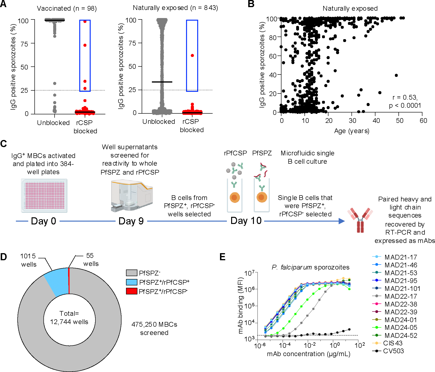
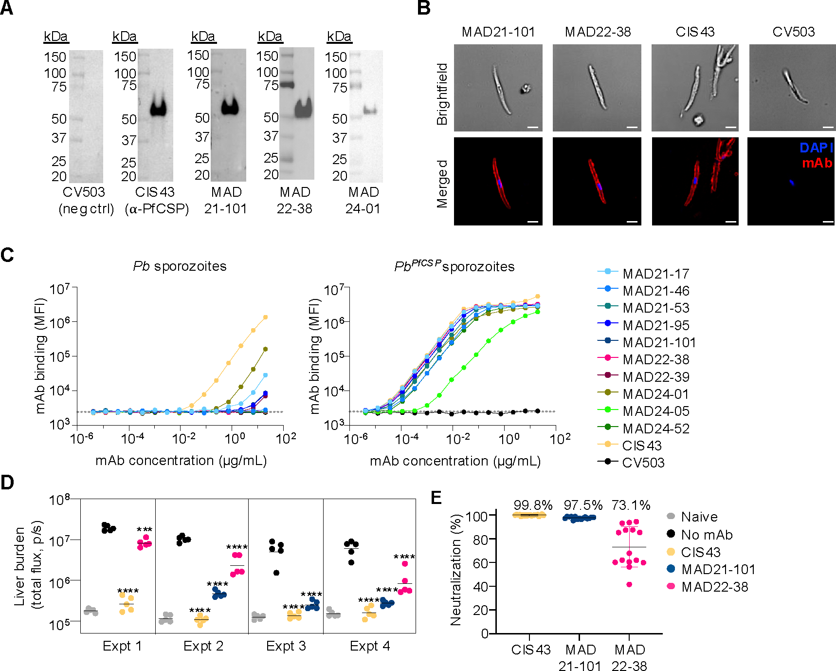
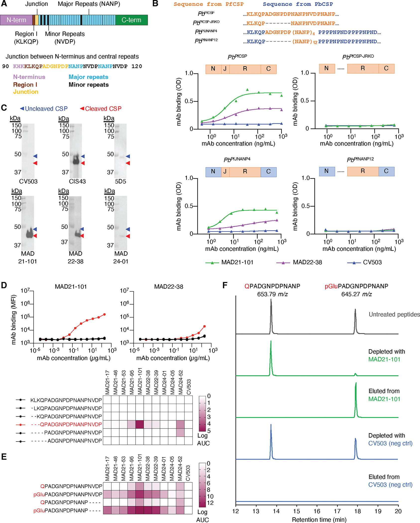
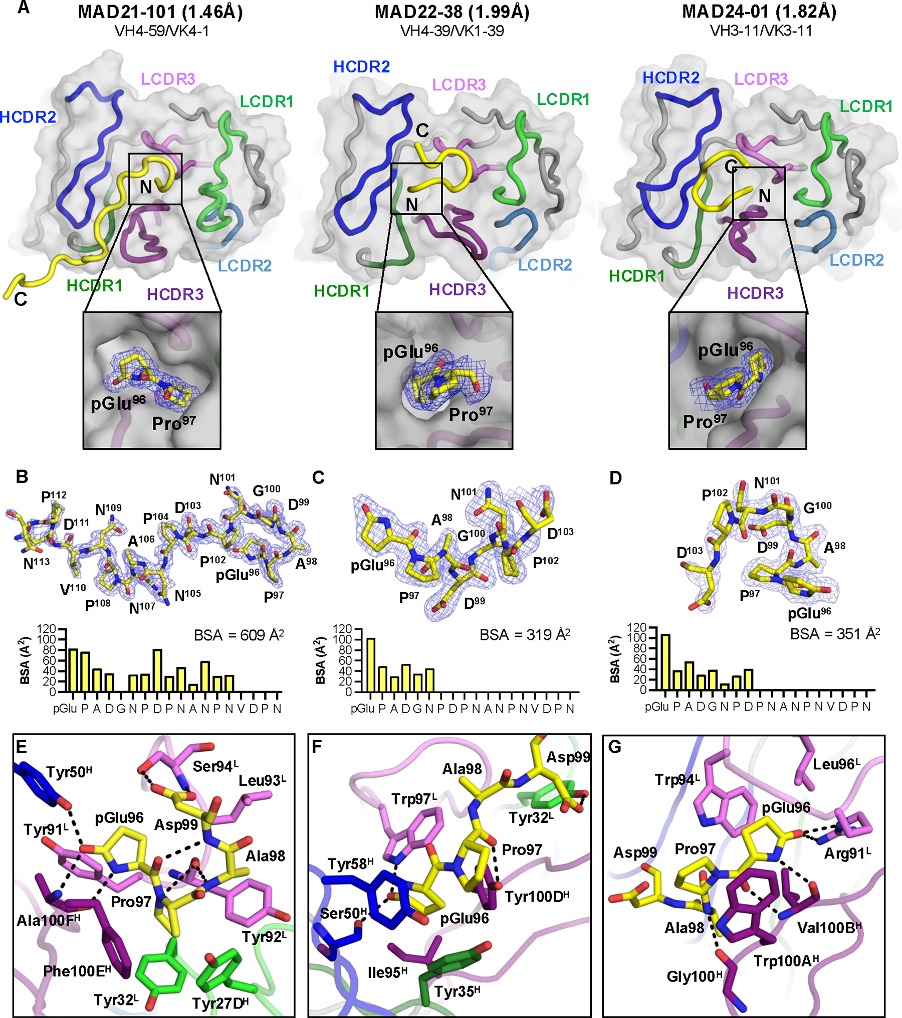
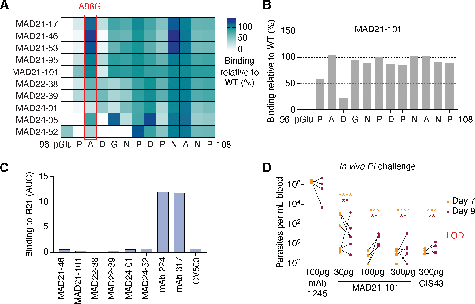
The most advanced monoclonal antibodies (mAbs) and vaccines against malaria target the central repeat region or closely related sequences within the Plasmodium falciparum circumsporozoite protein (PfCSP). Here, using an antigen-agnostic strategy to investigate human antibody responses to whole sporozoites, we identified a class of mAbs that target a cryptic PfCSP epitope that is only exposed after cleavage and subsequent pyroglutamylation (pGlu) of the newly formed N terminus. This pGlu-CSP epitope is not targeted by current anti-PfCSP mAbs and is not included in the licensed malaria vaccines. MAD21-101, the most potent mAb in this class, confers sterile protection against Pf infection in a human liver-chimeric mouse model. These findings reveal a site of vulnerability on the sporozoite surface that can be targeted by next-generation antimalarial interventions.


Conflict of interest statement

Competing interests: JT and CD are co-inventors on a provisional patent filed on the mAbs described in this study (PCT/US2023/074791). WW and KO are contractors for Henry M. Jackson Foundation for the Advancement of Military Medicine, Inc. and AB is a contractor for General Dynamics Information Technology, Inc working at the Agile Vaccines and Therapeutics Department, Naval Medical Research Command, Silver Spring. Contributions and experiments performed by LDSP do not bind or obligate the FDA. The content of this publication does not necessarily reflect the views or policies of the DHHS or of the institutions and companies with which the authors are affiliated. All other authors declare no competing interests.

Figures

Fig. 1.
Fig. 1.. Isolation of rare anti-sporozoite mAbs using an antigen-agnostic approach.
(A) Plasma IgG reactivity to Pf sporozoites with (red) and without (grey) pre-blocking of plasma samples by rPfCSP. Bars show mean. Donors with ≥8% reactivity to sporozoites after rPfCSP blocking in the initial screen were subjected to repeat analysis and these validated datapoints are shown in the plot for those donors. The dotted line shows the final 25% cutoff chosen to select positive donors, with down-selected donors enclosed in blue. (B) Correlation of plasma IgG reactivity to whole Pf sporozoites with donor age from the malaria-exposed cross-sectional cohort, p value and correlation coefficient were derived by Spearman’s rank correlation analysis. (C) Schematic of the antigen-agnostic workflow for isolation of mAbs against Pf sporozoites. IgG+ MBCs are sorted from PBMC samples and plated at a density of 25–100 cells per well in 384-well plates. The cells are activated and cultured for 9 d, followed by screening of supernatants for binding to Pf sporozoites and PfCSP. B cells of interest (PfSPZ+, PfCSP) are transferred to a microfluidics chip for single-cell screening against Pf sporozoites and PfCSP. Single B cells of interest (PfSPZ+, PfCSP) are selected for mAb production. PfSPZ, Pf sporozoites. Image produced using Biorender. (D) Number of MBC culture supernatants reactive towards whole Pf sporozoites and rPfCSP. (E) Titration curves for recombinant mAb binding to whole, freshly dissected Pf sporozoites. MAD22-17 bound more poorly to sporozoites than the other mAbs and was excluded from subsequent analysis. CIS43 is a control anti-PfCSP mAb and CV503 is a negative control anti-SARS-CoV-2 mAb. The dotted line shows the binding level with the buffer only control.
Fig. 2.
Fig. 2.. Isolated mAbs exclusively target Plasmodium-expressed PfCSP and reduce liver parasite burden in vivo.
(A) Western blot analysis of Pf sporozoite lysates. Lysates were analyzed by reducing SDS-PAGE and individual blots were probed with representative isolated mAbs (MAD21-101, MAD22-38 and MAD24-01), a control anti-PfCSP mAb (CIS43), and an anti-SARS-CoV-2 spike mAb as an isotype control (CV503). Lysate from 5 × 10⁴ sporozoites was loaded into each well. (B) Immunofluorescence images of Pf sporozoite surface staining with MAD21-101 and MAD22-38, an anti-PfCSP control mAb CIS43, and an isotype control anti-SARS-CoV-2 mAb CV503. Scale bar = 2 μm. (C) Flow cytometry analysis of mAb binding to freshly dissected sporozoites: wild-type P. berghei sporozoites and transgenic P. berghei sporozoites that express full-length PfCSP (PbPfCSP). CIS43 was included as a control anti-PfCSP mAb and the dotted line denotes MFI for the buffer control. WT, wild-type. (D and E) In vivo assay to evaluate mAb potency, as measured by liver parasite burden reduction. In this assay, 300 μg of each mAb was delivered 16 h pre-IV challenge with PbPfCSP sporozoites that express the luciferase reporter enzyme (PbPfCSP-GFP/FLuc). Liver parasite burden was measured by in vivo imaging and quantified as luminescence flux signals. The naïve group consisted of mice that were not infected with sporozoites and were used to determine the baseline flux signal, while the “no mAb” group was infected with sporozoites but not administered any mAb. (D) Liver burden reduction from four independent experiments. Data are shown with geometric mean for n = 5 mice per group and statistical significance was determined versus the no mAb condition using one-way ANOVA testing with Dunnett’s Test for multiple comparisons. *P < 0.05, **P < 0.01, ***P < 0.001, ****P < 0.0001 and ns, not significant. (E) Potency of the mAbs calculated based on sporozoite neutralization. Neutralization data are derived from at least 3 independent experiments. Bars show mean with standard deviation.
Fig. 3.
Fig. 3.. MAD21-101-type mAbs target the newly formed, pyroglutamate-modified N-terminus of PfCSP after sporozoite cleavage
(A) Schematic of PfCSP depicting the highly conserved region I (brown), the junction (beige) and central major NANP repeats (sky blue) interspersed with minor NVDP repeats (black), which are flanked by the N- and C-terminus (pink and green, respectively). Below, the sequence of region I, junction and initial repeats are shown. (B) ELISA analysis of mAb binding to lysate from Pb sporozoites that express full PfCSP (PbPfCSP) or PfCSP lacking key junction region residues (PbPfCSP-JRKO), and chimeric Pb sporozoites that express the PfCSP junction and NANP repeats (PbPfJNANP4) or PfCSP NANP repeats only (PbPfNANP12). Sequences and domains in orange are derived from PfCSP, while sequences and domains in blue are derived from PbCSP. SARS-CoV-2 spike mAb CV503 was included as an isotype control. (C) Western blot analysis of mAb binding to cleaved and uncleaved forms of PfCSP from Pf sporozoite lysate. Lysates were analyzed by SDS-PAGE under reducing conditions and individual blots were probed with MAD21-101, MAD22-38 and MAD24-01. Control mAbs that bind to uncleaved PfCSP only (5D5) or both cleaved and uncleaved PfCSP (CIS43) were included. CV503 IgG1 was included as an isotype control and lysate from 5 × 10⁴ sporozoites was loaded into each well. (D) Representative titration curves and AUC heat map showing mAb binding to N-terminally truncated PfCSP peptides. AUC, area under the curve. (E) AUC heat map showing mAb binding to PfCSP peptides with either an N-terminal Gln96 or pGlu96 residue. (F) An equimolar mixture of peptides with N-terminal Gln96 or pGlu96 was immunoprecipitated with MAD21-101 or CV503 (isotype control) and analyzed by LC-MS. The mAb-depleted supernatants were analyzed in addition to eluates. Shown are extracted ion chromatograms for the peptides. Retention times are aligned. Peptide abundance is normalized and offset for clarity. Peptide identity was confirmed from fragment spectra.
Fig. 4.
Fig. 4.. Structural basis of MAD21-101-like antibody epitope recognition and specificity.
(A) Overall view of X-ray structures of MAD21-101, MAD22-38, and MAD24-01 in complex with a peptide corresponding to PfCSP residues pGlu96-Asn113. The Fab surface shown in gray, bound peptide as a yellow backbone cartoon representation, framework regions in gray cartoons, and CDR1, CDR2, and CDR3 of Fab heavy and light chains are shown in dark and light green, blue, and purple, respectively. Inset showing the bound N-terminus in a pocket at the heavy-light chain interface, with pGlu-Pro motifs represented as yellow sticks with the electron density map (2Fo-Fc) in blue mesh (contoured to 1σ). Bound peptide of (B) MAD21-101, (C) MAD22-38, and (D) MAD24-01 shown in yellow sticks and electron density map (2Fo-Fc) in blue mesh (contoured to 1σ). Per-residue peptide buried surface area (Å2) at the peptide-Fab interface shown as yellow bars. (E-G) Binding interactions between the N-terminal peptide residues pGlu96-Asp99 to the hydrophobic pockets of (E) MAD21-101, (F) MAD22-38, and (G) MAD24-01, with interacting residues shown as sticks and hydrogen bonds in black dashed lines.
Fig. 5.
Fig. 5.. MAD21-101 binds to a new conserved epitope within PfCSP and provides sterile protection against Pf sporozoite infection.
(A) Heat map showing binding of MAD21-101-type mAbs to pGlu-PfCSP peptides bearing alanine or glycine (when original residue was alanine) substitutions. Colors are based on area under the curve (AUC) binding relative to binding to the wild-type peptide. The red rectangle highlights the common A98G substitution. (B) Binding of MAD21-101 to pGlu-CSP peptides bearing alanine or glycine (when original residue was alanine) substitutions. Bars represent binding in AUC versus binding to the wild-type peptide sequence and the red line denotes 50% binding versus the wild-type sequence. (C) Binding of MAD21-101-type and previously identified mAbs (317 and 224) to the PfCSP-based R21 vaccine. Analysis performed using an Meso Scale Discovery (MSD) assay. An anti-COV-2 spike IgG1 (CV503) was included as an isotype control. (D) Parasitemia in human liver-chimeric (FRG-huHep) mice after mosquito bite challenge with Pf sporozoites. An anti-Pfs25 IgG1 (mAb 1245) was included an isotype control and the red dotted line represents the limit of detection for the qRT-PCR assay (5 parasites/mL). Statistical significance was determined versus the isotype control using a mixed-effects model with Šídák’s multiple comparisons test. *P < 0.05, **P < 0.01, ***P < 0.001, ****P < 0.0001 and ns, not significant.

Similar articles

Cited by

References

    1. World Health Organisation, World malaria report 2023. https://www.who.int/teams/global-malaria-programme/reports/world-malaria... (2023).
    1. Sidjanski S, Vanderberg JP, Delayed migration of Plasmodium sporozoites from the mosquito bite site to the blood. Am J Trop Med Hyg 57, 426–429 (1997). - PubMed
    1. Boyd MF, Kitchen SF, The demonstration of sporozoites in human tissues. Am J Trop Med Hyg s1–19, 27–31 (1938).
    1. Amino R et al., Quantitative imaging of Plasmodium transmission from mosquito to mammal. Nat Med 12, 220–224 (2006). - PubMed
    1. Vanderberg JP, Frevert U, Intravital microscopy demonstrating antibody-mediated immobilisation of Plasmodium berghei sporozoites injected into skin by mosquitoes. Int J Parasitol 34, 991–996 (2004). - PubMed

Publication types

MeSH terms

LinkOut - more resources