. 2025 Jan 2;16(1):2.
doi: 10.1038/s41467-024-54791-0.

Remodelling of the immune landscape by IFNγ counteracts IFNγ-dependent tumour escape in mouse tumour models

Affiliations

Remodelling of the immune landscape by IFNγ counteracts IFNγ-dependent tumour escape in mouse tumour models

Vivian W C Lau et al. Nat Commun. .

Abstract

Loss of IFNγ-sensitivity by tumours is thought to be a mechanism enabling evasion, but recent studies suggest that IFNγ-resistant tumours can be sensitised for immunotherapy, yet the underlying mechanism remains unclear. Here, we show that IFNγ receptor-deficient B16-F10 mouse melanoma tumours are controlled as efficiently as WT tumours despite their lower MHC class I expression. Mechanistically, IFNγ receptor deletion in B16-F10 tumours increases IFNγ availability, triggering a remodelling of the immune landscape characterised by inflammatory monocyte infiltration and the generation of 'mono-macs'. This altered myeloid compartment synergises with an increase in antigen-specific CD8+ T cells to promote anti-tumour immunity against IFNγ receptor-deficient tumours, with such an immune crosstalk observed around blood vessels. Importantly, analysis of transcriptomic datasets suggests that similar immune remodelling occurs in human tumours carrying mutations in the IFNγ pathway. Our work thus serves mechanistic insight for the crosstalk between tumour IFNγ resistance and anti-tumour immunity, and implicates this regulation for future cancer therapy.


Conflict of interest statement

Competing interests: The authors declare no competing interests.

Figures

Fig. 1
Fig. 1. Presence of mutations in IFNγ signalling pathway genes does not preclude decrease in overall survival in clinical data.
A Frequency of alterations in IFNGR1, IFNGR2, JAK1, JAK2, or STAT1 (IFNγ pathway) across cancers in The Cancer Genome Atlas (TCGA), where cases in green represent gene mutations and purple are structural variants of the genes. For endometrial cancer, samples with POLE mutations have been excluded. Comparison of survival curves of endometrial (B, n = 462 without and n = 50 with mutation in the IFNγ pathway, exclusion of samples with POLE mutations), melanoma (C, n = 366 without and n = 57 with mutation in the IFNγ pathway), colorectal (D, n = 473 without and n = 49 with mutation in the IFNγ pathway), and esophagogastric (E, n = 1034 without and n = 103 with mutation in the IFNγ pathway) human cancers between unaltered cases and cases with mutations in IFNγ pathway genes. p values represent log-rank testing.
Fig. 2
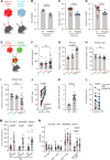
Fig. 2. B16F10 melanoma tumours with CRISPR-Cas9 knockout of IFNγR1 are efficiently controlled by the endogenous anti-tumour response.
AD B16-OVA WT (red) or IFNγRKO (blue) cells were engrafted subcutaneously into the flanks of C57Bl/6 WT mice and tumours were harvested after 11–16 days. A Diagram of the experimental setup. Graphics created with BioRender. Surface expression of IFNγR1 (B n[WT] = 3, n[IFNγRKO] = 6 for from two independent experiments), MHC-I H2-Db (C n[WT] = 17, n[IFNγRKO] = 10 from four independent experiments), and MHC-II I-A/I-E (D n[WT] = 17 for WT, n[IFNγRKO] = 10 from four independent experiments) expression on mCherry+ CD45 cells were analysed by flow cytometry. EI WT and IFNγRKO (KO) tumours expressing mCherry-OVA or ZsGreen-OVA transgenes were admixed 1:1 prior to engraftment in WT (F, G), IFNγKO (H) or CD8ɑKO mice (I) mice. E Experimental design. Graphics created with BioRender. F Tumour volumes of WT, IFNγRKO, or admixed tumours taken at endpoint on days 12–14 post-engraftment from three independent experiments for WT:WT (red; n = 11) and KO:KO (blue; n = 10), and admixed WT:KO tumours (purple; n = 37). GI Tumours were harvested and analysed by flow cytometry. Outgrowth of KO tumour cells relative to WT, expressed as percent selection of mCherry+ cells in control WT:WT (red; n = 6 (G, H), 7 (I)), KO:KO (blue; n = 7 (G, H), 8 (I)), or WT:KO (purple; n = 12 (G), 15 (H), 14 (I)) tumours at days 14–17 in WT (G), IFNγ KO (H) and CD8ɑKO (I) mice. Cells were gated on live CD45 mCherry+. J, K WT and IFNγRKO tumours expressing mCherry-OVA or ZsGreen-OVA transgenes were admixed 1:1 in vitro and treated with 10 ng/ml IFNγ when indicated. Ki67 staining (n = 16) (J) and the ratio between ZsGreen and mCherry (KO/WT) (n[Ctrl] = 3; n[IFNγ] = 17) (K) were assessed by flow cytometry after 2 days. Data are from three independent experiments, each n corresponds to a well (sample). L WT and IFNγRKO tumours expressing mCherry-OVA or ZsGreen-OVA transgenes were admixed 1:1 in vitro and treated with 10 ng/ml IFNγ for 10 h. Activated OTI T cells were added to tumour cells for 5 h (OTI:tumours = 2:1). The percentage of apoptotic cells was quantified by flow cytometry using Annexin V staining (n = 17). Data are from two independent experiments. M WT and IFNγRKO tumours expressing mCherry-OVA or ZsGreen-OVA transgenes were admixed 1:1 prior to engraftment in WT or CD8ɑKO mice. Tumour volumes of admixed tumours from WT (n[WT:WT] = 9, n[KO:KO] = 7, n[admix] = 13) or CD8ɑKO (n[WT:WT] = 7, n[KO:KO] = 8, n[admix] = 14) mice was measured on day 12/13 post-engraftment. Data are from two independent experiments (N) WT and IFNγRKO (KO) tumours expressing mCherry-OVA or ZsGreen-OVA transgenes were admixed 1:1 (WT:WT = red; KO:KO = blue, WT:KO = purple) prior to engraftment in WT mice and harvested at days 12–14 post-engraftment. Infiltration of lymphocyte populations as a percent of total CD45+ cells from admixed tumours in WT mice (n[WT:WT] = 11, n[KO:KO] = 10, n[admix] = 18) was analysed by flow cytometry. Each n is a tumour. Data are pooled from two or more independent experiments unless otherwise indicated. All data show mean ± SEM with p values by non-parametric two-sided Mann–Whitney t tests for comparisons between two groups, two-sided paired t tests for paired values, Kruskal–Wallis tests between three groups with multiple comparisons correction using Dunn’s method, and two-way ANOVA using Šídák’s test for multiple comparisons between multiple two or more groups of data. A, E Created in BioRender. Gerard (2024) https://BioRender.com/k40f729.
Fig. 3
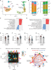
Fig. 3. Single-cell RNAseq analysis of CD45+ tumour-infiltrating cells reveals the presence of enhanced inflammatory milieu in IFNγRKO tumours.
A, B UMAP projection of CD45+ cells and relative abundance of distinct immune populations in WT and IFNγRKO tumours. Clusters show a combined 7014 cells, with 3004 cells from WT tumours, and 4010 cells from IFNγRKO tumours. Gene set enrichment analysis using hallmark (C) or Reactome (D) databases for identification of enriched signalling pathways, expressed as normalised enrichment scores (NES). Intra-tumoural concentrations of IFNγ (n[WT] = 25, n[KO] = 26) (E), IL-6 (n[WT] = 17, n[KO] = 22) (F), IL-4 (n[WT] = 21, n[KO] = 22) (G), IL-10 (n[WT] = 11, n[KO] = 6) (H) measured by ELISA or LegendPlex using supernatants of ex vivo WT (red) and IFNγRKO (blue) dissociated tumours, normalised to tumour weight. Data are pooled from four or more independent experiments. Data show mean ± SEM with p values by non-parametric two-sided Mann–Whitney t tests. I, J CellChat ligand–receptor inference analysis was performed on scRNAseq data from (A). I Heatmap of the differential number of interactions between sender (y-axis) and receiver (x-axis) populations. Bar plots on each axis represent the sum of all interactions in absolute values for each sender or receiver cell type. J Circle plot visualising strength of signalling interactions between immune populations from WT and IFNγRKO tumours. Vertices represent independent populations, and arrows indicate the direction of signals sent, where broader lines represent increased communication probability of signalling interactions.
Fig. 4
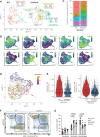
Fig. 4. Less differentiated monocyte–macrophage subsets with pro-inflammatory signatures are prominent in IFNγRKO tumours.
AE Myeloid cells from the scRNAseq data from Fig. 3A were subset and re-clustered. A UMAP of WT and IFNγRKO monocyte/macrophage subclusters with distinct myeloid subtypes highlighted by representative gene signatures. B Feature plots showing relative gene expression of key monocyte/macrophage genes. C Relative frequencies of each subcluster expressed as stacked bar plots. D Trajectory analysis overlaid on the UMAP projection of monocyte/macrophage cell clusters, coloured by pseudotime. E Violin plots comparing module scoring of angiogenic and regulatory TAMs gene signatures of macrophage subclusters of WT and IFNγRKO tumour samples. Box plots indicate median (middle line), 25th, 75th percentile (box) (n[WT] = 827 n[KO] = 1210 cells, 3 mice pooled). F, G WT, IFNγRKO or admix tumours were engrafted in WT mice and analysed by flow cytometry after 14 days. F Representative flow plots of tumour-infiltrating CD45+ CD11b+ cells from WT and IFNγRKO tumours, gated by Ly6C and MHC-II expression for delineation of monocyte, mono-mac and macrophage populations. Plots are representative of three or more independent experiments. G Relative frequencies of each gated population from WT (n = 14), IFNγRKO (n = 12) or admixed (n = 6) tumours. Data are pooled from three independent experiments. Data show mean ± SEM with p values by two-way ANOVA using Šídák’s test for multiple comparisons.
Fig. 5
Fig. 5. Control of IFNγRKO tumours is diminished in CCR2KO mice following lack of monocyte recruitment.
A, B WT (red) or IFNγRKO (blue) tumours were engrafted in WT (circle) or CCR2KO (triangle) mice. A Infiltration of myeloid populations relative to total CD45+ cells in WT or IFNγRKO tumours engrafted into WT (n[WT tumours] = 8, n[KO tumours] = 8) or CCR2KO (n[WT tumours] = 5, n[KO tumours] = 7) mice. B Tumour volumes of WT (n = 9) or IFNγRKO (n = 10) tumours in CCR2KO hosts measured at endpoint on day 13 post-engraftment. Data are from two independent experiments. C WT (red) or IFNγRKO (blue) tumours were engrafted in WT mice. Mice were treated with the INOS inhibitor L-NAME (INOS Inh) when indicated. Tumour volumes were measured on day 13 post-engraftment (n[Ctrl] = 7, n[L-NAME] = 8). D, E WT (red) or IFNγRKO (blue) tumours were engrafted in WT (circle; n[WT tumours] = 13, n[KO tumours] = 10) or CCR2KO (triangle; n[WT tumours] = 8, n[KO tumours] = 9) mice. D Infiltration of T cells and NK cells relative to total CD45+ cells. E Frequency of OVA-specific T cells as a proportion of CD8+ T cells in WT (n[WT tumours] = 12, n[KO tumours] = 10) and CCR2KO (n[WT tumours] = 8, n[KO tumours] = 10) mice. Data are from two independent experiments. FH Frozen sections from WT or IFNγRKO tumours engrafted in WT mice were stained with the indicated markers and imaged. Representative images indicating location of Ly6C (red) and F4/80 (Cyan) expressing cells relative to CD31+ (green) blood vessels. Scale bar = 40 µm (F), and 100 µm (G). H Graph shows the percentage of blood vessels that are in contact with Ly6C+ cells. Each dot is a tumour (n = 3). All data show mean ± SEM with p values by non-parametric two-sided Mann–Whitney t tests for comparisons between two groups, and two-way ANOVA using Šídák’s test for multiple comparisons between multiple two or more groups of data.
Fig. 6
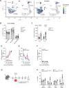
Fig. 6. Recruitment of monocytes is driven by CD8+ T cell-derived IFNγ.
A Module scoring of an IFNγ gene signature on the single-cell dataset from Fig. 3A. B Infiltration of myeloid populations relative to total CD11b+CD45+ cells in WT (red; n = 6), IFNγRKO (blue; n = 7) or admixed (purple; n = 14) tumours engrafted into IFNγKO mice. Data are pooled from two independent experiments. C Tumour volumes of WT (red) or IFNγRKO (blue) tumours measured on day 10 post-engraftment of WT (n[WT tumours] = 13, n[KO tumours] = 8) or IFNγKO (n[WT tumours] = 7, n[KO tumours] = 6) mice. Data are pooled from two independent experiments. DF WT or IFNγRKO tumours were engrafted in GREAT mice and harvested when indicated. Percentage of cells which are IFNγ+, as measured by EYFP expression by tumour-infiltrating CD8+ T cells (D), NK cells (E), and CD4+ T cells (F). Data are pooled from four independent experiments, with timepoints varying between experiments (WT tumours: n[day7/8] = 8, n[day9/10] = 16, n[day11/12] = 8, n[day14/16] = 4; KO tumours: n[day7/8] = 5, n[day9/10] = 13, n[day11/12] = 6, n[day14/16] = 5 day). G, H Antibody depletion of CD8+ T cells using anti-CD8β before and following tumour engraftment. G Experimental design. Graphics created with BioRender. H Frequency of specific Ly6Chi monocyte subsets following CD8+ T cell depletion was analysed by flow cytometry 13 days post-engraftment (n[WT-isotype] = 4, n[WT-anti-CD8β] = 3, n[KO-isotype] = 2, n[KO-anti-CD8β] = 3). All data show mean ± SEM with Kruskal–Wallis testing between three groups with multiple comparisons correction using Dunn’s method, and two-way ANOVA using Šídák’s test for multiple comparisons between multiple two or more groups of data. G Created in BioRender. Gerard (2024) https://BioRender.com/k40f729.
Fig. 7
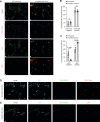
Fig. 7. CD8-monocyte crosstalk occurs close to blood vessels.
AC WT or IFNγRKO tumours were grown in WT mice. Frozen sections from WT or IFNγRKO tumours were stained with the indicated markers and imaged. A Representative immunofluorescence images taken at the core of WT and IFNγRKO tumours showing the location of Ly6C (red) and CD8 (magenta) cells relative to CD31+ blood vessels (green). Scale bar = 30 µm. B Bar graph shows the percentage of CD31+/Ly6C+ structures that contain CD8+ T cells Each dot is a tumour (n = 3). C Bar graph shows the percentage of CD31+/CD8+ T cell structures that contain Ly6C cells. Each dot is a tumour (n = 3). D, E IFNγRKO tumours were grown in GREAT mice. Frozen sections from IFNγRKO tumours were stained with the indicated markers and imaged. D Representative immunofluorescence images taken at the core of IFNγRKO tumours showing the location of CD8+ cells (red) and IFNγ expressing cells (green) relative to CD31+ blood vessels (cyan). E Representative immunofluorescence images taken at the core of IFNγRKO tumours showing the location of Ly6C+ cells (red) and IFNγ expressing cells (green) relative to CD31+ blood vessels (cyan). This is a representative example of three independent tumours. Scale bar = 30 µm All data show mean ± SEM with two-way ANOVA using Šídák’s test for multiple comparisons between two or more groups of data.
Fig. 8
Fig. 8. CD8-monocyte signatures are elevated in human tumours with identified IFNγ-pathway mutations, and spatially overlap with IFNγ response signatures.
A Enrichment scoring of CD8-monocyte gene signatures using single gene set enrichment analysis of human TCGA RNAseq datasets with (yellow) and without (blue) IFNγ-pathway mutations. Normalised gene counts from each tumour type were taken from samples which had moderate or high impact IFNGR1/2, JAK1/2, STAT1 mutations determined by whole-exome sequencing. Box plots indicate median (middle line), 25th, 75th percentile (box). Number of samples included for analysis are indicated for each sample set, and statistical testing using two-sided Wilcoxon signed-rank test with adjust p values by false discovery rate testing is shown. B Analysis of 10X Genomics Visium datasets for hallmark IFNγ response, CD8-monocyte, and endothelial cell gene signatures for human lung squamous cell carcinoma (B) or colon adenocarcinoma (C) samples. Gene set expression is indicated by heatmap, where colours represent log-normalised average expression.

References

    1. Kluger, H. M. et al. Defining tumor resistance to PD-1 pathway blockade: recommendations from the first meeting of the SITC Immunotherapy Resistance Taskforce. J. Immunother. Cancer8, e000398 (2020). - PMC - PubMed
    1. Sharma, P., Hu-Lieskovan, S., Wargo, J. A. & Ribas, A. Primary, adaptive, and acquired resistance to cancer immunotherapy. Cell168, 707–723 (2017). - PMC - PubMed
    1. Chin, Y. E. et al. Cell growth arrest and induction of cyclin-dependent kinase inhibitor p21WAF1/CIP1 mediated by STAT1. Science272, 719–722 (1996). - PubMed
    1. Shankaran, V. et al. IFNγ and lymphocytes prevent primary tumour development and shape tumour immunogenicity. Nature410, 1107–1111 (2001). - PubMed
    1. Zhou, F. Molecular mechanisms of IFN-γ to up-regulate MHC Class I antigen processing and presentation. Int. Rev. Immunol.28, 239–260 (2009). - PubMed

Publication types