Review
. 2025 Jan 3;10(1):2.
doi: 10.1038/s41392-024-02088-5.

In defence of ferroptosis

Affiliations
Review

In defence of ferroptosis

Francesca Alves et al. Signal Transduct Target Ther. .

Abstract

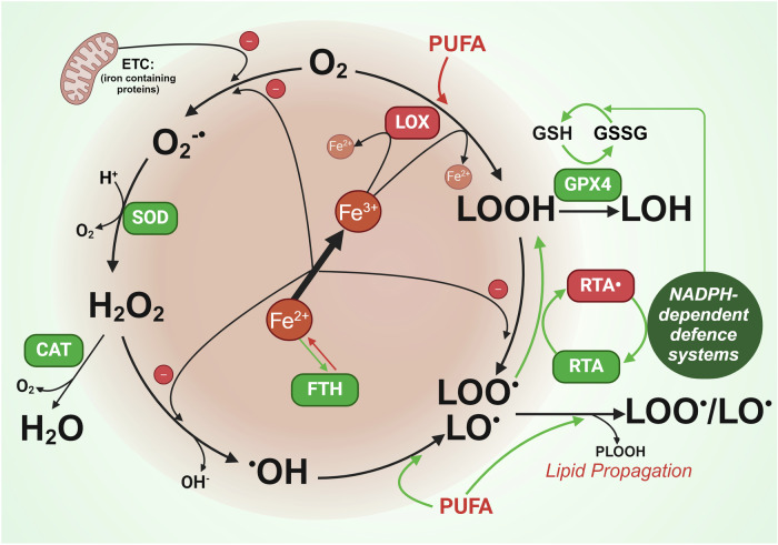
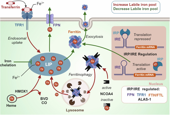
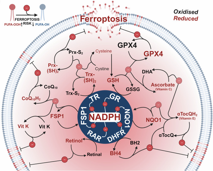
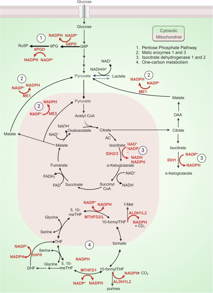
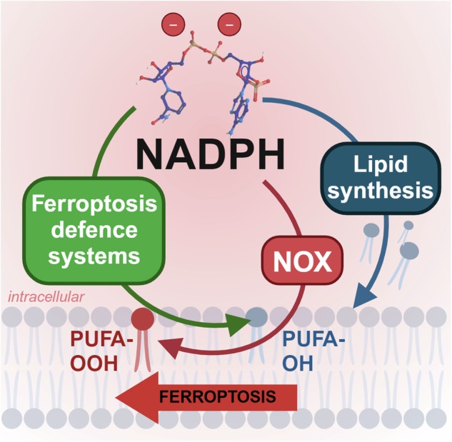
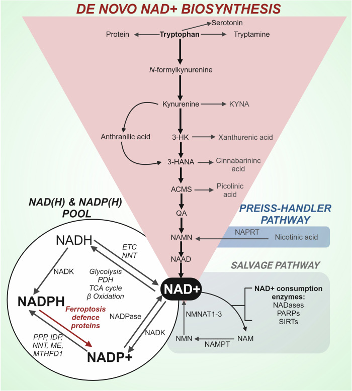
Rampant phospholipid peroxidation initiated by iron causes ferroptosis unless this is restrained by cellular defences. Ferroptosis is increasingly implicated in a host of diseases, and unlike other cell death programs the physiological initiation of ferroptosis is conceived to occur not by an endogenous executioner, but by the withdrawal of cellular guardians that otherwise constantly oppose ferroptosis induction. Here, we profile key ferroptotic defence strategies including iron regulation, phospholipid modulation and enzymes and metabolite systems: glutathione reductase (GR), Ferroptosis suppressor protein 1 (FSP1), NAD(P)H Quinone Dehydrogenase 1 (NQO1), Dihydrofolate reductase (DHFR), retinal reductases and retinal dehydrogenases (RDH) and thioredoxin reductases (TR). A common thread uniting all key enzymes and metabolites that combat lipid peroxidation during ferroptosis is a dependence on a key cellular reductant, nicotinamide adenine dinucleotide phosphate (NADPH). We will outline how cells control central carbon metabolism to produce NADPH and necessary precursors to defend against ferroptosis. Subsequently we will discuss evidence for ferroptosis and NADPH dysregulation in different disease contexts including glucose-6-phosphate dehydrogenase deficiency, cancer and neurodegeneration. Finally, we discuss several anti-ferroptosis therapeutic strategies spanning the use of radical trapping agents, iron modulation and glutathione dependent redox support and highlight the current landscape of clinical trials focusing on ferroptosis.


Conflict of interest statement

Competing interests: Ashley Bush is an Associate Editor of Signal Transduction and Targeted Therapy, but he has not been involved in the process of the manuscript handling. All the other authors declare no conflict of interest.

Figures

Fig. 1
Fig. 1
Central role of iron in reactive oxygen species generation and lipid peroxidation. Iron is involved directly and indirectly at several points to produce reactive oxygen species and lipid peroxidation. Indirectly, iron containing proteins in the electron transport chain (ETC) generate O2-• which is reduced to H2O2 by superoxide dismutase (SOD). H2O2 can either be quenched by catalase (CAT) or react with iron via the Fenton reaction, to generate hydroxyl radicals (•OH). The Fenton reaction can also catalyse the production of lipid peroxyl radicals (LOO• /LO•) from lipid hydroperoxide (LOOH). Radical trapping agents (RTAs) can quench lipid peroxyl radicals. Indirectly, iron contained in lipoxygenases (LOX) catalyse oxygenation of polyunsaturated fatty acids (PUFAs) and lipids to produce lipid hydroperoxide (LOOH). Glutathione peroxidase 4 (GPX4) can siphon lipid hydroperoxides away from fuelling lipid peroxidation and propagation by reducing PLOOH (high ferroptosis risk) to benign lipid alcohols (LOH). The reducing power of GPX4 is fuelled by reduced glutathione (GSH) which is dependent on NADPH to be recycled from its reduced form glutathione disulfide (GSSG). The breakdown of iron storage protein ferritin (FTH) can result in increased labile iron to facilitate these reactions. Figure created using Biorender.coms
Fig. 2
Fig. 2
Cellular labile iron pool regulation. The labile iron pool is regulated by several proteins including i.) transferrin receptor 1 (TFR1) that facilitates iron influx in the form of transferrin, ii.) ferroportin (FPN) an iron channel facilitating iron export, iii) Ferritin which can store labile iron or release iron after lysosomal degradation which is mediated by nuclear receptor activator 4 (NCOA4) and/or iv.) Heme degradation by heme oxygenase 1 (HMOX-1) which releases iron and produces byproducts Biliverdin (BVD) and carbon monoxide (CO). The iron response protein/Iron response element (IRP/IRE) system responds to labile iron concentrations and subsequently regulates the expression of several proteins involved in iron regulation. Figure created using Biorender.com
Fig. 3
Fig. 3
The reducing power of NADPH fuels ferroptosis defence. Each nicotinamide adenine dinucleotide phosphate (NADPH) molecule can donate two electrons. Electrons donated by NADPH reduce key anti-ferroptotic enzymes; glutathione reductase (GR), Ferroptosis suppressor protein 1 (FSP1), NAD(P)H Quinone Dehydrogenase 1 (NQO1), Dihydrofolate reductase (DHFR) and retinal dehydrogenases (RDH) and thioredoxin reductases (TR), which enable them to further propagate reduction reactions of multiple metabolites and proteins; retinol, retinal, tetrahydrobiopterin (BH4), dihydrobiopterin (BH2), α-tocopherol quinone (αTocQ), α-tocopherol quinol (αTocQH2), ascorbate, dehydroascorbate (DHA), glutathione (GSH), glutathione disulfide (GSSG), glutathione peroxidase 4 (GPX4), thioredoxin oxidised (Trx-S2), thioredoxin reduced (Trx-(SH)2), peroxiredoxin oxidised (Prx-S2), peroxiredoxin reduced (Prx-(SH)2), coenzyme Q10 (CoQ10), coenzyme Q10 reduced (CoQ10H2) and vitamin K (vit K), ultimately resulting in the prevention of lipid peroxidation. Figure created using Biorender.com
Fig. 4
Fig. 4
Key metabolic pathways fuelling NADPH generation. 1.) The pentose phosphate pathway, shunts from glucose-6-phosphate (G6P) to regenerate two nicotinamide adenine dinucleotide phosphates (NADPH) in two dehydrogenase steps i. G6P to 6-phosphogluconate (6PG) via glucose 6-phosphate dehydrogenase (G6PD) and ii. 6PG to ribose 5-phosphate (Ru5P) via 6 phosphogluconate dehydrogenase (6PGD). 2.) Malic enzymes 1, 2 and 3. Malic enzymes located within cytoplasm (ME1) and mitochondria (ME2 and ME3) catalyse the oxidative decarboxylation of malate to pyruvate while concurrently generating NADPH from NADP. 3.) Isocitrate dehydrogenases (IDHs) catalyse oxidative decarboxylation to produce NADPH. IDH1 localizes to varying extents to the cytoplasm, and IDH2/3 localise to the mitochondria. 4.) One-carbon (1 C) and folate metabolism which involves a series of 1 C transformations that generate and consume redox equivalents including the oxidisation of 10-Formyltetrahydrofolate (10-formyl-THF) to carbon dioxide (CO2) by cytosolic (1)/mitochondrial (2) 10-formyltetrahydrofolate dehydrogenase (ALDH1L1/2). NADPH can also be produced by reversible conversions of 5,10-methylene-tetrahydrofolate (5,10-meTHF) to 10-formylTHF by cytosolic (1) and mitochondrial (2) Methylenetetrahydrofolate Dehydrogenase (MTHFD1/2 L). Figure created using Biorender.com
Fig. 5
Fig. 5
Dimorphic roles for NADPH in ferroptosis. NADPH promotes lipid synthesis for phospholipid production and is used by enzymes like heme-containing NADPH oxidases (NOXs) that transfer electrons from cytosolic NADPH to generate ROS, which promote lipid peroxidation (PUFA-OOH). Yet, NADPH is also recruited by anti-ferroptotic enzymes to prevent lipid peroxidation and to generate ferroptosis-resistant phospholipids (PUFA-OH). The recruitment of NADPH for ferroptosis-defence appears to be dominant in homeostasis, potentially to check the pro-ferroptosis pathways it fuels. Figure created using Biorender.com
Fig. 6
Fig. 6
The NADP(H) pool and metabolism of NAD +. Mammalian cells use dietary tryptophan to synthesise nicotinic acid adenine dinucleotide (NAD + ) via the kynurenine pathway. The Kynurenine pathway has two key branches with the main path preferentially converting kynurenine into 3-hydroxykynurenine (3-HK) and then 3-hydroxyanthranilic acid (4-HANA), 2-amino 3-carboxymuconate 6-semialdehyde (ACMS), and quinolinic acid (QA), which is converted to nicotinic acid mononucleotide (NAMN), a common intermediate of the Preiss-Handler pathway. NAMN is subsequently metabolised to nicotinic acid adenine dinucleotide (NAAD) a direct precursor to NAD + . Several enzymes including NADases, Poly (ADP-ribose) polymerases (PARPs), and Sirtuins (SIRTs) utilise NAD+ as a substrate and generate nicotinamide (NAM). The salvage pathway regenerates NAD+ from the precursor NAM which is first converted by Nicotinamide phosphoribosyltransferase (NAMPT) to nicotinamide mononucleotide (NMN) and subsequently to NAD+ by Nicotinamide mononucleotide adenylyl transferase 1-3 (NMNAT 1-3). NAD+ contributes to the NAD(H) and NADP(H) pool via several metabolic pathways and enzymes; TCA cycle, tricarboxylic acid cycle; ETC, electron transport chain; NNT, nicotinamide nucleotide transhydrogenase; NADK, NAD kinase; PPP, pentose phosphate pathway; IDP, isocitrate dehydrogenase; ME, malic enzyme; MTHFD1, Methylenetetrahydrofolate Dehydrogenase; NAPRT, nicotinate phosphoribosyltransferase. Figure created using Biorender.com

References

    1. Dixon, S. J. et al. Ferroptosis: an iron-dependent form of nonapoptotic cell death. Cell149, 1060–1072 (2012). - PMC - PubMed
    1. Dixon, S. J. & Stockwell, B. R. The role of iron and reactive oxygen species in cell death. Nat. Chem. Biol.10, 9–17 (2014). - PubMed
    1. Feng, S. et al. The mechanism of ferroptosis and its related diseases. Mol. Biomed.4, 33 (2023). - PMC - PubMed
    1. Yang, W. S. et al. Peroxidation of polyunsaturated fatty acids by lipoxygenases drives ferroptosis. Proc. Natl. Acad. Sci. USA113, E4966–E4975 (2016). - PMC - PubMed
    1. Ayala, A., Muñoz, M. F. & Argüelles, S. Lipid Peroxidation: Production, Metabolism, and Signaling Mechanisms of Malondialdehyde and 4-Hydroxy-2-Nonenal. Oxid. Med. Cell. Longev.2014, 360438 (2014). - PMC - PubMed