. 2025 Jan 2;16(1):283.
doi: 10.1038/s41467-024-55217-7.

A proof-of-concept study in small and large animal models for coupling liver normothermic machine perfusion with mesenchymal stromal cell bioreactors

Collaborators, Affiliations

A proof-of-concept study in small and large animal models for coupling liver normothermic machine perfusion with mesenchymal stromal cell bioreactors

Umberto Cillo et al. Nat Commun. .

Abstract

To fully harness mesenchymal-stromal-cells (MSCs)' benefits during Normothermic Machine Perfusion (NMP), we developed an advanced NMP platform coupled with a MSC-bioreactor and investigated its bio-molecular effects and clinical feasibility using rat and porcine models. The study involved three work packages: 1) Development (n = 5): MSC-bioreactors were subjected to 4 h-liverless perfusion; 2) Rat model (n = 10): livers were perfused for 4 h on the MSC-bioreactor-circuit or with the standard platform; 3) Porcine model (n = 6): livers were perfused using a clinical device integrated with a MSC-bioreactor or in its standard setup. MSCs showed intact stem-core properties after liverless-NMP. Liver NMP induced specific, liver-tailored, changes in MSCs' secretome. Rat livers exposed to bioreactor-based perfusion produced more bile, released less damage and pro-inflammatory biomarkers, and showed improved mithocondrial function than those subjected to standard NMP. MSC-bioreactor integration into a clinical device resulted in no machine failure and perfusion-related injury. This proof-of-concept study presents a novel MSC-based liver NMP platform that could reduce the deleterious effects of ischemia/reperfusion before transplantation.


Conflict of interest statement

Competing interests: The authors declare no competing interests.

Figures

Fig. 1
Fig. 1. Set up of the NMP platform coupled with the MSC-bioreactor.
a Schematic diagram and b Picture of the NMP platform for small animal models coupled with the MSC-bioreactor. The bioreactor is perfused in a closed parallel circuit that includes a dedicated roller pump. A stopcocks for perfusion fluid collection is placed downstream the MSC-bioreactor and upstream the liver chamber. 1, Reservoir; 2, Waste; 3, Reinfusion; 4, Bioreactor peristaltic pump; 5, Bioreactor; 6, Pump MSCs side; 7, Liver peristaltic pump; 8, Membrane oxygenator; 9, Gas mixture; 10, Heating coil and bubble trap; 11, Pre-liver sampling; 12, Liver; 13, Post-liver sampling; 14; Bile collection. Abbreviations: MSCs mesenchymal stromal cells.
Fig. 2
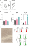
Fig. 2. MSCs remained metabolically active during liverless NMP and showed preserved stemness-specific characteristics at the end of the procedure.
a Perfusate gas analysis indicated glucose consumption and lactate production during liverless NMP. Estimation plots illustrate n = 5 liverless NMP experiments, with two measurements taken for each procedure (i.e., upstream and downstream of the MSCs-bioreactor). One-sided paired t-test, p-value: Glucose p = 0.085; Lactate: p < 0.0001. b The release of the apoptotic marker CK18 was stable over liverless NMP. Bars illustrate mean ± SEM, n = 5 biologically independent replicates. PRE denotes the supernatants immediately before bioreactors connection to the NMP circuit. One-way RM ANOVA, p = 0.682. c The MSCs seeded in the bioreactor secreted increasing amounts of IL-8 and IL-1ra during the liverless procedure. Bars illustrate mean ± SEM, n = 5 biologically independent replicates. PRE denotes the supernatants immediately before bioreactors connection to the NMP circuit. One-way ANOVA, p-value vs PRE: IL-8 p = 0.018; IL-1ra p = 0.002. Asterisks denote *p < 0.05. MSCs were harvested from the bioreactor at the end of liverless NMP and cultured in vitro under standard conditions until reaching 90% confluence. d Bioreactor-detached MSCs’ cultures displayed a spindle-shaped morphology and abundant production of extracellular matrix, which are well-recognized morphological characteristics of stem cells. A representative image captured at 40x magnification is shown; the bar scale on the bottom right denotes 100 micron. e After 9 ± 3 days of in vitro culturing, cells were enzymatically harvested for FACs analysis, which revealed a preserved expression of a set of MSC-specific markers (CD29, CD73, CD90, CD105). In contrast, the hematopoietic stem cell markers CD45, CD31, and CD34 were not detected. Five independent replicates were analyzed for each experimental condition. Abbreviations: cc-CK18, caspase-cleaved cytokeratin 18; IL-6, Interleukin 6; IL-1ra, Interleukin 1 receptor antagonist.
Fig. 3
Fig. 3. Changes in the MSC secretome after exposure to the rat liver-derived soluble factors during NMP.
a The concentration of selected MSC-related effectors was significantly different in the perfusates collected throughout liverless NMP compared with those withdrawn during liver NMP+bioreactor. Five independent replicates were analyzed for each experimental condition. Two-way RM ANOVA. FDR-adjusted p-values are shown for group comparison or interaction. The color-coded scale illustrates the different ranges of mediator release (pg). Abbreviations: ANGPTL4, Angiopoietin-like 4; CCL2/MCP-1, Chemokine C-C motif ligand 2/Monocyte Chemoattractant Protein-1; IDO, Indoleamine 2,3-Dioxygenase; IL-1ra, Interleukin 1 receptor antagonist; IL-4, Interleukin 4; IL-6, Interleukin 6;; IL-8, Interleukin 8; IL-10, Interleukin 10; IL-13, Interleukin 13; IL-18, Interleukin 18; IL-33, Interleukin 33; NA, not available.
Fig. 4
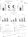
Fig. 4. The MSC-bioreactor-based NMP improved rat liver cell viability and function compared to the standard NMP procedure.
a The wash-out samples of the NMP+bioreactor group showed a greater donor-derived cell content relative to those collected during standard NMP. Bars denote mean ± SEM, n = 5 biologically independent replicates. Two-sided Mann-Whitney test, p-value vs NMP: **p = 0.008. Pie charts show, for each experimental group, the number of particles falling within the following gates: 1) debris, 3-4 μm (NMP+b-MSCs: 54.0% vs NMP: 50.2%); 2) cell fragments, 4.5-6.6 μm (39.7% vs 41.6%) 3) lymphocytes, 7.0-8.0 μm (5.4% vs 7.5%); 4) neutrophils/monocytes/endothelial cells/MSCs, 9-18 μm (1.0% vs 0.7%). b In the same wash-out samples, there was an increased concentration of NO metabolites in the bioreactor-based perfusion compared to the NMP group. Bars denote mean ± SEM, n = 5 biologically independent replicates. Two-sided Mann-Whitney test, p-value vs NMP: *p = 0.016. c Perfusate concentration of clinical hepatocellular damage markers was significantly lower in the NMP+bioreactor group relative to the NMP group. Raw data were adjusted based on circuit volume and liver weight. Points denote mean ± SEM, n = 4 biologically independent replicates. Two-way RM ANOVA, p-value of group comparison: AST p = 0.016; ALT p = 0.0085; LDH p = 0.0005. Tukey’s post hoc test was applied for multiple comparisons: asterisks denote *p < 0.05. Immunofluorescence-based analysis indicated that the NMP+bioreactor group showed d a reduced release of injury biomarkers and e an increased production of acute phase response proteins relative to the NMP group. Raw data were adjusted based on circuit volume and liver weight. Points denote mean ± SEM, n = 5 biologically independent replicates. Two-way RM ANOVA, p-value of group comparison: A2M p = 0.043; AGP p = 0.033; ARG1 p = 0.023 (p = 0.007 for interaction); GSTalpha p = 0.004 (p = 0.003 for interaction); Asterisks denote *p < 0.05, **p < 0.01. Abbreviations: A2M, Alpha-2-Macroglobulin; AGP, Alpha-1-acid glycoprotein; ALT, alanine aminotransferase; ARG1, Arginase-1; AST, aspartate aminotransferase; b-MSCs, MSC-bioreactor; GSTalpha, Glutathione Transferase Alpha; LDH, lactate dehydrogenase; NMP, normothermic machine perfusion.
Fig. 5
Fig. 5. The MSC-bioreactor-based perfusion ameliorated rat liver cell mitochondrial function.
a While the NMP group showed a decreased ATP content compared to both Native and SCS groups, the bioreactor-based perfusion exhibited a preserved energetic pool, with similar ATP concentration to that observed in the SCS group. Tissue ATP was measured in liver homogenates by bioluminescence-based assay. Raw data were adjusted based on liver weight. Data are expressed as mean ± SEM, n = 5 biologically independent replicates. One-way ANOVA, Tukey’s post hoc test. p-value: SCS vs Native 0.028; NMP vs Native p = 0.0001; NMP+b-MSCs vs Native p = 0.011; NMP vs SCS p = 0.033. Asterisks denote *p < 0.05, ***p < 0.001. b The bioreactor-based perfusion reversed the accumulation of succinate induced by cold ischemia. Results were adjusted based on perfusion fluid volume. Data are expressed as mean ± SEM, n = 5 biologically independent replicates. Two-way RM ANOVA, Tukey’s post hoc test, p-value: ***p = 0.0001. Abbreviations: b-MSCs, MSC-bioreactor; NMP, normothermic machine perfusion; SCS, static cold storage; W, wash-out.
Fig. 6
Fig. 6. Immunomodulating and pro-resolving effects associated with the MSC-bioreactor perfusion.
Perfusate profiling indicated that the bioreactor-based NMP induced a broad modulation of several factors involved in inflammation and leukocyte recruitment, inflammation resolution, and liver cell regeneration. Raw data were adjusted based on circuit volume and liver weight. Results are expressed as log2-transformed fold change between the NMP+bioreactor group and the NMP group at each time point. Five independent replicates were analyzed for each experimental condition. Two-way RM ANOVA, followed by Tukey’s post hoc test. The color-coded scale illustrates the diverse magnitudes of fold change. Abbreviations: b-MSCs, MSC-bioreactor; CCL2/MCP-1, Chemokine C-C motif ligand 2/Monocyte Chemoattractant Protein-1; CCL3/MIP-1α, Chemokine C-C motif ligand 3/Macrophage Inflammatory Protein-1α; CCL5/RANTES, Chemokine C-C motif ligand 5/regulated on activation normal T cell expressed and secreted; CTGF, Connective Tissue Growth Factor; CXCL-1/GRO α, C-X-C Motif Chemokine Ligand 1/Growth-regulated protein α; CXCL5/LIX, C-X-C Motif Chemokine Ligand 5/Lipopolysaccharide-induced CXC chemokine; CXCL10/IP-10, C-X-C Motif Chemokine Ligand 10/Interferon gamma-induced protein 10; FC, Fold change; HGF, Hepatocyte growth factor; IFN-γ, Interferon-γ; IL-4, Interleukin 4; IL-6, Interleukin 6; IL-10, Interleukin 10; IL-13, Interleukin 13; IL-18, Interleukin 18; NMP, normothermic machine perfusion; TIMP-1, Tissue Inhibitor of Metalloproteinase 1; TNF-α, Tumor Necrosis Factor-α; sICAM-1, soluble Intercellular Adhesion Molecule-1; VEGF, Vascular Endothelial Growth Factor.
Fig. 7
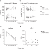
Fig. 7. MSC-bioreactor-based liver NMP in a large animal model.
In this pilot study, livers procured from donor swines were subjected to 4 h-NMP using the Liver Assist® (XVIVO Perfusion AB) device equipped with MSC-bioreactors (n = 3). The results were compared to those obtained from standard porcine liver NMP (n = 3). The collected data clearly indicate that the MSC-bioreactor can be safely integrated into a perfusion device currently available for the clinical setting. No difference in vascular a flows and b resistances was observed between livers subjected to the bioreactor-based perfusion compared to those exposed to the standard NMP. Points denote mean ± SEM, n = 3 biologically independent replicates. Two-way RM ANOVA; p-value between experimental groups: HA flow p = 0.151; HA resistance p = 0.285; PV flow p = 0.901; PV resistance p = 0.334. c Lactate clearance was similar across experimental groups. Points denote mean ± SEM, n = 3 biologically independent replicates. Two-way RM ANOVA; p = 0.563. d Perfusate LDH concentration tended to be lower in the NMP+bioreactor group. Points denote each experimental unit, n = 3 biologically independent replicates. Two-way ANOVA, p = 0.064. Abbreviations: b-MSCs, MSC-bioreactor; HA, hepatic artery; LDH, lactate dehydrogenase; NMP, normothermic machine perfusion; PV, portal vein.

References

    1. Parente, A. et al. Machine perfusion techniques for liver transplantation - A meta-analysis of the first seven randomized controlled trials. J. Hepatol. 10.1016/J.JHEP.2023.05.027 (2023). - PubMed
    1. Liew, B. et al. Liver transplant outcomes after ex vivo machine perfusion: a meta-analysis. Br. J. Surg.108, 1409–1416 (2021). - PubMed
    1. Lonati, C. et al. Influence of ex vivo perfusion on the biomolecular profile of rat lungs. FASEB J. fj.201701255R (2018). - PubMed
    1. Jassem, W. et al. Normothermic machine perfusion (nmp) inhibits proinflammatory responses in the liver and promotes regeneration. Hepatology70, 682–695 (2019). - PubMed
    1. Gatti, S. et al. Reduced expression of the melanocortin-1 receptor in human liver during brain death. Neuroimmunomodulation13, 51–55 (2006). - PubMed

Publication types

LinkOut - more resources