. 2025 Jan 2;16(1):268.
doi: 10.1038/s41467-024-55736-3.

PhpCNF-Y transcription factor infiltrates heterochromatin to generate cryptic intron-containing transcripts crucial for small RNA production

Affiliations

PhpCNF-Y transcription factor infiltrates heterochromatin to generate cryptic intron-containing transcripts crucial for small RNA production

Manjit Kumar Srivastav et al. Nat Commun. .

Abstract

The assembly of repressive heterochromatin in eukaryotic genomes is crucial for silencing lineage-inappropriate genes and repetitive DNA elements. Paradoxically, transcription of repetitive elements within constitutive heterochromatin domains is required for RNA-based mechanisms, such as the RNAi pathway, to target heterochromatin assembly proteins. However, the mechanism by which heterochromatic repeats are transcribed has been unclear. Using fission yeast, we show that the conserved trimeric transcription factor (TF) PhpCNF-Y complex can infiltrate constitutive heterochromatin via its histone-fold domains to transcribe repeat elements. PhpCNF-Y collaborates with a Zn-finger containing TF to bind repeat promoter regions with CCAAT boxes. Mutating either the TFs or the CCAAT binding site disrupts the transcription of heterochromatic repeats. Although repeat elements are transcribed from both strands, PhpCNF-Y-dependent transcripts originate from only one strand. These TF-driven transcripts contain multiple cryptic introns which are required for the generation of small interfering RNAs (siRNAs) via a mechanism involving the spliceosome and RNAi machinery. Our analyses show that siRNA production by this TF-mediated transcription pathway is critical for heterochromatin nucleation at target repeat loci. This study reveals a mechanism by which heterochromatic repeats are transcribed, initiating their own silencing by triggering a primary cascade that produces siRNAs necessary for heterochromatin nucleation.


Conflict of interest statement

Competing interests: The authors declare no competing interests.

Figures

Fig. 1
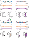
Fig. 1. The transcription factors Php5 and Moc3 bind to heterochromatic repeats.
a, b ChIP-seq analysis of TF distribution at the silent mat locus (a) and centromere 1 (b) in a wild-type background strain. The blue and small orange lines represent transcripts and small RNAs, respectively. Annotations: cenH refers to homology to centromeric dg/dh; IR signifies an Inverted Repeat. c The enlarged section from (a) shows the distribution of the specified TFs. CAS denotes Clr3-attracting sequence. d ChIP-qPCR analysis of the distribution of the indicated GFP-tagged transcription factors at cenH in wild-type cells. The data is expressed as the relative fold enrichment compared to the leu1 control locus and is normalized to the untagged strain. Significant enrichments are defined as those ≥ 2-fold (dotted line). Data from 3 independent biological experiments are presented as the mean ± SD. The positions of qPCR primers for dh and cenH are indicated by the black lines and arrows in (b) and (c), respectively. Source data are provided as a Source Data file.
Fig. 2
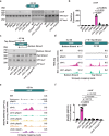
Fig. 2. Php5 forms a trimeric complex that localizes to heterochromatic repeats.
a Php5 immunopurified fractions were prepared from untagged and Php5-GFP expressing cells and were probed with anti-GFP antibodies. b Immunopurified fractions from untagged and Php5-GFP expressing cells were subjected to mass spectrometry analysis. The percentage of total peptide coverage for the indicated immunopurified proteins is displayed. Results from one biological replicate are shown; see also Supplementary Fig. 2a. c, d ChIP-qPCR analysis of the indicated GFP-tagged TFs was performed at cenH (c) and dh (d) in wild-type cells. Data from 3 independent biological experiments are presented as the mean ± SD of the relative fold enrichment compared to the leu1 control locus. The p-values were calculated using a two-tailed paired t-test. e, f ChIP-seq analysis to determine the localization of the indicated GFP-tagged TFs at the silent mat locus (e) and centromere 1 (f) in wild-type cells. Note that the Php5-GFP ChIP-seq data is the same as in Fig. 1a, b, as the experiments were performed simultaneously. g, h Euler diagrams represent the number of loci bound by Php5, Php3, and Php2 and contain a high (g) or low (h) abundance of CCAAT boxes. i, j Heat maps displaying ChIP-seq enrichments of PhpC subunits at loci containing high (i) or low (j) abundance of CCAAT boxes. Source Data are provided as a Source data file.
Fig. 3
Fig. 3. The binding of PhpC and Moc3 to heterochromatin is interdependent and requires CCAAT boxes.
a ChIP-seq analysis of GFP-tagged Php3 and Moc3 at the silent mat locus and centromere 1 in the indicated strains. b, c Fold enrichments of GFP-tagged Php3 and Moc3 were determined by ChIP-qPCR analysis at cenH and dh. d ChIP-seq analysis of GFP-tagged Php3 and Moc3 expressed in wild-type or CCAATmut strains. CCAAT boxes are indicated by vertical yellow lines in the schematics above, and the asterisk represents the location of two mutated CCAAT boxes. e, f Fold enrichments of GFP-tagged Php3 and Moc3 were determined by ChIP-qPCR analysis at cenH and dh. Data from 3 independent biological experiments are presented as the mean ± SD of the relative fold enrichment compared to the leu1 control locus. The p-values were calculated using a two-tailed paired t-test (b, c, e, f). WT Moc3-GFP and Php3-GFP ChIP-Seq data are replotted from Figs. 1a, b and  2e, f(a, d). Source data are provided as a Source Data file.
Fig. 4
Fig. 4. The histone fold domain of PhpC is pivotal for heterochromatin localization.
a, b ChIP-seq analysis of Php3 and Moc3 localization at the silent mat locus (a) and centromere 1 (b) in wild type, clr4∆, and nda3-KM311 strains. WT Moc3-GFP and Php3-GFP ChIP-seq data are replotted from Figs. 1a, b and 2e, f. Vertical yellow lines designate CCAAT boxes. Note that the highlighted CCAAT-enriched region is accessible to Php3 in clr4∆ cells. The small black line denotes the dg-annealing primer used in Supplementary Fig. 4c. c, d ChIP-qPCR analysis of GFP-tagged Php3 or Moc3 was performed at cenH (c) and dh (d) in the indicated strains. e, f ChIP-seq analysis of Php3 localization at the silent mat locus (e) and centromere 1 (f) in wild type and php3L12R strains. g ChIP-qPCR analysis of GFP-tagged Php3 was performed at cenH and dh in indicated strains. h Representative live-cell images of strains expressing GFP-tagged Php3 or Php3L12R from two independent experiments with similar results. Data from 3 independent biological experiments are presented as the mean ± SD. The p-values were calculated using a two-tailed paired t-test (c, d, g). Source data are provided as a Source Data file.
Fig. 5
Fig. 5. Cooperative function of PhpC and Moc3 promotes cenH transcription.
a RT-PCR was performed to analyze cenH transcripts in the indicated strains, with leu1 serving as the control for +RT and -RT reactions. The primer binding site at cenH is indicated by the black line. b RT-qPCR analysis of cenH transcripts in the indicated strains. Relative expression was compared to the leu1 control locus. c RT-PCR was performed to analyze cenH top and bottom strand transcripts in the indicated strains, with leu1 used as the control for +RT and -RT reactions. The top and bottom strand-specific primer binding site at cenH is indicated by the red and yellow lines, respectively. Representative results from two independent experiments are shown. d RNA-seq expression profile of the cenH top and bottom strand in the indicated strains. Reads were uniquely mapped to cenH. e, f RNA polymerase II (8WG16) occupancy at cenH was determined by ChIP-seq (e) and ChIP-qPCR (f) analyses in the indicated strains. Reads were uniquely mapped to cenH. For f, ChIP data is presented as relative fold enrichment compared to the tRNA control locus. Data from 3 independent biological experiments are presented as the mean ± SD. The p-values were calculated using a two-tailed paired t-test (b, f). Source data are provided as a Source Data file.
Fig. 6
Fig. 6. PhpC- and Moc3-mediated cenH transcription is crucial for producing Swi6HP1-independent siRNAs.
a, b siRNA-seq profiles at the cenH region in the indicated strains. Note that the reads were uniquely mapped to cenH. c, d ChIP-qPCR analysis of H3K9me3 enrichment at the silent mat locus in the indicated strains. Data is presented as the mean of 2 independent biological experiments. The numbered lines in the schematic denote the location of the primers. Source data are provided as a Source data file.
Fig. 7
Fig. 7. PhpC contributes to de novo heterochromatin establishment.
a 10-fold serial dilution assay of wild-type (WT) and CCAATmut strains on the indicated medium before and after trichostatin A (TSA) treatment and washout. Iodine staining was performed on non-selective (N/S) plates. b ChIP-qPCR analysis of H3K9me3 enrichment at the silent mat locus in the indicated TSA-treated strains. Data is presented as the mean of 3 independent biological experiments. Numbered lines in the schematic indicate the location of the primers. c Representative live-cell images of WT and CCAATmut strains harboring the mat2P::GFP reporter at the indicated time points (generations) after TSA treatment and washout. d Quantification of the cell fraction in the “ON” state (e.g., GFP-positive) at the indicated time points is shown. N = 260–651 cells for each data point. Source data are provided as a Source Data file.
Fig. 8
Fig. 8. Model showing the role of the histone fold domain-containing PhpCNF-Y in heterochromatic repeat transcription and siRNA generation.
The top panel illustrates how the histone fold domain (HFD) containing the trimeric TF complex PhpC, which recognizes underlying CCAAT boxes at heterochromatic regions such as cenH, binds to heterochromatic repeats in cooperation with Moc3. The bottom panel depicts how PhpC facilitates the transcription of the cenH bottom strand containing multiple cryptic introns (depicted as red rectangles). These introns likely stall the spliceosome that then recruits the RNAi machinery, particularly the RNA-dependent RNA polymerase Rdp1-containing protein complex RDRC, initiating siRNA production. The siRNAs are loaded onto the Ago1-containing RITS complex. Together with the protein Stc1, RITS recruits the Clr4Suv39h methyltransferase, promoting H3K9me and leading to de novo establishment of heterochromatin, independently of Swi6HP1. Once H3K9me is established, Swi6HP1 binds and also recruits the RNAi machinery via Ers1, maintaining a pool of siRNAs that further facilitate heterochromatin assembly. Consequently, the simultaneous disruption of both pathways results in the loss of siRNAs and H3K9me.

Similar articles

References

    1. Saksouk, N., Simboeck, E. & Dejardin, J. Constitutive heterochromatin formation and transcription in mammals. Epigenetics Chromatin8, 3 (2015). - PMC - PubMed
    1. Grewal, S. I. S. The molecular basis of heterochromatin assembly and epigenetic inheritance. Mol. Cell83, 1767–1785 (2023). - PMC - PubMed
    1. Nicetto, D. & Zaret, K. S. Role of H3K9me3 heterochromatin in cell identity establishment and maintenance. Curr. Opin. Genet. Dev.55, 1–10 (2019). - PMC - PubMed
    1. Janssen, A., Colmenares, S. U. & Karpen, G. H. Heterochromatin: guardian of the genome. Annu. Rev. Cell Dev. Biol.34, 265–288 (2018). - PubMed
    1. Jenuwein, T. & Allis, C. D. Translating the histone code. Science293, 1074–1080, (2001). - PubMed

Publication types

MeSH terms

Associated data