. 2025 Jan 2;15(1):15.
doi: 10.1038/s41598-024-84842-x.

Machine learning-based analysis of microfluidic device immobilized C. elegans for automated developmental toxicity testing

Affiliations

Machine learning-based analysis of microfluidic device immobilized C. elegans for automated developmental toxicity testing

Andrew DuPlissis et al. Sci Rep. .

Abstract

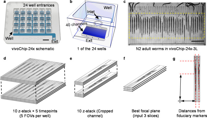
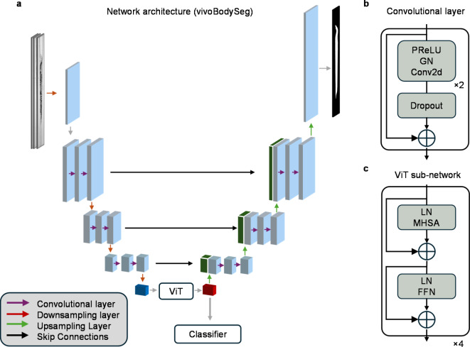
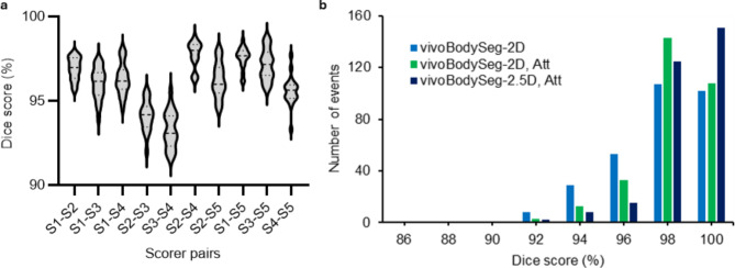
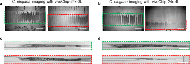
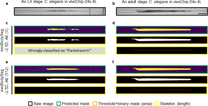
Developmental toxicity (DevTox) tests evaluate the adverse effects of chemical exposures on an organism's development. Although current testing primarily relies on large mammalian models, the emergence of new approach methodologies (NAMs) is encouraging industries and regulatory agencies to evaluate novel assays. C. elegans have emerged as NAMs for rapid toxicity testing because of its biological relevance and suitability to high throughput studies. However, current low-resolution and labor-intensive methodologies prohibit its application for sub-lethal DevTox studies at high throughputs. With the recent advent of the large-scale microfluidic device, vivoChip, we can now rapidly collect 3D high-resolution images of ~ 1000 C. elegans from 24 different populations. While data collection is rapid, analyzing thousands of images remains time-consuming. To address this challenge, we developed a machine-learning (ML)-based image analysis platform using a 2.5D U-Net architecture (vivoBodySeg) that accurately segments C. elegans in images obtained from vivoChip devices, achieving a Dice score of 97.80%. vivoBodySeg processes 36 GB data per device, phenotyping multiple body parameters within 35 min on a desktop PC. This analysis is ~ 140 × faster than the manual analysis. This ML approach delivers highly reproducible DevTox parameters (4-8% CV) to assess the toxicity of chemicals with high statistical power.

Keywords: C. elegans; Developmental toxicity; Few-shot learning; High-throughput screening; Microfluidics; U-Net.


Conflict of interest statement

Declarations. Competing interests: E.H., S.M., and A.B. are co-founders of vivoVerse, LLC and its Associates. A.D., A.S., A.L., E.H., S.M., and A.B. are inventors of several approved and pending patents. A.M. and S.G. at the time of their contribution are employed by vivoVerse, LLC and have no conflict of interest to declare.

Figures

Fig. 1
Fig. 1
High-resolution C. elegans body images are acquired using a vivoChip-24x platform. (a) Schematic of vivoChip-24x technology designed to immobilize C. elegans and capture high-resolution images from 24 different populations with up to 40 worms per population. Scale bar = 9 mm. (b) Detailed schematic of a single well from the 24-well device. Each well features an opening at the top and an inlet on the bottom, leading to 40 parallel immobilization channels beneath it. Worms are loaded into the well, flow through the inlet, and are immobilized within the 40 channels by the pressure of the fluid flow directed toward the exit. (c) Brightfield image of 40 adult C. elegans immobilized inside the microfluidic channels of the vivoChip-24x-3L device. Scale bar = 1 mm. (d) Graphic demonstrating a z-stack of 10 images collected for each FOV. Each FOV covers 8 channels using a 10 × , 0.4 NA objective. For each FOV, we collect a total of 50 images over 5 time points, with 10 z-slices (6 µm z-step size) captured at each time point (1-s interval). (e) We crop each channel for the first time point. (f) We identify the best focal plane for analysis and use 3 slices, comprising the focal plane, one slice above, and one slice below to train the network. (g) The location of the worm within the channel is used to determine the channel height by referencing its position with respect to the fiduciary cross mark present within each well of the vivoChip-24x.
Fig. 2
Fig. 2
The U-Net architecture of vivoBodySeg for automated analysis of C. elegans body. (a) Schematic of our 4-layer U-Net architecture. (b) Overview of our basic convolutional layer used in both the encoder and decoder components. (c) Overview of the vision transformer (ViT) integrated into the bottleneck layer.
Fig. 3
Fig. 3
Comparison of Dice scores for body segmentation by multiple scorers and different vivoBodySeg models. (a) Dice scores for all possible pairs of five individual scorers for a subset of 20 channels. (b) Bar plot showing Dice scores ranging from 86 to 100% (n = 301 C. elegans samples) for three U-Net models.
Fig. 4
Fig. 4
Images of C. elegans with developmental defects captured using two vivoChip-24x device designs. (a) Images of 40 trapping channels from the 3-layer chip (vivoChip-24x-3L) with immobilized D1 adult worms from a control population boxed in green (left panel) and a young adult worm population that was treated with 4 µM methylmercury in 0.2% DMSO boxed in red (right panel). (b) Images of 40 trapping channels from a 4-layer chip (vivoChip-24x-4L) with immobilized D1 adult worms from a control population boxed in green (left panel) and a young L4 stage worm population that was treated with 8 µM methylmercury in 0.2% DMSO (right panel). (c, d) High-magnification images of single control worms (green boxes) and methylmercury-treated worms (red boxes) from vivoChip-24x-3L (c) and vivoChip-24x-4L devices (d). Scale bars = 1 mm (a, b) and 200 µm (c, d).
Fig. 5
Fig. 5
Improved worm detection using few-shot learning with vivoBodySeg model. Images of a young L4 (a) and an adult stage (b) C. elegans, imaged in the vivoChip-24x-4L device. (c, d) Results from the baseline vivoBodySeg-2.5D, Att model with zero-shot learning. The model wrongly classified the channel as having a partial worm and thus discarded it from further analysis. The color in the predicted mask (green box) represents the pixel-wise probability of being a part of the worm mask. (e, f) Results from the vivoBodySeg-2.5D, Att model after few-shot learning using 512 images. The model correctly predicted the worm mask and accurately classified the channel images. Raw image, predicted mask, binary mask, and skeleton are compared. The scale bar = 200 μm.
Fig. 6
Fig. 6
Automated body parameter and autofluorescence analysis using the vivoBodySeg model. (a) Map showing exposure of 24 C. elegans populations to methylmercury (0.5 – 9.0 µM) and 0.2% DMSO. The experiment was repeated with five vivoChip-24x-4L devices. (b) The data from all five experiments were used to quantify average body length, body area, and body volume. The results are presented as mean ± SEM (n = 5 repeats). The solid lines represent the Hill-function fits to the experimental data, while the vertical dotted lines represent the EC10 values for each parameter, while the vertical solid lines represent the LOAEL value of 1.0 μM for all 3 parameters. Coefficients of variation (CV) are provided for the control wells. (c) Example images of worms from 0.5 µM and 9.0 µM methylmercury. Brightfield (BF) and autofluorescence (AF) images obtained using the GFP filter set, are shown. The scale bars = 200 µm. (d) Average autofluorescence intensity per unit body length for methylmercury and DMSO control populations. Data are presented as mean ± SEM (n ≥ 4 repeats). The vertical dotted line represents the EC10 value, while the vertical solid line represents the LOAEL value of 2.5 μM for mean intensity per length.

Update of

Similar articles

Cited by

References

    1. Kaletta, T. & Hengartner, M. O. Finding function in novel targets: C. elegans as a model organism. Nat. Rev. Drug. Discov.5, 387–398 (2006). - PubMed
    1. Silverman, G. A. et al. Modeling molecular and cellular aspects of human disease using the nematode Caenorhabditis elegans. Pediatr. Res.65, 10–18 (2009). - PMC - PubMed
    1. Hartman, J. H. et al. Xenobiotic metabolism and transport in Caenorhabditis elegans. J. Toxicol. Environ. Health B Crit. Rev.24(2), 51–94 (2021). - PMC - PubMed
    1. Leung, M. C. et al. Caenorhabditis elegans: An emerging model in biomedical and environmental toxicology. Toxicol. Sci.106, 5–28 (2008). - PMC - PubMed
    1. Corsi, A. K., Wightman, B. & Chalfie, M. A transparent window into biology: A primer on Caenorhabditis elegans. Genetics200(2), 387–407 (2015). - PMC - PubMed

MeSH terms

LinkOut - more resources