. 2025 Jan 2;15(1):447.
doi: 10.1038/s41598-024-84184-8.

Heterozygous mutation in BRCA2 induces accelerated age-dependent decline in sperm quality with male subfertility in rats

Affiliations

Heterozygous mutation in BRCA2 induces accelerated age-dependent decline in sperm quality with male subfertility in rats

Yashiro Motooka et al. Sci Rep. .

Abstract

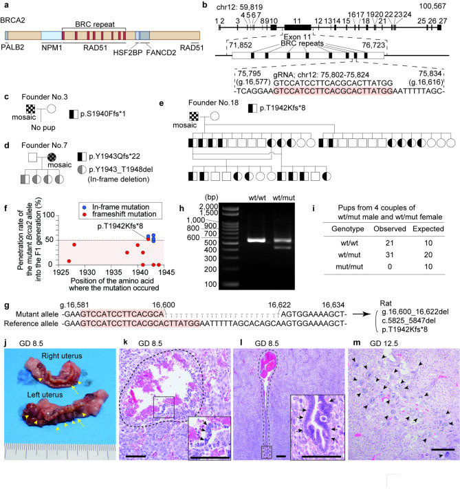
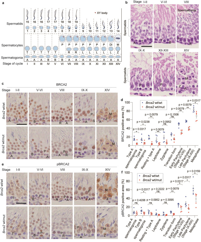
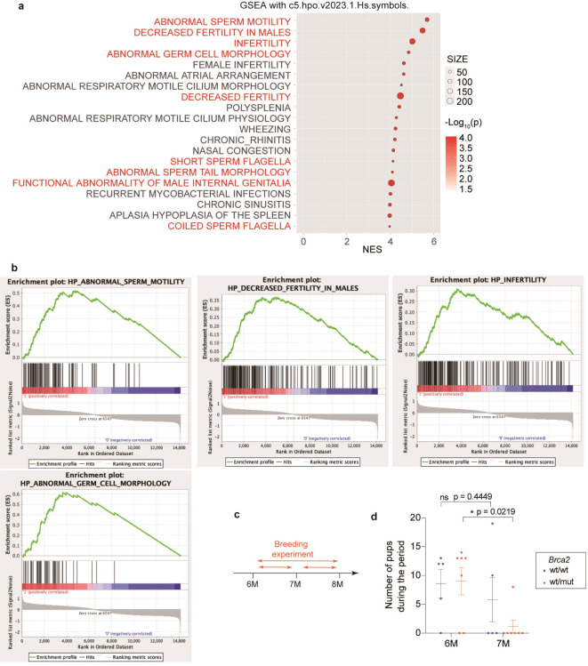
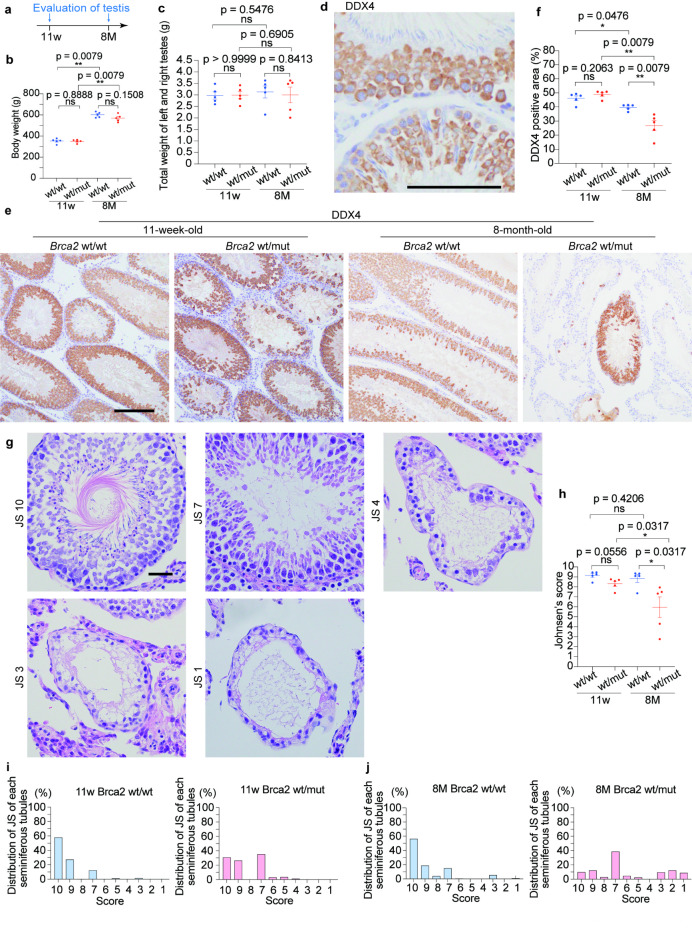
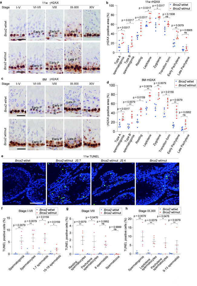
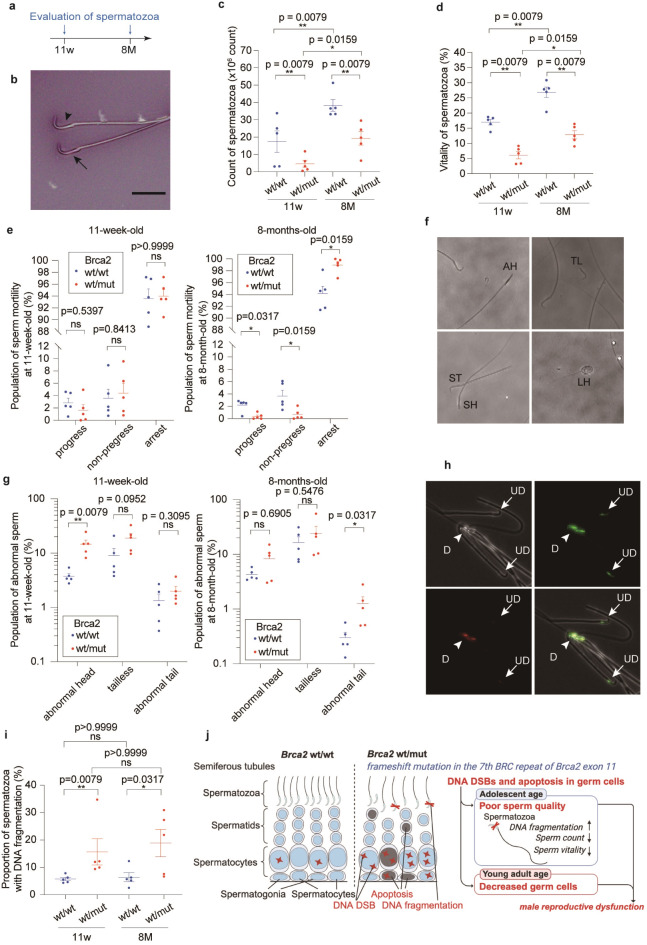
Tumor suppressor BRCA2 executes homologous recombination to repair DNA double-strand breaks in collaboration with RAD51, involving exon 11 and 27. Exon 11 constitutes a region where pathogenic variants (PVs) accumulate, and mutations in this region are known to contribute to carcinogenesis. However, the impact of the heterozygous PVs of BRCA2 exon 11 on the life quality beyond cancer risk, including male fertility, remains unclear. Here, we established a rat model with a frameshift on the seventh BRC repeat in Brca2 exon 11 (Brca2+/p.T1942fs), which is homologous to human BRCA2+/p.T1974fs, using CRISPR/Cas9 system. Our analyses revealed that the heterozygous rats with the PV in the BRCA2 exon 11 showed increased DNA double-strand breaks and apoptosis in spermatogonia and spermatocytes, accelerated testicular germ cell loss, and deterioration in sperm quality according with aging, ultimately resulting in early male reproductive dysfunction. Of note, these alterations in testes and sperm, including DNA fragmentation in spermatozoa, were observed from completion of sexual maturation. The present findings suggest that it is crucial to consider not only cancer risk but also potential declines in reproductive capacity in men carrying BRCA2 exon 11 PVs. Further investigation is warranted to determine whether similar traits appear in humans.

Keywords: Aging; BRCA2 exon 11; Heterozygote; Male subfertility; Pathogenic variant; Spermatogenesis.


Conflict of interest statement

Declarations. Competing interests: The authors declare no competing interests.

Figures

Fig. 1
Fig. 1
Design and the founders of Jcl: SD Brca2 (Here only Brca2 to be italic, please) mutant rats harboring a frameshift on exon 11 with embryonic lethality in the homozygotes. (a) Structure of rat BRCA2 protein. (b) Structure of rat Brca2 and guide RNA used for genome editing. (c-e) The pedigree of founder rats. (f) A graphical summary of the inheritance rate of the mutant Brca2 allele in the F1 generation, categorized by the site of the mutated amino acid. Founder rats that produced no pups are also plotted as 0%. This figure indicates that the frameshift mutation in Brca2 is less likely to be inherited to the subsequent generation. (g) The nucleotide sequence of the modified region within the Brca2 gene and their notation. (h) Representative result of genotyping PCR of the Brca2wt/wt and Brca2wt/mut. (i) Contingency table showing observed and expected counts of offspring with each genotype from 4 pairs of Brca2wt/mut male rats and Brca2wt/mut female rats. (j) Macroscopic image of uteri from a pregnant female taken at GD 8.5. Arrow heads, conceptuses; arrows, malformed conceptuses. (k, l) Representative histology of dying conceptuses (k) and live conceptuses (l) at GD 8.5 (arrows, embryonic components; bar = 200 μm). In the dying conceptus, cell detachment, loss of tissue continuity and hemorrhage are observed. (m) Microscopic image showing remnant of pregnancy observed at GD 12.5 (arrow heads, trophoblastic cells; bar = 200 μm). Fetal components are absent, and cluster of trophoblastic cells with large nuclei, some of which are syncytialized, are observed.
Fig. 2
Fig. 2
BRCA2 expression is decreased in Brca2wt/mut spermatocytes. (a) The schematic of the stages of the seminiferous epithelium and XY body formation in rats. A, type A spermatogonia; B, type B spermatogonia; R, resting spermatocytes; L, leptotene; T, transition form; P, pachytene; DI, diplotene and diakinesis; M, metaphase. (b) Typical histological images of each stage of seminiferous epithelium in rats (bar = 30 μm). (c, d, e, f) Typical immunohistochemistry images of BRCA2 (E2082-L2106) and phosphorylated BRCA2 (Ser2095) (bar = 30 μm), along with quantitative analysis (n = 5; with at least 10 fields taken randomly with x20 magnification were analyzed per individual), indicate that BRCA2 and phosphorylated BRCA2 were primarily expressed in spermatocytes from stages VI to XIV, labeled as early Pachytene (stage VI-VIII), late Pachytene (stage IX-XIII), and Metaphase (stage XIV). The expression of BRCA2 and phosphorylated BRCA2 tended to be weaker in Brca2wt/mut spermatogonia and spermatocytes compared to Brca2wt/wt across multiple stages.
Fig. 3
Fig. 3
Brca2wt/mut induces neither alterative splicing variant nor different-sized BRCA2 protein in testis. (a, b) Analysis of splicing variant based on RNA-seq with JunctionSeq analysis (Brca2wt/wt testes, n = 3; Brca2wt/mut testes, n = 3). The line graph describes the relative coverage of exons normalized by dividing by the length of each exon whereas the bar graph represents the relative coverage of junction of exons. This analysis revealed no statically significant changes in the splicing variants between Brca2wt/wt and Brca2wt/mut. (c) Diagram of the regions recognized by the anti-BRCA2 antibodies used in the immunoblot. We used an antibody against the first 100 amino acids of BRCA2; BRCA2(aa1-100), an antibody against the region from Glutamic acid at position 2070 to Serine at position 2120 on human BRCA2 (corresponding to positions 2082 to 2106 in rat BRCA2); BRCA2(E2070-2120), and an antibody against BRCA2 phosphorylated at Serine 2095; pBRCA2(Ser2095). In other words, one antibody recognizes the N-terminal side of the mutant point, while the other two recognize the C-terminal side. (d, e, f, g, h, i) Images of the immunoblot bands of testes lysates and their quantification results. The evaluation of BRCA2 (E2082-L2106) was conducted by reprobing the membrane used for the assessing of N-terminal domain of BRCA2 after stripping the antibody with an acidic pH protocol and confirming the removal of the antibody using Chemi-Lumi One Ultra. The predicted band size for BRCA2 (aa1-100) is 384 kDa. Uncropped images are available in Figure S2.
Fig. 4
Fig. 4
Brca2wt/mut male rats contribute to rapid decline of litter size according with aging after mating with a Brca2wt/wt female. (a) Top enriched gene sets in Brca2wt/mut testes compared to Brca2wt/wt by GSEA on testes from 10-week-old rats using gene set of “Human Phenotype Ontology” (n = 3). (b) Enrichment plots of the gene sets associated with male infertility. (c) Design of experiments evaluating the reproductive efficiency. (d) Trends of reproductive efficiency. Brca2wt/mut male rats exhibited significant decrease in reproductive efficiency at 7 months of age in comparison to those of 6 months old whereas Brca2wt/wt males did not show a significant decrease (Brca2wt/wt, n = 5; Brca2wt/mut, n = 7).
Fig. 5
Fig. 5
Brca2wt/mutmale rats reveal accelerated age-dependent decrease in testicular germ cells and Johnsen’s score. (a) Design of experiments regarding histological assessment of the testis. (b, c) Body weight and the weights of testes revealed no significant difference between Brca2wt/wt and Brca2wt/mut male rats. (d, e, f) Representative immunohistochemical images and quantitative analysis of DDX4 in seminiferous tubules (n = 5; bar = 100 μm). DDX4 is expressed in some population of germ cells, spermatocytes and stage 1–8 spermatids. IHC reveals a significant decrease of DDX4-positive germ cells in Brca2wt/mut rats at 8 months. (g) Representative histological images for Johnsen’s score 10, 7, 4, 3 and 1. Complete spermatogenesis with spermatozoa is classified as score 10, no spermatozoa but many spermatids as score 7, only few spermatocytes with no spermatids as score 4, spermatogonia without the other germ cells as score 3, and no cells in the seminiferous tubule as score 1. (h) Comparison of Johnsen’s score (n = 5) revealed that morphologically detectable spermatogenesis disorder is observed in Brca2wt/mut testes at 8 months. (i, j) Distribution of Johnsen’s score in each group. The bars represent the fraction of seminiferous tubules for each Johnsen’s score in a total of 500 tubules for each group (n = 5; 100 tubules per each rat). Seminiferous tubules scored as 7 are abundant in Brca2wt/mut at 11 weeks and 8 months. JS, Johnsen’s score; ns, not significant.
Fig. 6
Fig. 6
Brca2wt/mut testicular germ cells exhibit accumulation of DNA double-strand break and increased apoptosis. (a, b, c, d) Representative immunohistochemical images and quantitative analysis of γH2AX in spermatogonia and spermatocytes classified in type A spermatogonia (stage IX-XIV), type B spermatogonia (stage IV-VI), resting form (stage VIII), leptotene (stage IX-XIII), zygotene (stage XIV), transition form (stage I-V), early pachytene (stage VI-VIII) and late pachytene (stage IX-XIII) (n = 5, with at least 10 fields taken randomly with x20 magnification were analyzed per individual, bar = 30 μm, ). In spermatogonia and spermatocyte of Brca2wt/mut rats at 11 weeks and 8 months, accumulation of DNA double strand breaks is observed throughout the differentiation stages. R, resting spermatocytes; L, leptotene; T, transition form; P, pachytene; DI, diplotene and diakinesis; M, metaphase. (e) Representative images of TUNEL-staining in seminiferous tubules. Brca2wt/wt seminiferous tubules contained scarce TUNEL-positive cells (bar = 100 μm). On the other hand, Brca2wt/mut seminiferous tubules contained higher population of TUNEL-positive cells, especially in seminiferous tubules classified as JS-7 or lower. (f, g, h) Analysis of TUNEL-positive rate of germ cells within seminiferous tubules at each stage, excluding seminiferous tubules classified as JS-7 or lower. This analysis revealed that, compared to Brca2wt/wt, Brca2wt/mut exhibited increased apoptosis in spermatogonia, spermatocytes, and spermatids at all stages of seminiferous tubules.
Fig. 7
Fig. 7
Brca2wt/mut male rats reveal deteriorated sperm quality. (a) Design of experiments regarding the characterization of spermatozoa. (b) Representative images of Eosin Y-Nigrosin staining (bar = 20 μm; arrowhead, viable spermatozoa; arrow, dead spermatozoa). (c) Number of spermatozoa and (d) vitality of spermatozoa are decreased in Brca2wt/mut group both at 11 weeks and 8 months (n = 5). (e) Motility of sperms from Brca2wt/mut rats is not deteriorated at 11 weeks but decreased at 8 months (n = 5). (f) Representative morphology of abnormal sperms. AH, abnormal head sperm; TL, tailless sperm; ST, short tail sperm; SH, small head sperm; LH, large head sperm. (g) Abnormal sperm was more abundant in Brca2wt/mut than Brca2wt/wt both at 11 weeks and 8 months. (h) Representative images of the sperms stained with acridine orange (arrowhead, damaged sperm DNA as D; arrow, undamaged sperm DNA as UD). (i) Comparison of the proportion of spermatozoa with DNA fragmentation within a sperm sample detected from SCSA (n = 5). This proportion of spermatozoa with DNA fragmentation is commonly referred to as DNA fragmentation index, DFI. Both at 11 weeks and 8 months, the spermatozoa of the Brca2wt/mut rats show more DNA fragmentation than those of Brca2wt/wt. SCSA, sperm chromatin structure assay. (j) Summary of the findings.

Similar articles

Cited by

References

    1. McAllister, K. A. et al. Characterization of the rat and mouse homologues of the BRCA2 breast cancer susceptibility gene. Cancer Res.57, 3121–3125 (1997). - PubMed
    1. Roy, R., Chun, J. & Powell, S. N. BRCA1 and BRCA2: different roles in a common pathway of genome protection. Nat. Rev. Cancer. 12, 68–78. 10.1038/nrc3181 (2011). - PMC - PubMed
    1. Kwon, Y. et al. DNA binding and RAD51 engagement by the BRCA2 C-terminus orchestrate DNA repair and replication fork preservation. Nat. Commun.14, 432. 10.1038/s41467-023-36211-x (2023). - PMC - PubMed
    1. Davies, O. R. & Pellegrini, L. Interaction with the BRCA2 C terminus protects RAD51-DNA filaments from disassembly by BRC repeats. Nat. Struct. Mol. Biol.14, 475–483. 10.1038/nsmb1251 (2007). - PMC - PubMed
    1. Momozawa, Y. et al. Germline pathogenic variants of 11 breast cancer genes in 7,051 Japanese patients and 11,241 controls. Nat. Commun.9, 4083. 10.1038/s41467-018-06581-8 (2018). - PMC - PubMed

Publication types