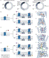
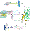
Review
. 2025 Apr;24(4):251-275.
doi: 10.1038/s41573-024-01083-3. Epub 2025 Jan 2.

Functional dynamics of G protein-coupled receptors reveal new routes for drug discovery

Affiliations
Review

Functional dynamics of G protein-coupled receptors reveal new routes for drug discovery

Paolo Conflitti et al. Nat Rev Drug Discov. 2025 Apr.

Abstract

G protein-coupled receptors (GPCRs) are the largest human membrane protein family that transduce extracellular signals into cellular responses. They are major pharmacological targets, with approximately 26% of marketed drugs targeting GPCRs, primarily at their orthosteric binding site. Despite their prominence, predicting the pharmacological effects of novel GPCR-targeting drugs remains challenging due to the complex functional dynamics of these receptors. Recent advances in X-ray crystallography, cryo-electron microscopy, spectroscopic techniques and molecular simulations have enhanced our understanding of receptor conformational dynamics and ligand interactions with GPCRs. These developments have revealed novel ligand-binding modes, mechanisms of action and druggable pockets. In this Review, we highlight such aspects for recently discovered small-molecule drugs and drug candidates targeting GPCRs, focusing on three categories: allosteric modulators, biased ligands, and bivalent and bitopic compounds. Although studies so far have largely been retrospective, integrating structural data on ligand-induced receptor functional dynamics into the drug discovery pipeline has the potential to guide the identification of drug candidates with specific abilities to modulate GPCR interactions with intracellular effector proteins such as G proteins and β-arrestins, enabling more tailored selectivity and efficacy profiles.


Conflict of interest statement

Competing interests: S.Y. is cofounder of AlphaMol Science Ltd. C.G.T. is a shareholder, consultant and member of the Scientific Advisory Board of Sosei Heptares. The remaining authors declare no competing interests.

Figures
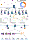
Figure 1.
Figure 1.
Classification of GPCR ligands and schematic representation of GPCR signalling pathways. A) Representation of different types of orthosteric and allosteric ligands and their effects on GPCR signaling. GPCRs are represented as blue helical bundles buried inside a lipid bilayer. Orthosteric ligands and allosteric modulators are represented as purple and orange spheres, respectively. G proteins are depicted as heterotrimeric complexes composed of a Gα (purple), Gβ (dark green), and Gγ (dark red) subunits. Arrestins are shown in light yellow. At the bottom, examples of pharmacological response curves for orthosteric ligands, biased ligands and allosteric modulators are shown. B) Distribution of FDA approved drugs targeting GPCRs categorized according to drug efficacy. C) Schematic representation of various signalling pathways activated by GPCR ligand binding. The details of pathways engaged by each G protein subtype are annotated.
Figure 2.
Figure 2.
Detailed comparison of the microswitch residues involved in allosteric communication between the OBS and the IBS among four class A GPCRs. In each panel, the receptor inactive structure is depicted as green cartoon, and the active structure is shown as yellow cartoon. Four GPCRs from different Class A subfamilies were selected to showcase the variability in microswitch conformations. The insets highlight the most important microswitches conserved across class A GPCRs: the P5.50I3.40F6.44/W6.48 motif (top left corner), the sodium binding pocket (top right corner) with the N7.49 residue belonging to the N7.49P7.50xxY7.53 motif, the E6.30-R3.50 ionic lock (bottom left corner), the E6.30/D3.49R3.50Y5.58 motif with the P and Y residues of the N7.49P7.50xxY7.53 motif (bottom right corner).
Figure 3.
Figure 3.
Mechanisms of allosteric modulation. A) Distribution of experimentally resolved allosteric modulators per GPCR class (left), modulation mechanism (center), and molecular type (right). B-D) Allosteric modulation by PAMs and NAMs binding a GPCR at the B) extracellular, C) intramembrane, D) and intracellular regions. Orthosteric ligands are represented as purple spheres and allosteric ligands are depicted as orange, red, and yellow spheres for extracellular, intramembrane, and intracellular-binding ligands, respectively. The sizes of the arrows showing the binding of the ligand, G protein, and formation of the active complex are proportional to the effects of the allosteric modulators. On the right, examples of PAMs and NAMs binding at the same regions are proposed. Structural details of E) LY3154207 binding to D1R (PDB ID 7X2F), F) avacopan to C5aR (6C1R), G) cinacalcet to CaSR (7M3F), H) SBI-553 to NSTR1 (ID 8JPB), and I) vercinon to CCR9 (ID 5LWE). The GPCR and its residues interacting with the ligand are represented as cyan cartoons and licorice, respectively. The allosteric modulators are shown as orange, red, or yellow licorice depending on their binding region. The G protein and its residues are represented as light green cartoons and licorice. Oxygens are coloured in red, nitrogens in blue, fluorine atoms in green and sodium cations in purple.
Figure 4.
Figure 4.
Action mechanism of biased, bivalent, and bitopic ligands. A) Example of a biased ligand (shown as purple sphere) to selectively promote G protein signalling while suppressing arrestin-mediated transduction. The inset shows the binding mode of oliceridine in MOR (PDB ID 8EFB) and the different stabilization of the GPCR active conformation, competent for the binding of the G protein but not the arrestin. B) Example of a biased ligand endowed with G protein subtype selectivity. The inset shows: (Upper) the binding mode of rivenprost (in purple) and L-902688 (in yellow) in the EP4-Gs complex, and (Lower) the binding pose of L-902688 in the EP4-Gs complex (ligand in yellow) compared to the binding pose of the same ligand in the EP4-Gi complex (ligand in orange) (10.1073/pnas.2216329120). C) Schematic representation of a bivalent ligand action mechanism in promoting dimerization of two different GPCRs. D) Example of GPCR binding by a bitopic ligand. In the inset, two different examples of bitopic ligands experimentally resolved are shown. On the left, salmeterol binding to β2 adrenergic receptor (PDB ID 6MXT) is reported. The horizontal solid line divides the exosite from the OBS. On the right, B6 binding to μ opioid receptor (PDB ID 7U2K) is depicted. Here, the horizontal solid line divides the OBS from the sodium binding pocket.
Figure 5.
Figure 5.
Experimental and computational pipeline for the rationalization of the GPCR activation mechanism and the allosteric network connecting the OBS and the IBS. NMR spectra were adapted from Abiko et al.
Figure 6.
Figure 6.
Example of GPCR dimerization leading to a change in the OBS and IBS conformations. At the bottom, the red arrows show the structural detail of the conformational rearrangement of the CCR5’s OBS (on the right) and IBS (on the left) in CCR5-CXCR4 heterodimer and CCR5 homodimer, respectively. In CCR5-CXCR4 dimer, the presence of CXCR4 induces a partial closure of the OBS, thus hampering ligand entry. On the other hand, in CCR5 homodimer, the volume of the IBS of one protomer increases due to the interaction with the other. Figures adapted from Di Marino et al.. The GPCRs forming the dimer are shown as light blue and green cartoons, whereas the corresponding inactive protomer is shown in yellow overlapped with the light blue protomer of the dimers for comparison.
Figure 7.
Figure 7.
Distinct lipid interaction mechanisms across the GPCR family. Structures of the Class A serotonin (5-HT1A, PDB: 7E2X) and neurotensin (NTSR1, PDB: 6UP7) receptors in complex with phosphatidylinositol-4-phosphate (PI(4)P) and the phosphatidylinositol-4,5-bisphosphate (PI(4,5)P2) headgroup, which aid binding of G protein and β-arrestin effector proteins., Interaction of the Class B1 glucagon receptor (GCGR, PDB: 5XEZ) with PI(4,5)P2, as observed in MD simulations (adapted from Ansell et al.).

References

    1. Lander ES et al. Initial sequencing and analysis of the human genome. Nature 409, 860–921 (2001). - PubMed
    1. Hauser AS, Attwood MM, Rask-Andersen M, Schiöth HB & Gloriam DE Trends in GPCR drug discovery: new agents, targets and indications. Nat. Rev. Drug Discov. 16, 829–842 (2017). - PMC - PubMed
    1. Santos R et al. A comprehensive map of molecular drug targets. Nat. Rev. Drug Discov. (2017) doi: 10.1038/NRD.2016.230. - DOI - PMC - PubMed
    1. Sriram K & Insel P G Protein-Coupled Receptors as Targets for Approved Drugs: How Many Targets and How Many Drugs? Mol. Pharmacol. (2018) doi: 10.1124/MOL.117.111062. - DOI - PMC - PubMed
    1. FDA. Summary of NDA Approvals & Receipts, 1938 to the present. https://www.fda.gov/about-fda/histories-fda-regulated-products/summary-n... (2024).

Publication types

LinkOut - more resources