. 2025 Jan 2;16(1):7.
doi: 10.1186/s13244-024-01884-5.

Multiparametric MRI and artificial intelligence in predicting and monitoring treatment response in bladder cancer

Affiliations

Multiparametric MRI and artificial intelligence in predicting and monitoring treatment response in bladder cancer

Yuki Arita et al. Insights Imaging. .

Abstract

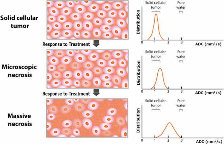
Bladder cancer is the 10th most common and 13th most deadly cancer worldwide, with urothelial carcinomas being the most common type. Distinguishing between non-muscle-invasive bladder cancer (NMIBC) and muscle-invasive bladder cancer (MIBC) is essential due to significant differences in management and prognosis. MRI may play an important diagnostic role in this setting. The Vesical Imaging Reporting and Data System (VI-RADS), a multiparametric MRI (mpMRI)-based consensus reporting platform, allows for standardized preoperative muscle invasion assessment in BCa with proven diagnostic accuracy. However, post-treatment assessment using VI-RADS is challenging because of anatomical changes, especially in the interpretation of the muscle layer. MRI techniques that provide tumor tissue physiological information, including diffusion-weighted (DW)- and dynamic contrast-enhanced (DCE)-MRI, combined with derived quantitative imaging biomarkers (QIBs), may potentially overcome the limitations of BCa evaluation when predominantly focusing on anatomic changes at MRI, particularly in the therapy response setting. Delta-radiomics, which encompasses the assessment of changes (Δ) in image features extracted from mpMRI data, has the potential to monitor treatment response. In comparison to the current Response Evaluation Criteria in Solid Tumors (RECIST), QIBs and mpMRI-based radiomics, in combination with artificial intelligence (AI)-based image analysis, may potentially allow for earlier identification of therapy-induced tumor changes. This review provides an update on the potential of QIBs and mpMRI-based radiomics and discusses the future applications of AI in BCa management, particularly in assessing treatment response. CRITICAL RELEVANCE STATEMENT: Incorporating mpMRI-based quantitative imaging biomarkers, radiomics, and artificial intelligence into bladder cancer management has the potential to enhance treatment response assessment and prognosis prediction. KEY POINTS: Quantitative imaging biomarkers (QIBs) from mpMRI and radiomics can outperform RECIST for bladder cancer treatments. AI improves mpMRI segmentation and enhances radiomics feature extraction effectively. Predictive models integrate imaging biomarkers and clinical data using AI tools. Multicenter studies with strict criteria validate radiomics and QIBs clinically. Consistent mpMRI and AI applications need reliable validation in clinical practice.

Keywords: Artificial intelligence; Biomarker; Multiparametric magnetic resonance imaging; Treatment response; Urinary bladder neoplasm.


Conflict of interest statement

Declarations. Ethics approval and consent to participate: Not applicable. Consent for publication: Not applicable. Competing interests: Y.A. is one of the scientific editorial board members of the ESR journal (European Radiology), but he was not involved in the manuscript handling process. The other authors of this manuscript declare no relationships with any companies, whose products or services may be related to the subject matter of the article.

Figures

Fig. 1
Fig. 1
VI-RADS scoring in representative bladder cancer patients. VI-RADS 1: Lesion size < 1 cm; VI-RADS 2: Exophytic tumor with stalk or sessile/broad-based tumor with thickened inner layer; VI-RADS 3: No category 2 findings, but no clear disruption of muscularis propria; VI-RADS 4: Extension of the tumor tissue into the muscularis propria; and VI-RADS 5; Extension of the tumor tissue into the extravesical fat. BCa, bladder cancer; DCE, dynamic contrast-enhanced; DW, diffusion-weighted; T2w, T2-weighted imaging; VI-RADS, Vesical Imaging Reporting and Data System
Fig. 2
Fig. 2
Flow chart of the current treatment planning process in MIBC patients. 5-FU, 5-fluorouracil; ddMVAC, dose-dense methotrexate, vinblastine, adriamycin, and cisplatin; EBRT, external beam radiation therapy; MIBC, muscle-invasive bladder cancer; MMC, mitomycin C; NAC, neoadjuvant chemotherapy; TURB, transurethral resection of bladder tumor
Fig. 3
Fig. 3
Mechanism of ADC from DW-MRI in response to treatment. The ADC map, representing the average movement of water molecules in tissues, is generated from DW-MRI using at least two b-values. Free water diffusion is restricted by barriers such as intact cell membranes, which are inversely related to the cellularity of tumor tissue. Reference: [37] ADC, apparent diffusion coefficient; DW, diffusion-weighted
Fig. 4
Fig. 4
Mechanism of DCE-MRI. The signal intensity time course of DCE-MRI data can be modeled using both semiquantitative and quantitative techniques. Semiquantitative parameters include signal enhancement, wash-in, and wash-out rates. Quantitative analysis involves modeling the time-course data of the contrast agent concentration curve to estimate the volume transfer constant, which indicates vascular perfusion and permeability. Reference: [37] DCE, dynamic-contrast-enhanced; EES,  extravascular extracellular space; Fp, flow blood plasma; Kep, rate constant of contrast agent from EES to blood plasma; Ktrans, volume transfer constant from blood plasma to EES; Ve, EES volume; Vp, plasma blood volume
Fig. 5
Fig. 5
QIBs and Radiomics that may be used to apply personalized care in bladder cancer patients. DCE, dynamic contrast-enhanced; DW, diffusion-weighted; QIB, quantitative imaging biomarker; T2w, T2-weighted imaging
Fig. 6
Fig. 6
NAC responder: a 76-year-old woman with a 24-mm urothelial carcinoma located in the left posterior bladder wall pre-NAC MRI shows a sessile and broad-based tumor with localized wall thickening and lack of clear disruption of the muscularis layer on T2WI (a) [T2WI VI-RADS score: 3], high-signal intensity on DW-MRI (b) [DW VI-RADS score: 3], and homogeneous low signal intensity on the ADC map with a mean ADC value of 0.579 × 10−3 mm2/s (c). The tumor is indicated by a white arrow. Post-NAC MRI demonstates reduced tumor volume with slight wall thickening on T2WI (d), linear high signal intensity on DW-MRI (e), and linear low signal intensity on ADC map with a mean ADC value of 0.982 × 10−3 mm2/s (f). The tumor is indicated by a white arrow. ADC, apparent diffusion coefficient; DW, diffusion-weighted; NAC, neoadjuvant chemotherapy; T2WI, T2-weighted imaging; VI-RADS, vesical imaging-reporting data system
Fig. 7
Fig. 7
NAC non-responder: a 71-year-old man with a 65-mm urothelial carcinoma with squamous differentiation the right posterior bladder wall Pre-NAC MRI reveals a lobulated tumor with partial disruption in the muscle layer on T2WI (a) [T2WI VI-RADS score: 4], high-signal intensity on DW-MRI indicating infiltration into the muscle propia (b) [DW VI-RADS score: 4], heterogeneous low signal intensity on the ADC map with a mean ADC value of 0.994 × 10−3 mm2/s (c), and DCE-MRI shows heterogeneous enhancement and muscle layer disruption (d) [DCE VI-RADS score: 4]. After one course of gemcitabine and cisplatin, and two courses of gemcitabine and docetaxel due to renal dysfunction, post-NAC MRI shows tumor expansion and invasion into the perivesical fat on T2WI (e) [T2WI VI-RADS: 5] and DW-MRI (f) [DW VI-RADS: 5], with a corresponding low signal intensity on the ADC map (a mean ADC value: 0.906 × 10−3 mm2/s) (g). DCE-MRI confirms perivesical infiltration (h) [DCE VI-RADS: 5]. The tumor is indicate white arrows, muscle infiltration by white arrowheads, and perivesical fat infiltration by yellow arrowheads. ADC, apparent diffusion coefficient; DCE, dynamic contrast-enhanced imaging; DW, diffusion-weighted neoadjuvant chemotherapy; T2WI, T2-weighted imaging; VI-RADS, vesical imaging-reporting and data system
Fig. 8
Fig. 8
Immunotherapy responder: a 76-year-old man with a 35-mm urothelial carcinoma in the anterior bladder wall pre-immunotherapy MRI shows a round-shaped tumor with no apparent muscularis disruption on T2WI (a) [T2WI VIRADS score: 3] and homogeneous high signal on DW-MRI (b) [DW VI-RADS score: 3]. Post-immunotherapy MRI reveals significant tumor reduction, with no apparent lesions on T2WI (c) or DW-MRI (d). The tumor is indicated by white arrow, and intact muscle layers by white arrowheads. ADC, apparent diffusion coefficient; DCE, dynamic contrast-enhanced; DW, diffusion-weighted; T2WI, T2-weighted imaging; VI-RADS, vesical imaging-reporting and data system
Fig. 9
Fig. 9
Post-BCG therapy: an 81-year-old man with multiple papillary nodules and broad wall thickening post-BCG therapy MRI shows intermediate signal intensity papillary nodules with broad wall thickening on T2WI (a), high signal intensity on DW-MRI (b), intermediate signal intensity on the ADC map with a mean ADC value of 1.272 × 10−3 mm2/s (c), and DCE-MRI shows multiple contrast-enhanced nodules (d). ADC, apparent diffusion coefficient; DW, diffusion-weighted; T2WI, T2-weighted imaging; VI-RADS, vesical imaging-reporting and data system
Fig. 10
Fig. 10
Trimodality therapy responder: a 74-year-old woman with a 53-mm urothelial carcinoma in the anterior bladder wall Pre-therapy MRI shows a lobulated tumor with partial disruption of the muscle layer on T2WI (a) [T2WI VI-RADS score: 4], high signal intensity on DW-MRI suggesting muscle infiltration (b) [DW VI-RADS score: 4], and homogeneous low signal intensity on the ADC map with a mean ADC value of 1.160 × 10−3 mm2/s (c). Post-therapy MRI, after trimodality therapy consisting of transurethral resection of bladder tumor (TURB), two courses of cisplatin treatment, and intensity-modulated radiation therapy (IMRT 60 Gy/30 fr), demonstrates significant tumor reduction with no apparent lesions on T2WI (d), DW-MRI (e), or the ADC map (f). The tumor is indicated by a white arrow, and disruptions or infiltrations by white arrowhead. ADC, apparent diffusion coefficient; DW-MRI, diffusion-weighted; IMRT, intensity-modulated radiation therapy T2-weighted imaging; TURB, transurethral resection of the bladder; VI-RADS, vesical imaging-reporting and data system
Fig. 11
Fig. 11
Deep learning-based segmentation and radiomics feature extraction. Deep learning-based segmentation is a technique using artificial intelligence models to automatically identify and delineate regions of interest within medical images, enhancing precision and efficiency in medical imaging analysis. Subsequent radiomics feature extraction is the process of converting medical images into high-dimensional data by extracting a large number of quantitative features. These features can be used for predicting disease outcomes, characterizing tumors, and aiding in personalized treatment planning. VOI, volume of interest
Fig. 12
Fig. 12
Mechanism of deep learning-based MRI reconstruction. Deep learning-based MRI reconstruction uses neural networks to improve the quality and speed of MR image reconstruction. a Data acquisition: Raw MR data, known as k-space data, is acquired using MR scanners. The k-space data represents the spatial frequency information of the scanned object. b Data Preprocessing: The acquired k-space data is preprocessed to normalize and prepare it for the neural network. This may include steps like zero-filling, undersampling, and noise reduction. c Neural Network Architecture: A deep learning model, typically a convolutional neural network or a variant like U-Net, is designed to handle the reconstruction task. The network is trained on paired datasets of undersampled k-space data and their corresponding fully sampled ground-truth images. d Training Phase: The model learns to map undersampled k-space data to high-quality reconstructed images. It minimizes the difference between the predicted images and the ground-truth images using a loss function, often involving mean squared error or other metrics relevant to image quality. e Inference Phase: The trained model is used to reconstruct new MR images from undersampled k-space data. The model processes the input data to produce high-quality, fully reconstructed images. f Postprocessing: The reconstructed images may undergo additional postprocessing steps to enhance visual quality and ensure clinical usability. This can include filtering, edge enhancement, and artifact correction. g Output: The final output is a high-quality MR image reconstructed faster and potentially with fewer artifacts than traditional methods. DL, deep learning; VI-RADS, Vesical Imaging Reporting and Data System

References

    1. Jubber I, Ong S, Bukavina L et al (2023) Epidemiology of Bladder Cancer in 2023: a systematic review of risk factors. Eur Urol 84:176–190 - PubMed
    1. Alfred Witjes J, Max Bruins H, Carrión A et al (2024) European Association of Urology Guidelines on Muscle-Invasive and Metastatic Bladder Cancer: Summary of the 2023 Guidelines. Eur Urol 85:17–31 - PubMed
    1. Herr H (2019) Preventable cancer deaths associated with bladder preservation for muscle invasive bladder cancer. Urology 130:20–21 - PubMed
    1. Panebianco V, Briganti A, Boellaard TN et al (2024) Clinical application of bladder MRI and the Vesical Imaging-Reporting and Data System. Nat Rev Urol 21:243–251 - PubMed
    1. Panebianco V, Narumi Y, Altun E et al (2018) Multiparametric magnetic resonance imaging for bladder cancer: development of VI-RADS (Vesical Imaging-Reporting And Data System). Eur Urol 74:294–306 - PMC - PubMed

LinkOut - more resources