Review
. 2025 Jan 3;10(1):1.
doi: 10.1038/s41392-024-02041-6.

Radiopharmaceuticals and their applications in medicine

Affiliations
Review

Radiopharmaceuticals and their applications in medicine

Siqi Zhang et al. Signal Transduct Target Ther. .

Abstract

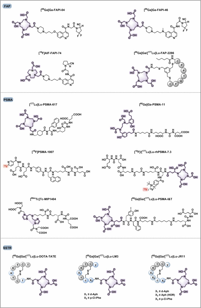
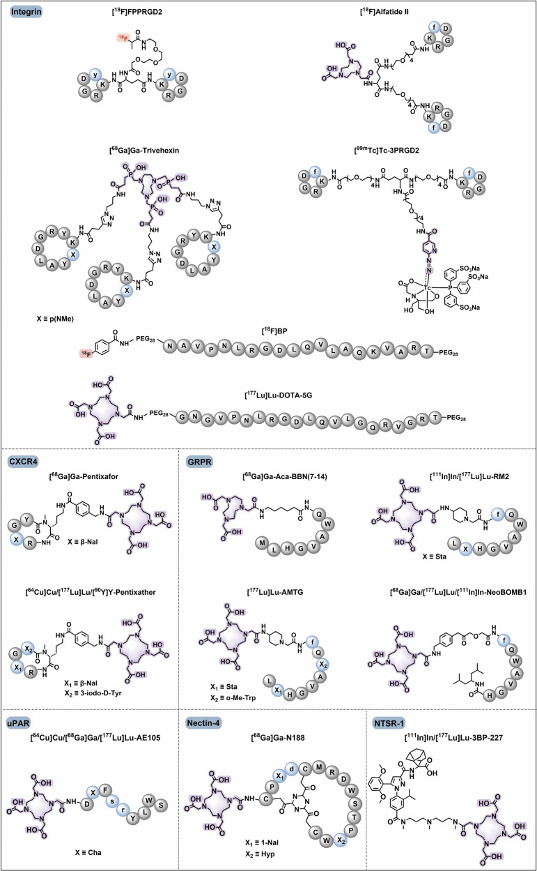
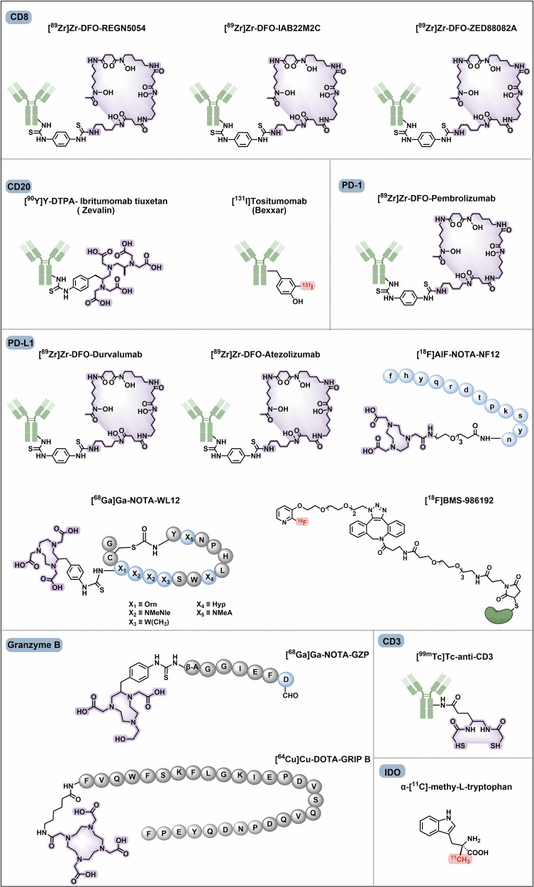
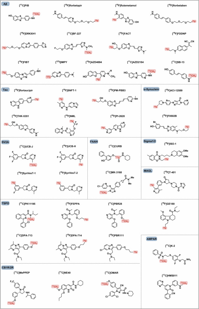
Radiopharmaceuticals involve the local delivery of radionuclides to targeted lesions for the diagnosis and treatment of multiple diseases. Radiopharmaceutical therapy, which directly causes systematic and irreparable damage to targeted cells, has attracted increasing attention in the treatment of refractory diseases that are not sensitive to current therapies. As the Food and Drug Administration (FDA) approvals of [177Lu]Lu-DOTA-TATE, [177Lu]Lu-PSMA-617 and their complementary diagnostic agents, namely, [68Ga]Ga-DOTA-TATE and [68Ga]Ga-PSMA-11, targeted radiopharmaceutical-based theranostics (radiotheranostics) are being increasingly implemented in clinical practice in oncology, which lead to a new era of radiopharmaceuticals. The new generation of radiopharmaceuticals utilizes a targeting vector to achieve the accurate delivery of radionuclides to lesions and avoid off-target deposition, making it possible to improve the efficiency and biosafety of tumour diagnosis and therapy. Numerous studies have focused on developing novel radiopharmaceuticals targeting a broader range of disease targets, demonstrating remarkable in vivo performance. These include high tumor uptake, prolonged retention time, and favorable pharmacokinetic properties that align with clinical standards. While radiotheranostics have been widely applied in tumor diagnosis and therapy, their applications are now expanding to neurodegenerative diseases, cardiovascular diseases, and inflammation. Furthermore, radiotheranostic-empowered precision medicine is revolutionizing the cancer treatment paradigm. Diagnostic radiopharmaceuticals play a pivotal role in patient stratification and treatment planning, leading to improved therapeutic outcomes in targeted radionuclide therapy. This review offers a comprehensive overview of the evolution of radiopharmaceuticals, including both FDA-approved and clinically investigated agents, and explores the mechanisms of cell death induced by radiopharmaceuticals. It emphasizes the significance and future prospects of theranostic-based radiopharmaceuticals in advancing precision medicine.


Conflict of interest statement

Competing interests: The authors declare no competing interests.

Figures

Fig. 1
Fig. 1
Overview of significant milestones and regulatory approvals for the discovery of radiopharmaceuticals. The journey began in 1896 with Henri Becquerel’s accidental discovery of “rays” emitted from uranium, a phenomenon later termed “radioactivity” by Marie Curie. Curie’s research further proceeded in the field by discovering the radioactive elements polonium and radium in 1898. In 1936, John H. Lawrence first used phosphorus-32 for the treatment of leukaemia which was the first example of the use of radionuclides in medicine. The commercial and regulatory landscape for radiopharmaceuticals began to take shape with the sale of the first commercial radiopharmaceutical, iodine-131 human serum albumin (RISA), by Abbott Laboratories. This regulatory evolution continued with the FDA’s decision to phase out the exemption for radiopharmaceuticals and regulate them as generic drugs. [131I]Sodium iodide was approved by FDA for treating thyroid disease. Technological and clinical advances further accelerated with the FDA’s approval of thallium-201 for myocardial perfusion imaging and the introduction of the first technetium-99m labelled radiopharmaceutical ([99mTc]Tc-exametazime) for stroke diagnosis. [18F]FDG was first approved for identifying regions of abnormal glucose metabolism associated with foci of epileptic seizures in 1989. In 1999, the PET/CT scanner was invented by Dr. Townshend, who combined precise imaging with detailed anatomical information and improved diagnostic accuracy. On March 12, 2000, the FDA published a notice that expanded the approval of [18F]FDG for new indications. The therapeutic scope of radiopharmaceuticals was further expanded with the approval of the radioimmunotherapy drugs Zevalin and Bexxar for treating NHL. [18F]Florbetapir, the first Aβ-specific PET radiotracer approved by the FDA in 2012, was used for the evaluation of patients with cognitive impairment. Bayer’s [223Ra]RaCl2, the first α-particle radiopharmaceutical, markedly improved the treatment of metastatic cancers. Lutathera was the first FDA-approved radiopharmaceutical for targeted RPT in 2018. Another targeted RPT, Pluvicto, was approved for prostate cancer and achieved near-blockbuster status in 2023
Fig. 2
Fig. 2
List of radioactive elements in the periodic table. Elements that emit radiation are called radionuclides. They are classified as positron emitters, α-emitters, β-emitters, γ-emitters, Auger electron emitters and hybrid nuclides according to their different forms of decay and have different clinical roles. In this figure, they are presented in different colours. Due to the difference in mass numbers, these radionuclides often exhibit significant variations in their physical properties and applications. There are 11 elements used for β-therapy, whereas α-therapy is less common, with five elements currently used in clinical settings. A small number of auger electron emitters, such as platinum-191, indium-111, and iodine-125, can also be used for therapy.β-imaging comprises the majority, with 17 elements used, followed by γ-imaging, which involves four elements. In addition, hybrid nuclides include several elements that can emit different types of radiation. These radionuclides have mixed applications that can simultaneously provide imaging and therapeutic functions. The rich variety of medical isotopes provides infinite possibilities for radiopharmaceuticals
Fig. 3
Fig. 3
Summary of approved radiopharmaceuticals. (a) Approval radiopharmaceuticals used in diagnosis and therapy for different diseases. Diagnostic agents are categorized into seven categories on the basis of indications. Several agents are used in multiple-diseases ([18F]FDG, for example), and they are preferentially categorized into their primary indications. All therapeutic radiopharmaceuticals are applied for oncology. (b) Radionuclides used in PET (37.0%) and SPECT (63.0%) scanning. Fluorine-18, gallium-68, carbon-11, nitrogen-13, copper-64, and rubidium-82 labelled agents are approved for PET/CT diagnosis. For SPECT/CT imaging, technetium-99m, iodine-123, indium-111, gallium-67, iodine-125, and thallium-201 are used. (c) Radionuclides used in cancer therapy, including iodine-131, yttrium-90, lutetium-177, phosphorus-32, strontium-89, samarium-153, and radium-223. (d) Numbers of approved diagnostic radiopharmaceuticals used in various diseases catalogued by radionuclides. Technetium-99m is mostly used in clinical imaging for multiple diseases. Fluorine-18 is used mainly in oncology and neurodegenerative disorders. (e) Targeting vectors for diagnostic and therapeutic radiopharmaceuticals. Small molecules are used as the major vectors for radiopharmaceuticals discovery. Peptides play a distinct role both in diagnosis and therapy, particularly after the FDA approval of [68Ga]/[177Lu]Ga-DOTA-TATE for NETs. Antibodies play essential roles in both imaging and therapy because of their strong binding affinity in vivo. Others indicate protein and serum albumin-based radiopharmaceuticals. The number of approved radiopharmaceuticals in each catalogue is presented
Fig. 4
Fig. 4
Widely studied targets for radiopharmaceuticals in tumour, neurodegenerative disorders and cardiovascular diseases. Radiopharmaceuticals are mainly used in the diagnosis and treatment of tumours, neurodegenerative disorders, and cardiovascular diseases. The TME contains tumour cells, immune cells, CAFs, and vascular endothelial cells, which play essential roles in cancer progression. Tumour targets for radiopharmaceutical development include GPCR-based transmembrane proteins (SSTR, GRPR, NTSR-1, CXCR4, and mGluR1), transmembrane proteins with four-pass domains (CLDN18.2), heterodimeric receptors (HER2 and the integrin family), other receptor (uPAR with no transmembrane and intracellular domains), immune checkpoints (PD-L1), and tumour antigens or other kinds of tumour biomarkers (PSMA, CD38, CAIX, GPC3, and Nectin-4). FAPs are expressed on both CAFs and tumour cells. VEGFRs are crucial tumour targets expressed by vascular endothelial cells. Immune cells that express checkpoints (PD-1, CTLA4, OX40, and ICOS), antigens (CD8, CD3, CD4, CD20, and CD30), and other biomarkers (IDO and Granzyme B) also serve as critical targets for cancer radiotheranostics. Aβ, tau, and α-synuclein plaques are the main causes of neurodegenerative disorders. The critical proteins expressed on synapses involved in neurotransmitter regulation include AMPAR and VMAT2 (transporter); FAAH and MAGL (signalling); SV2A, CB1R/21 R, sigma-1/2, and TSPO (transmembrane proteins), which have emerged as attractive targets for neurodegenerative disorders. Radiopharmaceuticals are currently used for the diagnosis of cardiovascular diseases. Owing to the important role of macrophages in disease progression, biomarkers that are expressed mainly on macrophages (TSPO, integrins) are potent imaging markers for cardiovascular pathology. Moreover, the FAP and VEGFR also showed potential in cardiovascular imaging. Part of this figure was created with Biorender.com
Fig. 5
Fig. 5
Chemical structures of clinically evaluated tumour-direct FAP, PSMA, and SSTR targeting radiopharmaceuticals. Representative clinically evaluated tumour-directed FAP-, PSMA- and SSTR-targeting radiopharmaceuticals. PSMA-targeting radiopharmaceuticals with a glutamate-urea-lysine structural motif, including PSMA-11, PSMA-1007, PSMA-617, and rhPSMA-7.3, have been approved. PSMA-targeting ligands that enable simultaneous diagnosis and therapy, including PSMA-I&T and rhPSMA, are of high value. SSTR-targeting radiopharmaceuticals play essential roles in the radiotheranostics of NETs. The antagonists, including LM3 and JR11, which have greater safety and affinity, are promising in SSTR-targeting imaging agents. FAP-targeting radiotracers may prove advantageous over [18F]FDG in the localization and visualization of solid tumours, such as FAPI-04, FAPI-46, and FAPI-74. Additionally, FAP-2286 has been shown to facilitate radiotheranostics. Grey circles: natural amino acids; blue circles: unnatural amino acids; highlighting in red: labelling with fluorine-18; highlighting in purple: chelators for metal radionuclide labelling
Fig. 6
Fig. 6
Chemical structures of clinically evaluated tumour-direct Integrin, CXCR4, GRPR, UPAR, NTSR-1, Nectin-4 targeting radiopharmaceuticals. Representative clinically evaluated tumour-directed promising radiopharmaceuticals targeting Integrin, CXCR-4, GRPR, uPAR, NTSR-1, and Nectin-4. In the Integrin family, RGD motif-based αvβ3-targeting radiopharmaceuticals, particularly [99mTc]Tc-3PRGD2, may be the next widely implemented diagnostic agent in clinical applications. Integrin αvβ6 targeting ligand 5 G has also demonstrated promising results in clinical trials. The efficacy of CXCR4-targeting ligands, including pentixafor and pentixather, as well as uPAR-targeting ligand AE105, has been demonstrated in numerous clinical studies. GRPR antagonists based on the BBN-like peptides, such as BBN(7–14), RM2, AMTG, and NeoBOMB1, have demonstrated remarkable therapeutic efficacy in the RPT of GRPR-positive tumours. A non-peptide NTSR-1 antagonist, 3BP-227, has been demonstrated to exhibit great receptor affinity and diminished normal organ uptake, making it a promising NTSR-1-targeted radiopharmaceutical for clinical investigation and translation. The bicyclic-peptide-based radiotracer, [68Ga]Ga-N188, has been demonstrated to be efficient for imaging tumour Nectin-4. Grey circles: natural amino acids; blue circles: unnatural amino acids; highlighting in red: labelling with fluorine-18; highlighting in purple: chelators for metal radionuclide labelling
Fig. 7
Fig. 7
Chemical structures of clinically evaluated radiopharmaceuticals involving immune-related targets. Representative clinically evaluated immune-related radiopharmaceuticals, such as CD8, CD3, CD20, PD-1, PD-L1, IDO and Granzyme B, CD20 and PD-L1/PD-1 are well-established in clinical settings. Two CD20-targeting radiopharmaceuticals have been approved ([90Y]Y-DTPA-Ibritumoma btiuxetan and [131I]Tositumomab), while emerging targets such as CD8 and Granzyme B are gaining attraction for their potential for clinical translation. Although CD3 and IDO have fewer clinical applications, they represent promising areas for future exploration. Antibody-based radiopharmaceuticals are highly specific but can present challenges in terms of PK and tumour penetration, while peptide-based drugs provide faster clearance and better tissue penetration and are ideal for imaging. Small molecules, while cleared quickly, require careful design to ensure specificity. Grey circles: natural amino acids; blue circles: unnatural amino acids; highlighting in red: labelling with fluorine-18, carbon-11, or iodine-131; highlighting in purple: chelator for metal radionuclide labelling
Fig. 8
Fig. 8
Chemical structures of clinically evaluated radiopharmaceuticals involved in neurodegenerative disorders. Representative clinically evaluated radiopharmaceuticals for neurodegenerative disorders. For AD diagnosis, the 18F-fluorinated radiotracers are gaining more focus with the approval of [18F]florbetapir, [18F]flutemetamol, [18F]florbetaben, and [18F]flortaucipir (Tau-targeting). Although [11C]PiB is mostly used in AD diagnosis, [18F]AZD4694 (Aβ-targeting) and [18F]PI-2620 (Tau-targeting) are most promising approved radiotracers. TSPO-targeting diagnosis has been well investigated in brain imaging. On the basis of [11C]PK11195, many radiopharmaceuticals have emerged and [18F]GE180 was investigated in phase II. For Sigma1/2, effective and promising imaging results with the discovery of [18F]ISO-1 and other high binding affinity tracers are under developed. Highlighting in red: labelling with fluorine-18 and carbon-11
Fig. 9
Fig. 9
The potential biological mechanism of radiopharmaceutical-induced cell death. Radiopharmaceuticals induce SSBs (β-emitters) or DSBs (α-emitters and Auger electron emitters), resulting in cancer cell senescence via the SASP, mitochondrial apoptosis, and STING-NLRP3 axis-dependent pyroptosis. Radiopharmaceuticals reshape the tumour immune microenvironment through three mechanisms. First, radiopharmaceuticals induce the release of “danger signals” (also known as danger-associated molecular patterns), including HSP70 and HMGB1, which are essential for the activation of DCs. Second, radiation-induced cell death stimulates the release of tumour antigens, which are presented by activated DCs. The cGAS-STING pathway also activates cytotoxic T cells through the interferon response. Therefore, T cells are recruited to the TME and activated to induce immunogenic cancer cell death. Radiopharmaceuticals also induce the production of ROS, leading to cell apoptosis and an inflammatory response. The mechanisms are concluded from studies of radiopharmaceuticals or learned from the research of radiotherapy. Part of this figure was created with Biorender.com

Similar articles

Cited by

References

    1. Bodei, L., Herrmann, K., Schoder, H., Scott, A. M. & Lewis, J. S. Radiotheranostics in oncology: Current challenges and emerging opportunities. Nat. Rev. Clin. Oncol.19, 534–550 (2022). - PMC - PubMed
    1. Bollineni, V. R., Collette, S. & Liu, Y. Functional and molecular imaging in cancer drug development. Chin. Clin. Oncol.3, 17 (2014). - PubMed
    1. Bolcaen, J. et al. A perspective on the radiopharmaceutical requirements for imaging and therapy of glioblastoma. Theranostics11, 7911–7947 (2021). - PMC - PubMed
    1. Aboagye, E. O., Barwick, T. D. & Haberkorn, U. Radiotheranostics in oncology: Making precision medicine possible. CA-Cancer J. Clin.73, 255–274 (2023). - PubMed
    1. Boros, E. & Packard, A. B. Radioactive transition metals for imaging and therapy. Chem. Rev.119, 870–901 (2018). - PubMed

Substances