. 2025 Jan 2;15(1):434.
doi: 10.1038/s41598-024-84462-5.

FSTL1 aggravates high glucose-induced oxidative stress and transdifferentiation in HK-2 cells

Affiliations

FSTL1 aggravates high glucose-induced oxidative stress and transdifferentiation in HK-2 cells

Baoyuan Zhang et al. Sci Rep. .

Abstract

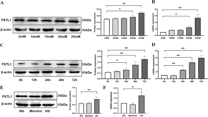
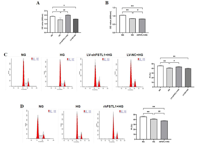
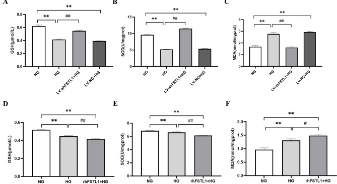
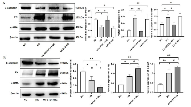
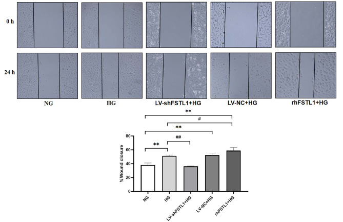
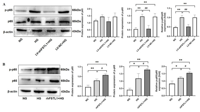
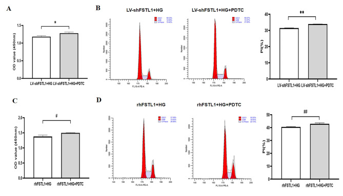
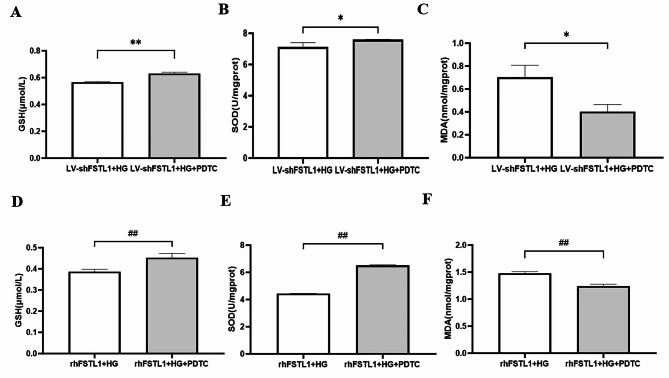
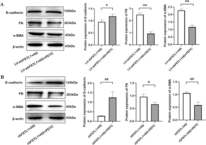
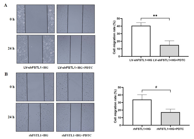
Chronic hyperglycemia, a hallmark of diabetes, can trigger inflammatory responses in the kidney, leading to diabetic nephropathy (DN). Follistatin-like protein 1 (FSTL1) has emerged as a potential therapeutic target in various kidney diseases. This study investigated the effect of high glucose on FSTL1 expression and its role in oxidative stress and cellular transdifferentiation injury in HK-2 human proximal tubule epithelial cells, a model of DN. We investigated FSTL1's level in HK-2 cells exposed to high glucose using Western blotting and quantitative real-time polymerase chain reaction (qRT-PCR). FSTL1 was manipulated using recombinant human FSTL1 (rhFSTL1) or lentiviral shFSTL1. We then analyzed proliferation, oxidative stress, transdifferentiation, cell migration, and the nuclear factor kappa-B (NF-κB) signaling pathway potentially involved in FSTL1 effects. Finally, we blocked the NF-κB pathway to see its influence on these cellular processes. High glucose exposure significantly increased FSTL1 in HK-2 cells, with longer/higher glucose further amplifying this effect. Silencing of FSTL1 ameliorates cellular damage by promoting proliferation, enhancing superoxide dismutase (SOD) and glutathione (GSH) activity, and reducing malondialdehyde (MDA) production, inhibiting cell migration. Furthermore, it prevented the harmful conversion of HK-2 cells from epithelial to myofibroblast-like phenotypes, evidenced by decreased fibronectin (FN) and α-smooth muscle actin (α-SMA) and preserved E-cadherin. Notably, silencing FSTL1 also inhibited the NF-κB signaling pathway. Conversely, rhFSTL1 exhibited opposite effects. Importantly, blocking NF-κB reversed the detrimental effects of FSTL1. These findings suggest that FSTL1 contributes to high glucose-induced kidney injury by promoting oxidative stress and cellular transdifferentiation potentially via the NF-κB pathway. Targeting FSTL1 may represent a novel therapeutic strategy for preventing or mitigating DN progression.

Keywords: Diabetic nephropathy; Follistatin-like protein 1; Oxidative stress; Transdifferentiation.


Conflict of interest statement

Competing interests: The authors declare no competing interests.

Figures

Fig. 1
Fig. 1
Effect of high glucose induction on FSTL1 expression in HK-2 cells. (A, B) Effect of high glucose concentration on FSTL1 expression in HK-2 cells. (A) FSTL1 protein expression (Western blotting); (B) FSTL1 mRNA expression (qPCR). (C, D) Effect of high glucose duration of action on FSTL1 expression in HK-2 cells. (C) FSTL1 protein expression (Western blotting); (D) FSTL1 mRNA expression (qPCR). (E, F) Effect of osmotic pressure on FSTL1 expression in HK-2 cells. (E) FSTL1 protein expression (Western blotting); (F) FSTL1 mRNA expression (qPCR). *P < 0.05 and **P < 0.01. NG, normal glucose; HG, high glucose; Mannitol, high osmolarity.
Fig. 2
Fig. 2
Effect of FSTL1 on proliferation of high glucose-induced HK-2 cells. (A, B) CCK-8 assays for cell proliferation. (C, D) Flow cytometry assays for cell proliferation. (A, C) Silencing FSTL1 expression; (B, D) Giving rhFSTL1. Compared to the NG group, *P < 0.05 and **P < 0.01. Compared to the HG group, #P < 0.05 and ##P < 0.01. shFSTL1, silencing of FSTL1; rhFSTL1, recombinant human FSTL1.
Fig. 3
Fig. 3
Effect of FSTL1 on oxidative stress in high glucose-induced HK-2 cells. (A, B, C) Silencing of FSTL1 by shRNA attenuated HG-induced oxidative stress. (A) GSH level; (B) SOD level; (C) MDA level. (D, E, F) rhFSTL1 aggravated HG-induced oxidative stress. (D) GSH level; (E) SOD level; (F) MDA level. Compared to the NG group, **P < 0.01. Compared to the HG group, #P < 0.05 and ##P < 0.01. GSH, glutathione; SOD, superoxide dismutase; MDA, malondialdehyde.
Fig. 4
Fig. 4
Effect of FSTL1 on transdifferentiation of high glucose-induced HK-2 cells. (A) Silencing FSTL1 expression; (B) Giving rhFSTL1. Compared to the NG group, *P < 0.05 and **P < 0.01. Compared to the HG group, #P < 0.05 and ##P < 0.01.
Fig. 5
Fig. 5
Effect of FSTL1 on cell migration of high glucose-induced HK-2 cells. Compared to the NG group, **P < 0.01. Compared to the HG group, #P < 0.05 and ##P < 0.01.
Fig. 6
Fig. 6
Effect of FSTL1 on NF-κB expression in high glucose-induced HK-2 cells. (A) Silencing FSTL1 expression; (B) Giving rhFSTL1. Compared to the NG group, *P < 0.05 and **P < 0.01. Compared to the HG group, #P < 0.05 and ##P < 0.01.
Fig. 7
Fig. 7
Effect of FSTL1 on HK-2 cell proliferation after blocking the NF-κB signaling pathway. (A, C) CCK-8 assays for cell proliferation. (B, D) Flow cytometry assays for cell proliferation. Compared to the LV-shFSTL1 + HG group, *P < 0.05. Compared to the rhFSTL1 + HG group, #P < 0.05. PDTC, NF-κB signaling pathway inhibitor.
Fig. 8
Fig. 8
Effect of FSTL1 on oxidative stress in HK-2 cells after blocking the NF-κB signaling pathway. (A, B, C) Silencing FSTL1 expression. (A) GSH level; (B) SOD level; (C) MDA level. (D, E, F) Giving rhFSTL1. (D) GSH level; (E) SOD level; (F) MDA level. Compared to the LV-shFSTL1 + HG group, *P < 0.05 and **P < 0.01. Compared to the rhFSTL1 + HG group, ##P < 0.01.
Fig. 9
Fig. 9
Effect of FSTL1 on transdifferentiation in HK-2 cells after blocking the NF-κB signaling pathway. (A) Silencing FSTL1 expression; (B) Giving rhFSTL1. E-cadherin, FN, and α-SMA expression levels were examined by Western blotting. Compared to the LV-shFSTL1 + HG group, *P < 0.05 and **P < 0.01. Compared to the rhFSTL1 + HG group, #P < 0.05 and ##P < 0.01.
Fig. 10
Fig. 10
Effect of FSTL1 on cell migration in HK-2 cells after blocking the NF-κB signaling pathway. (A) Silencing FSTL1 expression; (B) Giving rhFSTL1. Compared to the LV-shFSTL1 + HG group, **P < 0.01. Compared to the rhFSTL1 + HG group, #P < 0.05.

Similar articles

Cited by

References

    1. Samsu, N. Diabetic nephropathy: Challenges in pathogenesis, diagnosis, and treatment. BioMed Res. Int.2021, 1–17 (2021). - PMC - PubMed
    1. Hu, Q. et al. Diabetic nephropathy: Focusing on pathological signals, clinical treatment, and dietary regulation. Biomed. Pharmacother.159, 114252 (2023). - PubMed
    1. Matoba, K. et al. Unraveling the role of inflammation in the pathogenesis of diabetic kidney disease. Int. J. Mol. Sci.20, 3393 (2019). - PMC - PubMed
    1. Navarro-González, J. F., Mora-Fernández, C., De Fuentes, M. M. & García-Pérez, J. Inflammatory molecules and pathways in the pathogenesis of diabetic nephropathy. Nat. Rev. Nephrol.7, 327–340 (2011). - PubMed
    1. Zhang, Y. & Xia, Y. Role of follistatin-like 1 (Fstl1) in chronic kidney disease. Kidney Int. Rep.4, S387 (2019).

Publication types

MeSH terms

LinkOut - more resources