Review
. 2025 Jan;329(1):e13435.
doi: 10.1111/imr.13435.

Recognition of Self and Viral Ligands by NK Cell Receptors

Affiliations
Review

Recognition of Self and Viral Ligands by NK Cell Receptors

Roy A Mariuzza et al. Immunol Rev. 2025 Jan.

Abstract

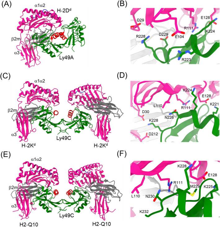
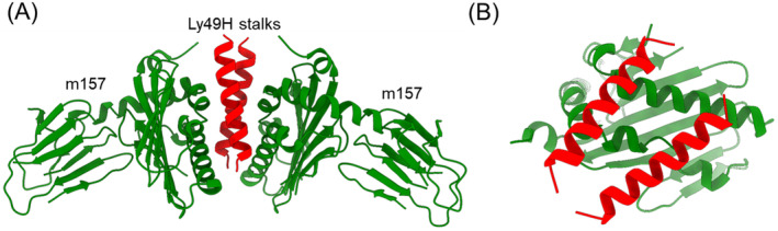
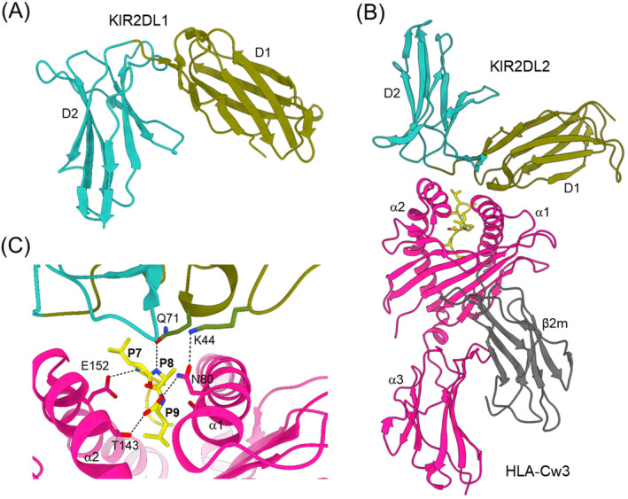
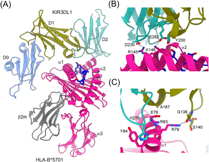
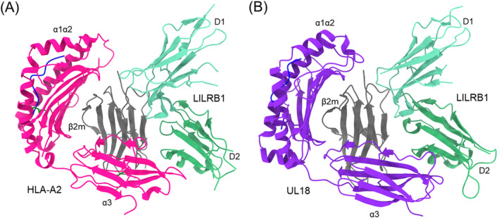
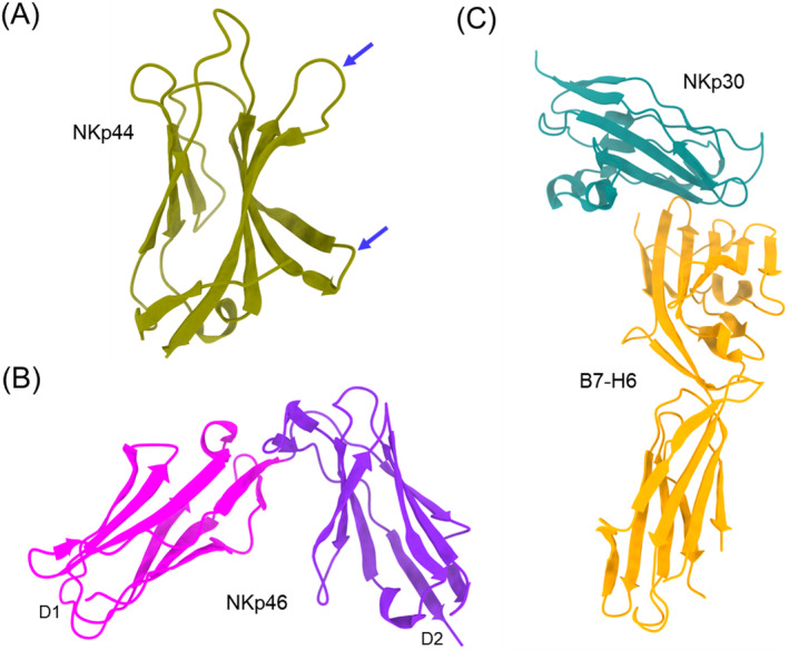
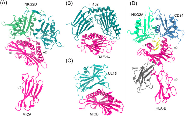
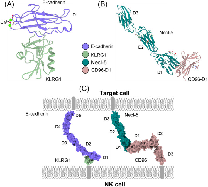
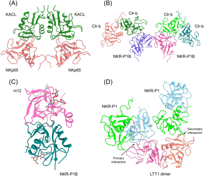
Natural killer (NK) cells are essential elements of the innate immune response against tumors and viral infections. NK cell activation is governed by NK cell receptors that recognize both cellular (self) and viral (non-self) ligands, including MHC, MHC-related, and non-MHC molecules. These diverse receptors belong to two distinct structural families, the C-type lectin superfamily and the immunoglobulin superfamily. NK receptors include Ly49s, KIRs, LILRs, and NKG2A/CD94, which bind MHC class I (MHC-I) molecules, and NKG2D, which binds MHC-I paralogs such MICA and ULBP. Other NK receptors recognize tumor-associated antigens (NKp30, NKp44, NKp46), cell-cell adhesion proteins (KLRG1, CD96), or genetically coupled C-type lectin-like ligands (NKp65, NKR-P1). Additionally, cytomegaloviruses have evolved various immunoevasins, such as m157, m12, and UL18, which bind NK receptors and act as decoys to enable virus-infected cells to escape NK cell-mediated lysis. We review the remarkable progress made in the past 25 years in determining structures of representatives of most known NK receptors bound to MHC, MHC-like, and non-MHC ligands. Together, these structures reveal the multiplicity of solutions NK receptors have developed to recognize these molecules, and thereby mediate crucial interactions for regulating NK cytolytic activity by self and viral ligands.

Keywords: KIR; Ly49; MHC; NK receptor; structure; virus.


Conflict of interest statement

The authors declare no conflicts of interest.

Figures

FIGURE 1
FIGURE 1
Structures of Ly49–MHC complexes. (A) Ly49A bound to H‐2Dd (PDB accession code 1QO3) [15]. The α1, α2, and α3 domains of the MHC‐I heavy chain are deep pink; β2m is gray; the MHC‐bound peptide is blue; the Ly49A dimer is green; the α2 helices of the Ly49A dimer are red. The complex is oriented with the NK cell at the top and the target cell at the bottom. (B) The Ly49A–H‐2Dd interface. The side chains of contacting residues are drawn in stick representation. (C) Structure of Ly49C bound to H‐2Kb (1P4L) [17]. (D) The Ly49C–H‐2Kb interface. (E) Structure of Ly49C in complex with H2‐Q10 (5J6G) [19]. (F) The Ly49C–H2‐Q10 interface.
FIGURE 2
FIGURE 2
Structure of the MCMV immunoevasin m157 in complex with Ly49H stalks. (A) Side view of the Ly49H–m157 complex (4JO8) [21]. Two m157 monomers (green) bind the α‐helical Ly49H stalks (red). (B) Top view of the Ly49H–m157 complex. The Ly49H stalks straddle the α1/α2 platform of m157.
FIGURE 3
FIGURE 3
Structures of KIR2DL and a KIR2DL–HLA‐C complex. (A) Structure of unbound KIRDL1 (1NKR) [23], which comprises Ig‐like D1 and D2 domains. (B) Structure of KIR2DL2 bound to HLA‐Cw3 (1EFX) [35]. The α1, α2, and α3 domains of the MHC‐I heavy chain are deep pink; β2m is gray; the MHC‐bound peptide is yellow. (C) Close‐up of the KIR2DL2–HLA‐Cw3 interface. Hydrogen bonds are drawn as black dotted lines.
FIGURE 4
FIGURE 4
Structure of KIR3DL1 bound to HLA‐B*5701. (A) Structure of the KIR3DL1–HLA‐B*5701 complex (3VH8) [38]. The α1, α2, and α3 domains of the MHC‐I heavy chain are deep pink; β2m is gray; the MHC‐bound peptide is dark blue. (B) Contacts between KIR3DL1 and the α2 helix of HLA‐B*5701. Dotted black lines represent hydrogen bonds. (C) Contacts between KIR3DL1 and the α1 helix of HLA‐B*5701.
FIGURE 5
FIGURE 5
Interaction of LILRB1 with HLA‐A2 and the HCMV immunoevasin UL18. (A) Structure of LILRB1 in complex with HLA‐A2 (1P7Q) [9]. The α1, α2, and α3 domains of the MHC‐I heavy chain are deep pink; β2m is gray; the MHC‐bound peptide is blue. (B) Structure of LILRB1 bound to the HCMV MHC‐I mimic UL18 (3D2U) [10]. The α1, α2, and α3 domains of UL18 are violet; β2m is gray; the UL18‐bound peptide is blue.
FIGURE 6
FIGURE 6
Structures of human natural cytotoxicity receptors. (A) Structure of NKp44 (1HKF) [16]. The blue arrows point to loops forming a positively charged groove that may bind anionic ligands. (B) Structure of NKp46 (1P6F) [18]. (C) Structure of NKp30 bound to B7‐H6 (3PV6) [20], which is selectively expressed on tumors.
FIGURE 7
FIGURE 7
Structures of NKG2D and NKG2A/CD94 complexes. (A) Complex between NKG2D and MICA (1HYR) [24]. (B) Complex between the MCMV immunoevasin m157 NKG2D ligand RAE‐1γ (1JSK) [26]. (C) Structure of the HCMV immunoevasin UL16 bound to MICB (2WY3) [99]. (D) Structure of human NKG2A/CD94 bound to HLA‐E (3CDG) [30].
FIGURE 8
FIGURE 8
Recognition of cell–cell adhesion proteins by KLRG1 and CD96. (A) Structure of KLRG1 bound to the membrane‐distal D1 domain of E‐cadherin (3FF8) [34]. Bound Ca2+ ions are shown as green spheres. (B) Structure of the membrane‐distal D1 domain of CD96 bound to necl‐5 (6ARQ) [37]. N‐linked glycans are drawn in stick representation. (C) (left) Space‐filling model of the KLRG1–E‐cadherin complex. Ig‐like domains of E‐cadherin are labeled D1–D5. The model was constructed by superposing the structure of KLRG1 bound to D1 of E‐cadherin onto the structure of full‐length E‐cadherin (D1–D5) (3Q2V) [124]. (right) Space‐filling model of the CD96–necl‐5 complex. CD96 comprises three Ig‐like domains, but only D1 is present in the crystal structure. CD96 D2 and D3 were homology‐modeled using SWISS‐MODEL.
FIGURE 9
FIGURE 9
C‐type lectin‐like receptor–ligand pairs in the NK gene complex. (A) Structure of the human NKp65–KACL complex (4IOP) [39]. The NKp65 and KACL homodimers bind in a head‐to‐head orientation. (B) Structure of the mouse NKR‐P1B–Clr‐b complex (6E7D) [42]. Each Clr‐b dimer binds one monomer of the NKR‐P1B dimer. (C) Structure of the mouse NKR‐P1B–m12 complex (5TZN) [44]. N‐linked glycans are shown as sticks. The m12 immunoevasin straddles NKR‐P1B in a claw‐like manner. (D) Structure of the human NKR‐P1–LLT1 complex (5MGT) [41]. The LLT1 dimer (pink/orange) contacts the NKR‐P1 dimer formed by the green and light blue monomers. The light blue NKR‐P1 monomer binds LLT1 in the primary interaction mode. The green NKR‐P1 monomer binds LLT1 in the secondary interaction mode.

Similar articles

Cited by

References

    1. Di Santo J. P., “Natural Killer Cells: Diversity in Search of a Niche,” Nature Immunology 9 (2008): 473–475. - PubMed
    1. Vivier E., Tomasello E., Baratin M., Walzer T., and Ugolini S., “Functions of Natural Killer Cells,” Nature Immunology 9 (2008): 503–510. - PubMed
    1. Caliguiri M. A., “Human Natural Killer Cells,” Blood 112 (2008): 461–469. - PMC - PubMed
    1. Cerwenka A. and Lanier L. L., “Natural Killer Cell Memory in Infection, Inflammation and Cancer,” Nature Reviews. Immunology 16 (2016): 112–123. - PubMed
    1. Long E. O., Sik K. H., Liu D., Peterson M. E., and Rajagopalan S., “Controlling Natural Killer Cell Responses: Integration of Signals for Activation and Inhibition,” Annual Review of Immunology 31 (2013): 227–258. - PMC - PubMed