. 2025 Jan;15(1):e70152.
doi: 10.1002/ctm2.70152.

Exosome-derived long non-coding RNA AC010789.1 modified by FTO and hnRNPA2B1 accelerates growth of hair follicle stem cells against androgen alopecia by activating S100A8/Wnt/β-catenin signalling

Affiliations

Exosome-derived long non-coding RNA AC010789.1 modified by FTO and hnRNPA2B1 accelerates growth of hair follicle stem cells against androgen alopecia by activating S100A8/Wnt/β-catenin signalling

Shaojun Chu et al. Clin Transl Med. 2025 Jan.

Abstract

Background: The increased incidence of androgenic alopecia (AGA) causes adverse physiological and psychological effects on people of all genders. The hair follicle stem cells (HFSCs) have displayed clinical improvements on AGA. However, the molecular mechanism of HFSCs against AGA remains elusive.

Methods: The expression and prognosis of lncRNA AC010789.1 in AGA hair follicle tissues were assessed by qRT-PCR analysis. CCK-8, EdU and Transwell analysis were utilized to assess cell growth. The specific binding between AC010789.1 and FTO mediated m6A modification or the effect of AC010789.1 on hnRNPA2B1, S100A8 and Wnt/β-catenin signaling expression was confirmed by bioinformatic analysis, RIP, RNA pull-down and Western blot assay. The effects of Exosome-loaded AC010789.1 prompted HFSCs proliferation and hair follicle regeneration were confirmed in hairless mice.

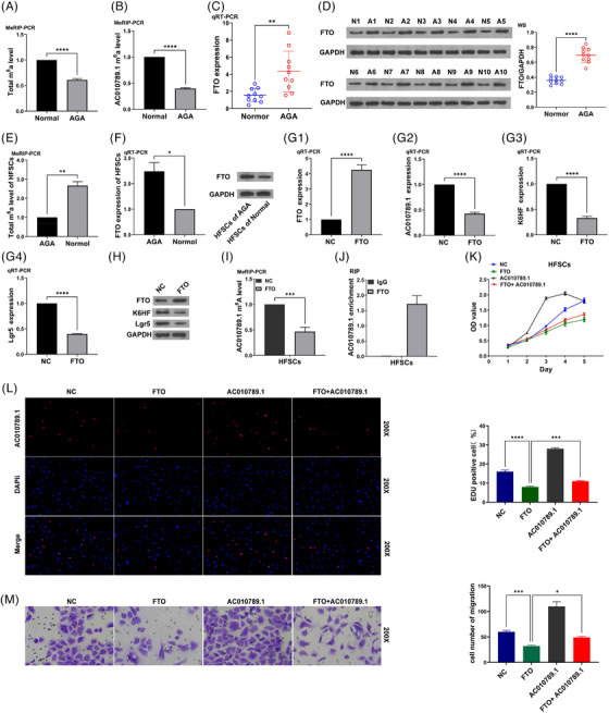
Results: We herein found that the mRNA levels of lncRNA AC010789.1 were decreased in AGA tissue samples but increased in HFSCs of surrounding normal tissue samples. Overexpression (OE) of AC010789.1 promoted HFSC proliferation, DNA synthesis and migration as well as K6HF and Lgr5 upregulation, whereas knockdown of AC010789.1 showed the opposite effects. The total or AC010789.1 m6A levels were reduced and FTO demethylase was upregulated in AGA tissue samples, but these indicated the reverse results in HFSCs of surrounding normal tissue samples. FTO OE decreased AC010789.1 m6A levels and its mRNA levels in HFSCs and abolished AC010789.1-induced HFSCs proliferation. In addition, AC010789.1 was identified to bind to m6A reader hnRNPA2B1, which was downregulated in AGA but upregulated in HFSCs of surrounding normal tissue samples. hnRNPA2B1 OE attenuated AC010789.1 knockdown-induced inhibition of HFSCs proliferation. Moreover, AC010789.1 could bind to and enhance downstream S100A8 protein expression, which mediated Wnt/β-catenin signaling to accelerate HFSCs proliferation. Exosome-loaded AC010789.1 prompted HFSCs proliferation and hair follicle regeneration in mice.

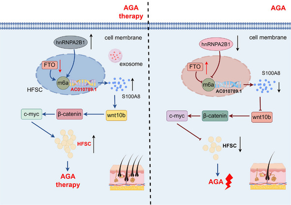
Conclusions: Our findings demonstrated that exosome-derived lncRNA AC010789.1 modified by FTO and hnRNPA2B1 facilitated the proliferation of human HFSCs against AGA by activating S100A8/Wnt/β-catenin signaling.

Key points: Long non-coding RNA (lncRNA) AC010789.1 was downregulated in hair follicle tissues from androgenic alopecia (AGA) and upregulated in hair follicle stem cells (HFSCs). LncRNA AC010789.1 promoted the proliferation and migration of HFSCs. FTO/hnRNPA2B1-mediated m6A modification of lncRNA AC010789.1 promoted HFSCs growth by activating S100A8/Wnt/β-catenin signalling. Exosome-derived AC010789.1 accelerated HFSCs proliferation.

Keywords: FTO; HFSCs; S100A8; hnRNPA2B1; lncRNA AC010789.1; proliferation.


Conflict of interest statement

The authors declare no conflict of interest.

Figures

FIGURE 1
FIGURE 1
Long non‐coding RNA (lncRNA) AC010789.1 was downregulated in hair follicle tissues from androgenic alopecia (AGA) and upregulated in hair follicle stem cells (HFSCs). (A) Real‐time quantitative polymerase chain reaction (qRT‐PCR) analysis of the expression levels of AC010789.1 in 10 pairs of AGA hair follicle tissues. (B) qRT‐PCR analysis of the expression levels of AC010789.1 in HFSCs. (C) 5‐ethynyl‐2'‐deoxyuridine assay (EdU) flow cytometry analysis of the molecular markers CD29 and CD71 of HFSCs. (D) qRT‐PCR analysis of the expression levels of AC010789.1 after the transfection with AC010789.1 OE lentiviruses into HFSCs and K6HF and Lgr5 after the transfection with AC010789.1 OE lentiviruses into HFSCs. (E) Western blot analysis of the expression levels of AC010789.1, K6HF and Lgr5 after the transfection with AC010789.1 OE lentiviruses into HFSCs. (F) qRT‐PCR analysis of the expression levels of AC010789.1 after transfection with si‐AC010789.1 into HFSCs and K6HF and Lgr5 after transfection with si‐AC010789.1 into HFSCs. (G) Western blot analysis of the expression levels of AC010789.1, K6HF and Lgr5 after transfection with si‐AC010789.1 into HFSCs. Data shown are the mean ± SEM of three experiments. *< .05, **< .01 and ***< .001.
FIGURE 2
FIGURE 2
LncRNA AC010789.1 promoted the proliferation and migration of hair follicle stem cells (HFSCs) (A) CCK8 analysis of the cell proliferation viability after the transfection with AC010789.1 OE lentiviruses into HFSCs. (B) 5‐ethynyl‐2'‐deoxyuridine assay (EdU) assays of the DNA synthesis after the transfection with AC010789.1 OE lentiviruses into HFSCs. (C) Transwell analysis of cell migration capabilities after the transfection with AC010789.1 OE lentiviruses into HFSCs. (D) CCK8 analysis of the cell proliferation viability after the transfection with si‐AC010789.1 into HFSCs. (E) EdU assays of the DNA synthesis after the transfection with si‐AC010789.1 into HFSCs. (F) Transwell analysis of cell migration capabilities after the transfection with si‐AC010789.1 into HFSCs. Data shown are the mean ± SEM of three experiments. **< .01, ***< .001 and ****p < .0001.
FIGURE 3
FIGURE 3
FTO mediated m6A modification of long non‐coding RNA (lncRNA) AC010789.1 in hair follicle stem cells (HFSCs) (A, B) MeRIP‐PCR analysis of the total m6A levels and AC010789.1 m6A levels in androgenic alopecia (AGA) hair follicle tissues. (C, D) Real‐time quantitative polymerase chain reaction (qRT‐PCR) and Western blot analysis of the expression levels of FTO in AGA hair follicle tissues. (E) MeRIP‐PCR analysis of the total m6A levels in AGA HFSCs. (F) qRT‐PCR and Western blot analysis of the expression levels of FTO in HFSCs. (G, H) qRT‐PCR and Western blot analysis of the expression levels of FTO, AC010789.1, K6HF and Lgr5 after the transfection with FTO OE plasmids into HFSCs. (I) MeRIP‐PCR analysis of the effects of FTO OE on AC010789.1 m6A levels in HFSCs. (J) RIP analysis of the endogenous interaction of AC010789.1 mRNAs with FTO protein in HFSCs. (K‐M) CCK‐8, 5‐ethynyl‐2'‐deoxyuridine assay (EdU) and Transwell analysis of the cell proliferation and migration viability after the co‐transfection with FTO and AC010789.1 OE lentiviruses into HFSCs. Data shown are the mean ± SEM of three experiments. *< .05, **< .01, ***p < .001 and ****p < .0001.
FIGURE 4
FIGURE 4
hnRNPA2B1 interacted with long non‐coding RNA (lncRNA) AC010789.1 to promote hair follicle stem cells (HFSCs) growth. (A) Silver impregnation and Mass spectrum analysis of AC010789.1‐interacting proteins in HFSCs. (B, C) Real‐time quantitative polymerase chain reaction (qRT‐PCR) analysis of the expression levels of hnRNPA2B1 in 10 pairs of androgenic alopecia (AGA) hair follicle tissues. (D) qRT‐PCR and Western blot analysis of the expression levels of hnRNPA2B1 in HFSCs. (E) RNA pull‐down verification of the interaction of hnRNPA2B1 protein with the AC010789.1 mRNAs in HFSCs. (F, G) qRT‐PCR and Western blot analysis of the expression levels of hnRNPA2B1, AC010789.1, K6HF and Lgr5 after the transfection with hnRNPA2B1 OE plasmids into HFSCs. (H) MeRIP‐PCR analysis of the effects of hnRNPA2B1 OE on AC010789.1 m6A levels in HFSCs. (I) RIP analysis of the enrichment of the endogenous AC010789.1 in hnRNPA2B1 protein in HFSCs. (K‐M) CCK8, 5‐ethynyl‐2'‐deoxyuridine assay (EdU) and Transwell analysis of the cell proliferation and migration viability after the co‐transfection with hnRNPA2B1 OE plasmids and si‐AC010789.1 into HFSCs. Data shown are the mean ± SEM of three experiments. *< .05, **< .01, ***p < .001 and ****p < .0001.
FIGURE 5
FIGURE 5
S100A8 was identified as a downstream regulator of AC010789.1 to promote hair follicle stem cells (HFSCs) growth by activating Wnt/β‐catenin signalling (A) RNA pull‐down analysis of the interaction of S100A8 protein with the AC010789.1 mRNAs in HFSCs. (B) RIP analysis of the endogenous enrichment of AC010789.1 mRNAs in S100A8 protein in HFSCs. (C, D) Real‐time quantitative polymerase chain reaction (qRT‐PCR) and Western blot analysis of the expression levels of S100A8 after the transfection with AC010789.1 OE lentiviruses or si‐AC010789.1 into HFSCs. (E, F) qRT‐PCR and Western blot analysis of the expression levels of S100A8, K6HF and Lgr5 after the transfection of si‐AC010789.1 into HFSCs. (G‐I) CCK8, 5‐ethynyl‐2'‐deoxyuridine assay (EdU) and Transwell analysis of the cell proliferation and migration viability after the co‐transfection with AC010789.1 OE lentiviruses and si‐S100A8 into HFSCs. (J) Western blot analysis of the expression levels of S100A8, Wnt10b, β‐catenin and c‐myc after the co‐transfection with AC010789.1 OE lentiviruses and si‐S100A8 into HFSCs. Data shown are the mean ± SEM of three experiments. **< .01, ***p < .001 and ****p < .0001.
FIGURE 6
FIGURE 6
Exo‐AC010789.1 facilitated hair follicle stem cells (HFSCs) proliferation through the Wnt/β‐catenin signalling. (A) Real‐time quantitative polymerase chain reaction (qRT‐PCR) analysis of the expression levels of AC010789.1 in extracellular vesicles Exo‐AC010789.1 and Exo‐NC. (B) qRT‐PCR analysis of the stability of Exo‐AC010789.1. (C) Western blot analysis of the expression levels of extracellular vesicle markers ALIX and CD9 in Exo‐AC010789.1. (D) Microflow analysis of extracellular vesicle particle size. (E) Fluorescence microscopy of HFSCs absorbing exosomes. (F–H) 5‐ethynyl‐2'‐deoxyuridine assay (EdU), CCK8 and Transwell analysis of the effects of Exo‐AC010789.1 on HFSCs proliferation and migration. (I) Western blot analysis of the effects of Exo‐AC010789.1 on Wnt signalling transduction. Data shown are the mean ± SEM of three experiments. ns p ≥ .05,*< .05, **< .01 and ****p < .0001.
FIGURE 7
FIGURE 7
Exo AC010789.1 enhanced hair growth in hairless mice. (A) Observations of the area covered by hair on the back of mice. (B) Hematoxylin and Eosin (HE) analysis of skin tissue morphology (×200, scale 50µm). (C) Immunohistochemistry (IHC) analysis of the protein expression of K6HF, Lgr5, S100A8, Wnt10b and c‐myc. (D) IF detection of β‐catenin and Ki67 protein expression in skin tissues (×200, scale 50µm).
FIGURE 8
FIGURE 8
Schematic diagram of the molecular mechanism of long non‐coding RNA (lncRNA) AC010789.1 in hair follicle stem cells (HFSCs) against androgenic alopecia (AGA). FTO/hnRNPA2B1 mediates m6A modification of lncRNA AC010789.1 to activate S100A8/Wnt/β‐catenin signalling, contributing to HFSCs growth and exosome‐derived AC010789.1 in HFSCs could be applied for treatment of AGA.

Similar articles

Cited by

References

    1. Piraccini BM, Alessandrini A. Androgenetic alopecia. G Ital Dermatol Venereol. 2014;149(1):15‐24. - PubMed
    1. Devjani S, Ezemma O, Kelley KJ, et al. Androgenetic Alopecia: Therapy Update. Drugs. 2023;83(8):701‐715. - PMC - PubMed
    1. Leirós GJ, Ceruti JM, Castellanos ML, et al. Androgens modify Wnt agonists/antagonists expression balance in dermal papilla cells preventing hair follicle stem cell differentiation in androgenetic alopecia. Mol Cell Endocrinol. 2017;439:26‐34. - PubMed
    1. Hu XM, Li ZX, Zhang DY, et al. A systematic summary of survival and death signalling during the life of hair follicle stem cells. Stem Cell Res Ther. 2021;12(1):453. - PMC - PubMed
    1. Dong C, Du J, Yu Z, et al. Global research status and trends in hair follicle stem cells: a bibliometric analysis. Stem Cell Rev Rep. 2022;18(6):2002‐2015. - PubMed

Substances

LinkOut - more resources