

Removal of a fractured file beyond the apical foramen using robot-assisted endodontic microsurgery: a clinical report
- PMID: 39748344
- PMCID: PMC11697827
- DOI: 10.1186/s12903-024-05329-9
Removal of a fractured file beyond the apical foramen using robot-assisted endodontic microsurgery: a clinical report
Abstract
Background: Endodontic file fractures are common complications of root canal treatment, and requires removal via specialized techniques such as endodontic microsurgery when the file beyond the apical foramen. It is often challenging to precisely and minimally remove a fractured file. Recently the use of dental autonomous robotic system (ATR) has shown promise in precisely and minimally in dental surgery. Therefore, this case details a technique for using the ATR system to precisely and minimally guide the removal of a fractured file beyond the apical foramen.
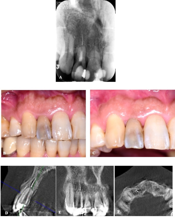
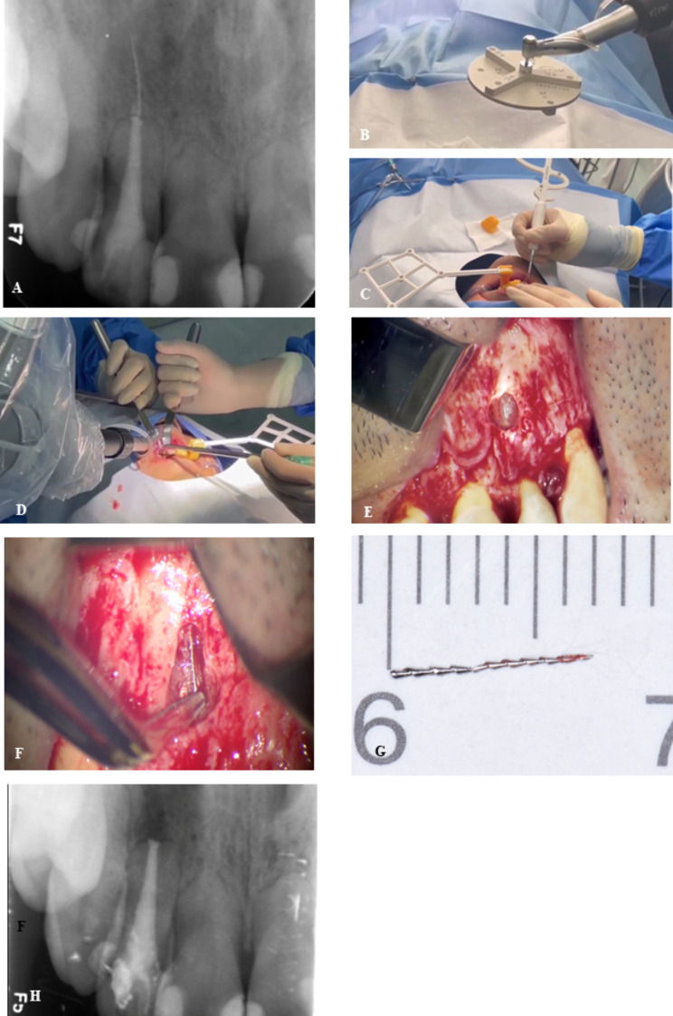
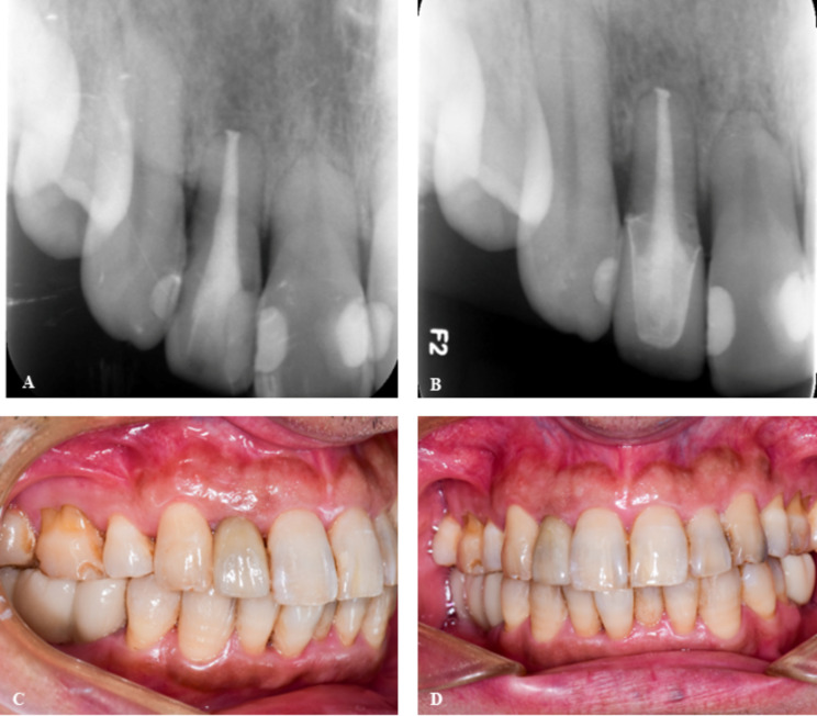
Case presentation: A 48-year-old male patient, with no evidence of bone defects, was diagnosed with a file fractured completely beyond the apical foramen during root canal treatment of the right maxillary lateral incisor. Patient information was used to incorporate a digital model into preoperative planning software to develop a surgical strategy. The ATR system employs spatial alignment methods for registration, directing the robotic arm to independently locate the fractured file in accordance with the surgical plan. To maximize minimally invasive surgery, the long fractured file was removed in two stages. After removing the bone and fracture file, the clinician performed suturing under a microscope. No complications were observed during the surgery, and the treatment appeared to be successful based on the 9-month follow-up evaluation.
Conclusions: The ATR system enables precise localization of the fractured file beyond the apical foramen with intact cortical plates. This technology has the potential to improve positioning accuracy, minimize the need for invasive bone removal, reduce intraoperative time, and facilitate successful endodontic microsurgical procedures.
Keywords: Apical microsurgery; Autonomous; Fractured file removal; Osteotomy; Robotic system.
© 2024. The Author(s).
Conflict of interest statement
Declarations. Ethics approval and consent to participate: This study did not involve human or animal subjects’ experiment, and thus no ethical approval was required. The case report adhered to the guidelines established by the journal. Consent for publication: Patient gave their written informed consent for their personal or clinical details along with any identifying images to be published in this case report. Competing interests: The authors declare no competing interests.
Figures

Similar articles
-
Utilizing spiral computerized tomography during the removal of a fractured endodontic instrument lying beyond the apical foramen.Int Endod J. 2010 Dec;43(12):1143-51. doi: 10.1111/j.1365-2591.2010.01780.x. Int Endod J. 2010. PMID: 21039622
-
Endodontic Microsurgery With an Autonomous Robotic System: A Clinical Report.J Endod. 2024 Jun;50(6):859-864. doi: 10.1016/j.joen.2024.02.005. Epub 2024 Feb 17. J Endod. 2024. PMID: 38369101
-
Minimally invasive guided endodontic microsurgery with Zekrya bur: A 48-month follow-up case report.Aust Endod J. 2024 Dec;50(3):687-692. doi: 10.1111/aej.12884. Epub 2024 Aug 20. Aust Endod J. 2024. PMID: 39162044
-
Clinical decision-making after endodontic instrument fracture.Br Dent J. 2013 Apr;214(8):395-400. doi: 10.1038/sj.bdj.2013.379. Br Dent J. 2013. PMID: 23619858 Review.
-
Establishing Apical Patency: To be or not to be?J Contemp Dent Pract. 2017 Apr 1;18(4):326-329. doi: 10.5005/jp-journals-10024-2040. J Contemp Dent Pract. 2017. PMID: 28349913 Review.
Cited by
-
Expert consensus on management of instrument separation in root canal therapy.Int J Oral Sci. 2025 Jun 9;17(1):46. doi: 10.1038/s41368-025-00372-w. Int J Oral Sci. 2025. PMID: 40484859 Free PMC article. Review.
References
-
- Madarati AA, Hunter MJ, Dummer PM. Management of intracanal separated instruments. J Endod. 2013;39(5):569–81. - PubMed
-
- Coaguila-Llerena H, Lazo-Quezada G, Teves A, Zevallos-Chávez M, Faria G. Removal of separated instruments from unfavourable locations: Case reports using the HBW ultrasonic ring or a surgical approach. Aust Endod J. 2023;49(2):358–64. - PubMed
-
- Terauchi Y, Ali WT, Abielhassan MM. Present status and future directions: Removal of fractured instruments. Int Endod J. 2022;55(Suppl 3):685–709. - PubMed
-
- Benjamin G, Ather A, Bueno MR, Estrela C, Diogenes A. Preserving the Neurovascular Bundle in Targeted Endodontic Microsurgery: A Case Series. J Endod. 2021;47(3):509–19. - PubMed
-
- Strbac GD, Schnappauf A, Giannis K, Moritz A, Ulm C. Guided Modern Endodontic Surgery: A Novel Approach for Guided Osteotomy and Root Resection. J Endod. 2017;43(3):496–501. - PubMed
Publication types
MeSH terms
Grants and funding
LinkOut - more resources
Full Text Sources
Miscellaneous
