. 2025 May;93(5):1054-1066.
doi: 10.1002/prot.26794. Epub 2025 Jan 2.

Characterization of the E26H Mutant Schistosoma japonicum Glutathione S-Transferase

Affiliations

Characterization of the E26H Mutant Schistosoma japonicum Glutathione S-Transferase

János András Mótyán et al. Proteins. 2025 May.

Abstract

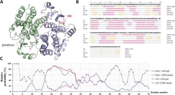
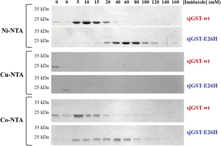
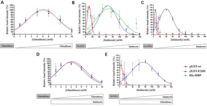
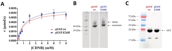
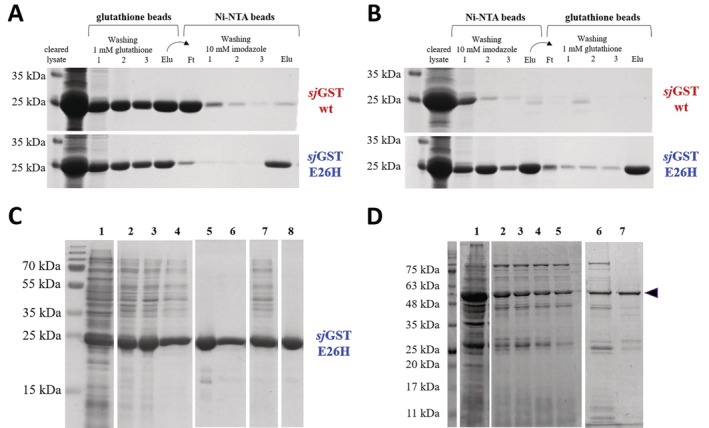
Glutathione-S-transferase, such as that of Schistosoma japonicum (sjGST) belongs to the most widely utilized fusion tags in the recombinant protein technology. The E26H mutation of sjGST has already been found to remarkably improve its ability for binding divalent ions, enabling its purification with immobilized metal affinity chromatography (IMAC). Nevertheless, most characteristics of this mutant remained unexplored to date. In this study, we performed a comparative analysis of the wild-type and the E26H mutant sjGST by using in vitro as well as in silico approaches. We confirmed that the sjGST(E26H) protein exhibits significantly increased affinity for binding nickel ions as compared to the wild-type. In addition, we proved that the sjGST(E26H) can be purified efficiently either with glutathione- or immobilized metal ion-affinity chromatography, even in consecutive purification steps. The human retroviral-like aspartic protease 1 (ASPRV1) conjugated with the sjGST(E26H) fusion tag was also successfully purified by using both of these affinity chromatographic approaches. Our studies revealed that the E26H mutant sjGST can be used as a versatile affinity tag because the modified protein retains the kinetic features of the wild-type and its affinity towards glutathione, while can be purified efficiently by IMAC, as well.

Keywords: Schistosoma japonicum; GST; affinity chromatography; fusion tag; glutathione S‐transferase; protein purification; recombinant protein.


Conflict of interest statement

The authors declare no conflicts of interest.

Figures

FIGURE 1
FIGURE 1
Quaternary and secondary structure of sjGST. (A) The overall structure of the sjGST homodimer is represented based on its crystal structure (5GZZ.pdb) [17]. The monomers are shown by light green and blue colors. The glutathione molecules binding to the active sites are shown by sticks, the E26 residue is red. (B) Arrangements of the secondary structural elements are shown based on the crystal structure of wild‐type (wt) sjGST (1M99.pdb) and based on in silico predictions (using GO4R4 and JPred4 online tools) for the wild‐type and E26H mutant proteins. “E”: strand, “H”: helix, “–”: coil. The sequence of the wild‐type sjGST was downloaded from the UniProt database (UniProt ID: P08515). (C) The graph represents the relative probabilities of the helices and coils (for the 1–70 region) predicted by GOR4.
FIGURE 2
FIGURE 2
Binding of wild‐type and E26H mutant sjGSTs to Ni‐, Cu‐, and Co‐NTA surfaces. The beads were washed in consecutive steps with buffers of increasing eluent (imidazole) concentration, the collected fractions were analyzed by SDS‐PAGE. Representative gel images are shown based on the results of three independent experiments.
FIGURE 3
FIGURE 3
Binding of wild‐type and E26H mutant sjGSTs to affinity surfaces. (A) The beads were washed in consecutive steps with buffers of increasing eluent (imidazole) concentration, the collected fractions were analyzed by SDS‐PAGE. Representative gel images are shown based on the results of three independent experiments. (B) The Ni‐NTA beads were washed in consecutive steps with buffers of increasing eluent (imidazole) concentration, the collected fractions were analyzed by SDS‐PAGE. Representative gel images are shown based on the results of three independent experiments. (C) The Co‐NTA beads were washed in consecutive steps with buffers of increasing eluent (imidazole) concentration, the collected fractions were analyzed by SDS‐PAGE. Error bars represent SD (n = 3). Representative gel images are shown based on the results of three independent experiments. (D) The effect of glutathione on the binding of sjGST to glutathione affinity surface. The binding affinities of wild‐type and E26H mutant sjGSTs to glutathione affinity surface were studied in the presence of soluble imidazole (200 mM final concentration). (E) The binding affinities of wild‐type and E26H mutant sjGSTs to Ni‐NTA affinity surface were studied in the presence of soluble glutathione (10 mM final concentration). The applied affinity beads are indicated in boxes of gray background, the increasing and constant eluent (glutathione and/or imidazole) concentrations are represented schematically below the graphs by white triangles and boxes, respectively. Error bars represent SD (n = 3). Representative gel images are shown in Figure S1.
FIGURE 4
FIGURE 4
Effect of E26H mutation of sjGST on the kinetic parameters, dimerization and detection by Western‐blot. (A) The activity measurements were performed by using CDNB colorimetric substrate. Kinetic parameters were determined by fitting the data to the Michaelis–Menten equation, the calculated kinetic values are shown in Table 3. Error bars represent SD (n = 6). (B) Separation of the monomeric and dimeric sjGSTs is shown in a representative Coomassie‐stained native polyacrylamide gel. The figure shows different parts of the same gel, separated by white spaces. (C) Detection of purified wild‐type and E26H mutant sjGSTs by Western‐blot using a polyclonal anti‐GST antibody, a representative blot image is shown.
FIGURE 5
FIGURE 5
Ni‐NTA and glutathione affinity purification of sjGST proteins. (A) Purification of wild‐type and E26H mutant sjGSTs from cleared cell lysates with glutathione affinity beads, followed by IMAC. (B) Purification of wild‐type and E26H mutant sjGSTs with IMAC, followed by glutathione affinity beads. The eluates from the first purification steps (containing the corresponding eluent) were directly transferred to the second affinity surface, independently from the order of the two applied methods (A, B). Representative gel images show SDS‐PAGE analysis of cleared lysates, samples from washing steps (1–3), the flowthrough (Ft) and eluate (Elu) fractions. The GST‐fused proteins were present in the fractions after washing steps, as they were present in the cleared lysates in excessive amount. (C) Purification of E26H sjGST using two affinity chromatography steps. A representative SDS‐PAGE gel image is shown. The proteins were purified with chromatography using affinity columns, the samples are numbered as it follows: Sample 1, total cell lysate; sample 2–4, purified sjGST(E26H), eluate fractions from Ni‐NTA affinity chromatography; sample 5–6, purified sjGST(E26H), eluate fractions from glutathione affinity chromatography; sample 7, pooled 2–4. eluate fractions; sample 8, purified sjGST(E26H), purified in two steps (the pooled eluate fractions from Ni‐NTA purification (sample 7) were further purified with glutathione affinity chromatography). The figure shows different parts of the same gel, separated by white spaces. (D) SDS‐PAGE analysis of the samples from the expression and purification of sjGST(E26H)‐ASPRV1‐28(D212A). Sample 1. Total cell lysate; Sample 2–5. Eluate fractions from the purification of sjGST(E26H)‐ASPRV1‐28(D212A) with Ni‐NTA affinity chromatography (in total 25 × 1 mL fractions were collected during the linear gradient elution (from 0 to 400 mM imidazole), eluate fraction 14–17 are represented); Sample 6. Pooled eluate fractions from Ni‐NTA purification (2–5) that were subjected for glutathione affinity chromatography; Sample 7. Eluate fraction of sjGST(E26H)‐ASPRV1‐28(D212A) from glutathione affinity chromatography. The figure shows different parts of the same gel, separated by white spaces. The arrowhead indicates the GST‐fused ASPRV1‐28 protein.

Similar articles

References

    1. Terpe K., “Overview of Tag Protein Fusions, From Molecular and Biochemical Fundamentals to Commercial Systems,” Applied Microbiology and Biotechnology 60, no. 5 (2003): 523–533. - PubMed
    1. Kimple M. E., Brill A. L., and Pasker R. L., “Overview of Affinity Tags for Protein Purification,” Current Protocols in Protein Science 73 (2013): 9.9.1–9.9.23. - PMC - PubMed
    1. Zhao X., Li G., and Liang S., “Several Affinity Tags Commonly Used in Chromatographic Purification,” Journal of Analytical Methods in Chemistry 2013 (2013): 581093. - PMC - PubMed
    1. Costa S., Almeida A., Castro A., and Domingues L., “Fusion Tags for Protein Solubility, Purification and Immunogenicity in Escherichia coli : The Novel Fh8 System,” Frontiers in Microbiology 5 (2014): 63. - PMC - PubMed
    1. Schäfer F., Seip N., Maertens B., Block H., and Kubicek J., “Purification of GST‐Tagged Proteins,” Methods in Enzymology 559 (2015): 127–139. - PubMed

MeSH terms

LinkOut - more resources