. 2025 Jan 1;18(1):DMM052012.
doi: 10.1242/dmm.052012. Epub 2025 Jan 24.

Prostatic Escherichia coli infection drives CCR2-dependent recruitment of fibrocytes and collagen production

Affiliations

Prostatic Escherichia coli infection drives CCR2-dependent recruitment of fibrocytes and collagen production

Brandon R Scharpf et al. Dis Model Mech. .

Abstract

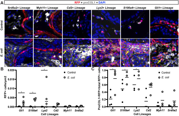
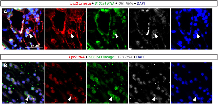
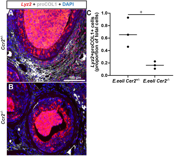
Prostate fibrosis contributes to lower urinary tract dysfunction (LUTD). To develop targeted treatments for prostate fibrosis, it is necessary to identify the cell types and molecular pathways required for collagen production. We used a genetic approach to label and track potential collagen-producing cell lineages in mouse prostate through a round of Escherichia coli UTI89-mediated prostate inflammation. E. coli increased collagen density and production in Gli1+, S100a4+, Lyz2+ and Cd2+ cell lineages, but not in Myh11+ or Srd5a2+ cell lineages, in the mouse prostate. Molecular phenotyping revealed GLI1+LYZ+S100A4+ cells (fibrocytes) in histologically inflamed human prostate. These fibrocytes colocalized with regions of increased collagen in men with LUTD. Fibrocyte recruitment and collagen synthesis was impaired in Ccr2 null mice but restored by allotransplantation of Rosa-GFP donor bone marrow-derived cells. These results suggest that bone marrow-derived fibrocytes are a mediator of prostatic collagen accumulation.

Keywords: CCR2; Fibrocyte; Fibrosis; LUTD; Myofibroblast; Prostate.


Conflict of interest statement

Competing interests The authors declare no competing or financial interests.

Figures

Fig. 1.
Fig. 1.
The periurethral collagen band is thicker in men who had simple prostatectomy to treat lower urinary tract dysfunction (LUTD) than in young male organ donors. Whole prostate sections were obtained from men aged 66-82 years who had their prostates removed by simple prostatectomy to treat LUTD (‘LUTD’) and from (young) male organ donors aged 18-47 years (‘Donor’). (A) Prostate sections were stained with Picrosirius Red (PSR) and imaged using fluorescent microscopy. A 5.0 mm×1.0 mm region of interest (ROI) was selected adjacent to the center of the urethra, as indicated by a rectangle labeled 'b1' in the micrograph for the representative donor prostate and 'b2' in the micrograph for the LUTD prostate. Scale bar: 5 mm. (B) Representative plots showing brightness of PSR stain versus distance from the urethra in an ROI from one donor and one LUTD sample. (C) Average PSR brightness across all prostate samples. Donor samples have abundant PSR stain immediately adjacent to the urethra that decreases with distance from the urethral edge. LUTD patient prostates maintain significantly greater PSR stain abundance with distance from urethra. Results are shown from ten donor and 17 LUTD prostates. Linear regression analysis was performed to determine the coefficient of determination (r2) and whether slopes of linear regression lines significantly differed (P<0.05).
Fig. 2.
Fig. 2.
Collagen-producing (proCOL1+) cells in the inflamed mouse and human prostate are ACTA2 (non-myofibroblasts). Human prostate sections were obtained as described in the Materials and Methods. Eight-week-old WT mice were transurethrally catheterized and administered two bolus doses, spaced 24 h apart, of E. coli UTI89 [optical density (OD) 0.80 in 100 µl PBS]. Mice were euthanized 7 days after the first bolus dose of E. coli. Lower urinary tracts were collected, fixed, embedded in paraffin and sectioned. Inflamed mouse and human prostate samples were immunostained with antibodies against alpha-smooth muscle actin (ACTA2) and procollagen 1 (proCOL1). Nuclei were stained with DAPI. Arrowheads indicate ACTA2, proCOL1+ cells. Images were taken using an Eclipse E600 compound microscope at 20× magnification. Results are representative of three individuals per group. Scale bars: 50 µm (bottom) and 100 µm (top).
Fig. 3.
Fig. 3.
Prostate inflammation expands Gli1+, Lyz2+, Cd2+ and S100a4+ cell lineages but not Srd5a2+ or Myh11+ cell lineages. The Ai14 reporter allele was bred to each of the following strains to create reporter mice: Cd2-icre, Gli1tm3(cre/ERT2), Lyz2tm1(cre), Myh11-cre, S100a4-cre and Srd5a2-G2aCE. Seven-week-old Gli1 and Srd5a2 reporter mice were administered 200 mg/kg/day of tamoxifen for 4 days to activate cre; the other cre alleles are constitutively active. At 8 weeks of age, reporter mice were transurethrally catheterized and administered two bolus doses, spaced 24 h apart, of E. coli UTI89 (OD 0.80 in 100 µl PBS) or sterile PBS. Mice were euthanized 7 days after the first bolus dose of E. coli. (A) Lower urinary tracts were collected, fixed, embedded in paraffin, sectioned and immunostained with antibodies against proCOL1 and RFP to visualize the Ai14 reporter, and with DAPI to visualize nuclei. Yellow arrows indicate the locations of RFP+ and proCOL1+ co-expressing cells. Images were taken using an Eclipse E600 compound microscope at 20× magnification. Scale bar: 50 µm. (B,C) Densities (B) and proportions (C) of RFP+ cells that produce collagen were determined. Results show three to five mice per group. Individual lineages were compared to their own respective control using a paired two-tailed Student's t-test. ‘+’ indicates a significant difference (P<0.05) from the control group.
Fig. 4.
Fig. 4.
Molecular phenotyping two distinct mouse strains demonstrates the presence of Gli1, Lyz2 and S100a4 triple-positive cells in E. coli-infected mouse dorsal prostate. Eight-week-old male mice were anesthetized and E. coli (100 µl; OD 0.8) was introduced via a transurethral catheter. Prostate tissue was collected 1 week later, fixed and paraffin embedded. (A) Lyz2tm1(cre); R26R-tdtomato mouse dorsal prostate sections were labeled with hybridization chain reaction (HCR) probes against Gli1 and S100a4, and immunolabeled with an antibody against RFP. (B) S100a4-cre; R26R-tdtomato mouse dorsal prostate sections were labeled with HCR probes against Lyz2 and Gli1 mRNA, and immunolabeled with an antibody against RFP. Arrowheads indicate triple-positive cells. Images were taken using an Eclipse E600 compound microscope at 20× magnification. Results are representative of three mice per group. Scale bar: 50 µm.
Fig. 5.
Fig. 5.
LYZ+/GLI1+/S100A4+ triple-positive cells are present in the transition zone of the inflamed human prostate and produce collagen. Human prostate samples, obtained by simple prostatectomy from men with LUTD (Table S1), were cut into sections and categorized as having high-grade inflammation based on the total number of CD45+ immune cells (Fig. S2). RNAscope™ assay was used to visualize LYZ and GLI1 mRNA, and immunofluorescence (IF) was used to visualize S100A4 and proCOL1 protein. Tissues were imaged using a Keyence BZ-X700 microscope, with images stitched and compressed into whole images [magnification at 4× (A), 10× (B) or 20× (C)], or an Eclipse E600 compound microscope at 20× magnification (D; Fig. S4). Scale bars: 50 mm (A,C,D) and 5 mm (B). Results are representative of five individuals per group.
Fig. 6.
Fig. 6.
Ccr2 is required for recruitment and infiltration of collagen-producing cells to E. coli-infected mouse prostate. Eight-week-old control (Ccr2+/−) and Ccr2 null (Ccr2−/−) male mice were transurethrally catheterized and administered two bolus doses, spaced 24 h apart, of E. coli UTI89 (OD 0.80 in 100 µl PBS) or sterile PBS. Mice were euthanized 7 days after the first bolus dose of E. coli. (A,B) Ccr2+/− (A) and Ccr2−/− (B) mouse dorsal prostate sections were stained with an antibody against the intracellular collagen precursor, proCOL1. Images were taken in the dorsal prostate lobe using an Eclipse E600 compound microscope at 20× magnification. Scale bar: 100 µm. Individual channels are provided in Fig. S5 for further emphasis of co-expression. (C) RNAscope™ assay was used to visualize Lyz2 mRNA. Lyz2+proCOL1+ double-positive cells were significantly less frequent in Ccr2−/− than in genetic control Ccr2+/−mouse prostates (three mice per experimental group). Groups were compared using an unpaired two-tailed Student's t-test. ‘+’ indicates a significant difference (P<0.05) between groups.
Fig. 7.
Fig. 7.
Ccr2+ circulating bone marrow-derived cells drive the fibrotic response to prostate inflammation. Eight-week-old control (Ccr2+/−) and Ccr2 null (Ccr2−/−) male mice were anesthetized with isoflurane and retro-orbitally injected with bone marrow cells from Rosa-GFP donor mice (27G needle, 5 million cells in 100 µl PBS) or PBS alone. Mice were then immediately transurethrally, catheterized and administered the first of two bolus doses, spaced 24 h apart, of E. coli UTI89 (OD 0.80 in 100 µl PBS) or sterile PBS. Eight-week-old wild-type mice were similarly infected with E. coli UTI89. Mice were euthanized 7 days after the first bolus dose of E. coli. (A) The expression of C-C motif chemokine ligand 2 (Ccl2) mRNA, which encodes the protein ligand of CCR2, was measured in dorsal prostate and normalized to that of Ppia. (C-E) Lower urinary tracts of Ccr2−/+ and Ccr2−/− male mice were collected, fixed, embedded in paraffin, sectioned, stained with PSR, illuminated, and imaged using a Texas Red filter to reveal collagen fibers on an Eclipse E600 compound microscope at 20× magnification. Scale bar: 50 µm. (B) Collagen fibers per image were quantified with CT-FIRE software. Results are from nine to ten (A) or three to five (B-E) mice per group. Groups were compared using an unpaired two-tailed Student's t-test (A) or a two-way ANOVA that passed (P>0.05) Spearman's test for heteroscedasticity and the Shapiro–Wilk test for normality of residuals followed by Sidak's multiple comparisons test (B). Treatment (*P=0.0002) and genotype (+P=0.004) had significant effects; interaction was not significant. P<0.05 was considered statistically significant.

Similar articles

References

    1. Ahn, S. and Joyner, A. L. (2004). Dynamic changes in the response of cells to positive hedgehog signaling during mouse limb patterning. Cell 118, 505-516. 10.1016/j.cell.2004.07.023 - DOI - PubMed
    1. Alluri, L. S. C., Paes Batista Da Silva, A., Verma, S., Fu, P., Shen, D. L., MacLennan, G., Gupta, S. and Bissada, N. F. (2021). Presence of specific periodontal pathogens in prostate gland diagnosed with chronic inflammation and adenocarcinoma. Cureus 13, e17742. 10.7759/cureus.17742 - DOI - PMC - PubMed
    1. Andersson-Sjöland, A., De Alba, C. G., Nihlberg, K., Becerril, C., Ramírez, R., Pardo, A., Westergren-Thorsson, G. and Selman, M. (2008). Fibrocytes are a potential source of lung fibroblasts in idiopathic pulmonary fibrosis. Int. J. Biochem. Cell Biol. 40, 2129-2140. 10.1016/j.biocel.2008.02.012 - DOI - PubMed
    1. Bauman, T. M., Nicholson, T. M., Abler, L. L., Eliceiri, K. W., Huang, W., Vezina, C. M. and Ricke, W. A. (2014). Characterization of fibrillar collagens and extracellular matrix of glandular benign prostatic hyperplasia nodules. PLoS ONE 9, e109102. 10.1371/journal.pone.0109102 - DOI - PMC - PubMed
    1. Bell-Cohn, A., Mazur, D. J., Hall, C. C., Schaeffer, A. J. and Thumbikat, P. (2019). Uropathogenic Escherichia coli-induced fibrosis, leading to lower urinary tract symptoms, is associated with type-2 cytokine signaling. Am. J. Physiol. Renal. Physiol. 316, F682-F692. 10.1152/ajprenal.00222.2018 - DOI - PMC - PubMed