

Low-density scalp electrical source imaging of the ictal onset zone network using source coherence maps
- PMID: 39748857
- PMCID: PMC11693594
- DOI: 10.3389/fneur.2024.1483977
Low-density scalp electrical source imaging of the ictal onset zone network using source coherence maps
Abstract
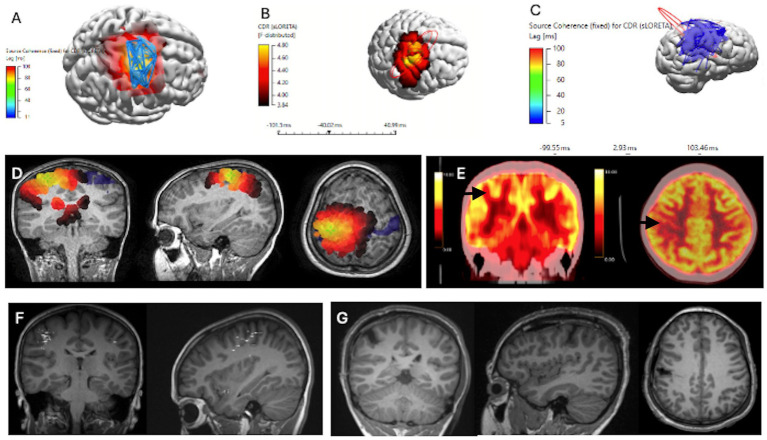
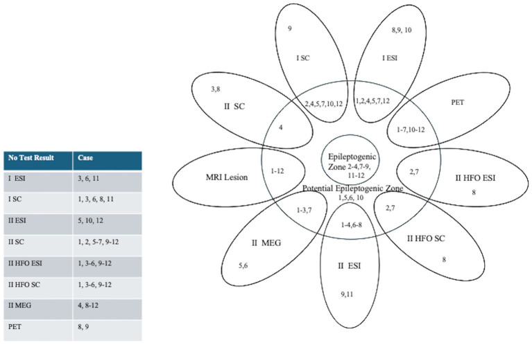
Introduction: This study investigated low-density scalp electrical source imaging of the ictal onset zone and interictal spike ripple high-frequency oscillation networks using source coherence maps in the pediatric epilepsy surgical workup. Intracranial monitoring, the gold standard for determining epileptogenic zones, has limited spatial sampling. Source coherence analysis presents a promising new non-invasive technique.
Methods: This was a retrospective review of 12 patients who underwent focal resections. Source coherence maps were generated using standardized low-resolution electromagnetic tomography and concordance to resection margins was assessed, noting outcomes at 3 years post-surgery.
Results: Ictal source coherence maps were performed in 7/12 patients. Six of seven included the surgical resection. Five of seven cases were seizure free post-resection. Interictal spike ripple electrical source imaging and interictal spike ripple high-frequency oscillation networks using source coherence maps were performed for three cases, with two of three included in the resection and all three were seizure free.
Discussion: These findings may provide proof of principle supporting low-density scalp electrical source imaging of the ictal onset zone and spike ripple network using source coherence maps. This promising method is complementary to ictal and interictal electrical source imaging in the pediatric epilepsy surgical workup, guiding electrode placement for intracranial monitoring to identify the epileptogenic zone.
Keywords: electrical source imaging; epilepsy surgery; ictal onset zone; low density; source coherence maps.
Copyright © 2024 Sadeghzadeh, Freibauer, RamachandranNair, Whitney, Al Nassar, Jain, Donner, Ochi and Jones.
Conflict of interest statement
The authors declare that the research was conducted in the absence of any commercial or financial relationships that could be construed as a potential conflict of interest.
Figures
References
-
- Jehi L, Jette N, Kwon CS, Josephson CB, Burneo JG, Cendes F, et al. Timing of referral to evaluate for epilepsy surgery: expert consensus recommendations from the surgical therapies Commission of the International League against epilepsy. Epilepsia. (2022) 63:2491–506. doi: 10.1111/epi.17350, PMID: - DOI - PMC - PubMed
LinkOut - more resources
Full Text Sources