. 2024 Dec 28:19:14059-14074.
doi: 10.2147/IJN.S497346. eCollection 2024.

Mitochondria-Targeting Virus-Like Gold Nanoparticles Enhance Chemophototherapeutic Efficacy Against Pancreatic Cancer in a Xenograft Mouse Model

Affiliations

Mitochondria-Targeting Virus-Like Gold Nanoparticles Enhance Chemophototherapeutic Efficacy Against Pancreatic Cancer in a Xenograft Mouse Model

Youshuai Meng et al. Int J Nanomedicine. .

Abstract

Background: The dense and fibrotic nature of the pancreatic tumor microenvironment significantly contributes to tumor invasion and metastasis. This challenging environment acts as a formidable barrier, hindering effective drug penetration and delivery, which ultimately limits the efficacy of conventional cancer treatments. Gold nanoparticles (AuNPs) have emerged as promising nanocarriers to overcome the extracellular matrix barrier; however, their limited targeting precision, poor delivery efficiency, and insufficient photothermal conversion present challenges.

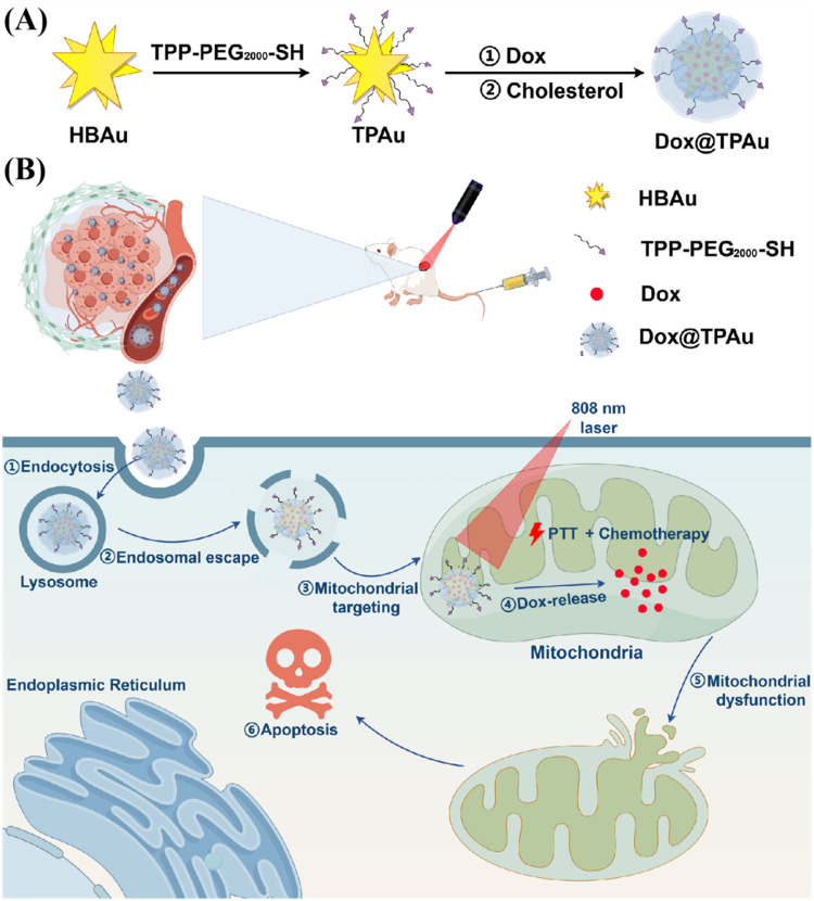
Methods: We developed triphenyl phosphonium-functionalized high-branch gold nanoparticles, denoted as Dox@TPAu, to enhance drug delivery and targeting capabilities. The targeted penetration, biopharmaceutical and pharmacokinetic properties of Dox@TPAu were characterized, and the synergistic therapeutic effect was evaluated by the BxPC-3 xenograft tumor mouse model.

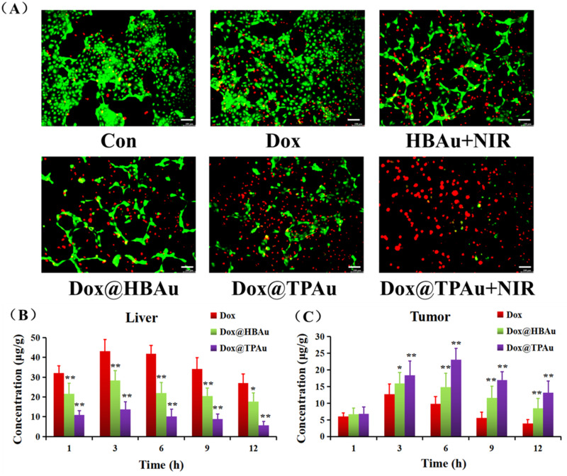
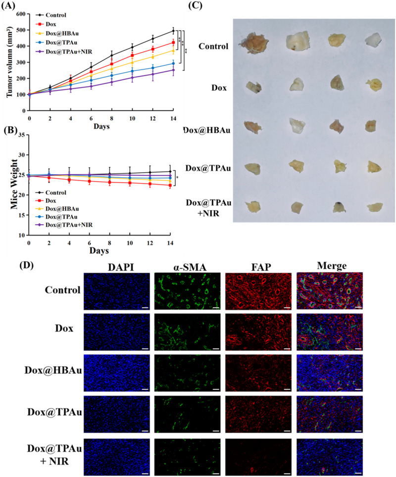
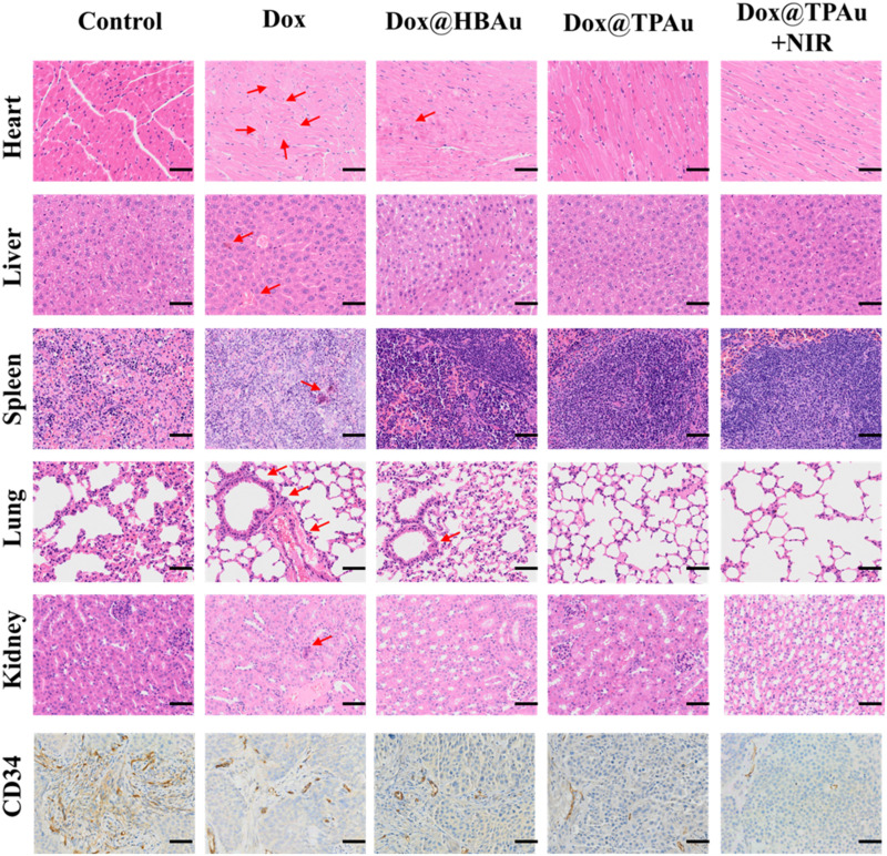
Results: Dox@TPAu exhibits superior photothermal conversion efficiency (91.0%) alongside a high drug loading efficiency (26%) and effective photo-triggered drug-release potential. This Dox@TPAu drug delivery system adeptly accumulates at tumor sites due to its unique properties, enabling targeted localization within cancer cells and the mitochondria of stromal fibroblasts. This localization disrupts mitochondrial function and transfer-processes crucial for energy production, metabolism, and cell signaling within the tumor microenvironment. Pharmacokinetic analyses revealed an optimal spatiotemporal distribution of Dox@TPAu at the tumor site. This strategic accumulation enables precise disruption of both the physical barrier and cancer cells, enhancing treatment efficacy through near-infrared light-triggered local chemo-photothermal synergistic therapy.

Conclusion: Our findings demonstrate that this innovative strategy effectively leverages the unique properties of mitochondria-targeting, virus-like AuNPs for precise and efficient stromal depletion, presenting a promising approach to enhance the efficacy of pancreatic cancer treatment.

Keywords: chemo-photothermal therapy; gold nanoparticle; mitochondria-targeting; pancreatic cancer; stromal depletion.


Conflict of interest statement

The authors report no conflicts of interest in this work.

Figures

Scheme 1
Scheme 1
Schematic diagram of photo-triggered high-branch Au nanoparticles (HBAu) via functional modification for mitochondria-targeted drug delivery. (A) The design and Preparation of Dox@TPAu. Dox@TPAu is composed of TPP-functionalized HBAu encapsulating Dox. (B) Anticancer mechanism of Dox@TPAu augmented chemophototherapeutic efficacy. Enhanced synergistic therapy relies on loosened extracellular matrix (ECM) and therapeutic sensitization due to in-situ hyperthermia therapy and drug release triggered by near-infrared (NIR) irradiation.
Figure 1
Figure 1
Preparation and characterization of Dox@TPAu. Transmission electron microscopy (TEM) images of (A) gold nanoseed (Scale bar, 50 nm) and (D) Dox@TPAu (Scale bar, 100 nm). UV-vis spectra of (B) gold nanoseed and (C) HBAu at concentrations of 0, 12.5, 25, 50, and 100 μg/mL (E) Size distribution and (F) zeta potential of Dox@TPAu.
Figure 2
Figure 2
Photothermal properties and mitochondrial targeting of Dox@TPAu (A) Evaluation of photothermal effect of Dox@TPAu. The Dox@TPAu suspension (containing HBAu 0, 12.5, 25, 50, and 100 μg/mL) was exposed to an 808-nm laser (1 W/cm2 for 5 min), and the temperature was monitored at an interval of 30s for 5 min. (B) Assessment of photothermal stability of Dox@TPAu through three cycles of laser on/off (C) Investigation of mitochondrial specificity of Dox@HBAu and Dox@TPAu. BxPC-3 cells were treated with Dox-loaded gold nanosystem (HBAu or TPAu) for 2 h or 6 h, followed by staining and imaging with MitoTracker@Red and Hoechst 33342 (Scale bar: 25 μm).
Figure 3
Figure 3
In vitro cytotoxicity assessment of Dox, Dox@HBAu, and Dox@TPAu on BxPC-3 cells. FACS analysis of mitochondrial dysfunction markers (JC-1, mitochondrial potential) in BxPC-3 cells following treatment with (A) PBS, (B) Dox, (C) HBAu plus 808-nm laser, (D) Dox@HBAu, (E) Dox@TPAu and (F) Dox@TPAu plus 808-nm laser (1 W/cm2 for 5 min) (G and H) Evaluation of cytotoxic effects of various concentrations of Dox, HBAu, Dox@HBAu, and Dox@TPAu with/without irradiation on BxpC-3 cells for 48 h, with cell viability determined using the MTT assay. Data are presented as mean ± SD (n=5).
Figure 4
Figure 4
(A) Live/dead staining of BxPC-3 cells post various treatments BxPC-3 cells underwent treatment with PBS, Dox, HBAu plus 808-nm laser, Dox@HBAu, Dox@TPAu, and Dox@TPAu plus 808-nm laser (1 W/cm2 for 5 min), followed by evaluating cell status (Scale bar: 100 μm). Biodistribution patterns of Dox in (B) liver and (C) tumor of BxPC-3 tumor-bearing mouse post intravenous administration of Dox, Dox@HBAu, Dox@TPAu at Dox dose of 3 mg/kg over different time points. Data are presented as mean ± SD (n=3), *P < 0.05 and **P < 0.01, (one-sample t-test) compared to the Dox group.
Figure 5
Figure 5
Investigating the synergistic therapeutic effect of Chemo-PTT with Dox@TPAu in vivo (A) tumor growth curves and (B) body weight changes of BxPC-3 tumor-bearing mice over a 2-week period post-treatment with PBS, Dox, Dox@HBAu, Dox@TPAu and Dox@TPAu plus 808-nm laser (1 W/cm2 for 5 min) (C) Representative photograph of excised BxPC-3 tumors from mice treated with PBS, Dox, Dox@HBAu, Dox@TPAu, and Dox@TPAu plus 808-nm laser (1 W/cm2 for 5 min), and tumors were collected for further analysis. (D) Representative immunofluorescence (IF) staining images of α-SMA and FAP positive regions in mouse tumor (Scale bar: 100 µm). Data are presented as mean ± SD (n=3), *P < 0.05, and **P < 0.01 (one-sample t-test) compared to the Dox group.
Figure 6
Figure 6
Representative H&E and immunohistochemical (IHC) staining images of CD34 positive areas in mouse tissues including heart, liver, spleen, lung, kidney, and tumor from BxPC-3 xenograft-bearing mouse after intravenous administration of PBS, Dox, Dox@HBAu, Dox@TPAu, and Dox@TPAu plus 808-nm laser (Scale bar: 100 µm).

Similar articles

Cited by

References

    1. Bray F, Laversanne M, Jemal A, et al. Global cancer statistics 2022: GLOBOCAN estimates of incidence and mortality worldwide for 36 cancers in 185 countries. CA Cancer J Clin. 2024;74:229–263. doi:10.3322/caac.21834 - DOI - PubMed
    1. Niu N, Shen X, Xue J, et al. Tumor cell-intrinsic epigenetic dysregulation shapes cancer-associated fibroblasts heterogeneity to metabolically support pancreatic cancer. Cancer Cell. 2024;42:869–884. doi:10.1016/j.ccell.2024.03.005 - DOI - PubMed
    1. Kolbeinsson HM, Chandana S, Chung M, et al. Pancreatic cancer: a review of current treatment and novel therapies. J Invest Surg. 2023;36:2129884. doi:10.1080/08941939.2022.2129884 - DOI - PubMed
    1. Grünwald BT, Devisme A, Khokha R, et al. Spatially confined sub-tumor microenvironments in pancreatic cancer. Cell. 2021;184:5577–5592. doi:10.1016/j.cell.2021.09.022 - DOI - PubMed
    1. Sherman MH, Beatty GL. Tumor microenvironment in pancreatic cancer pathogenesis and therapeutic resistance. Ann Rev Paleopathol Mech Dis. 2023;18:123–148. doi:10.1146/annurev-pathmechdis-031621-024600 - DOI - PMC - PubMed

MeSH terms