

Distinct brain systems are involved in subjective minute estimation with eyes open or closed: EEG source analysis study
- PMID: 39748918
- PMCID: PMC11693652
- DOI: 10.3389/fnins.2024.1506987
Distinct brain systems are involved in subjective minute estimation with eyes open or closed: EEG source analysis study
Abstract
Introduction: Time perception is a fundamental cognitive function, the brain mechanisms of which are not fully understood. Recent electroencephalography (EEG) studies have shown that neural oscillations in specific frequency bands may play a role in this process. In the current study, we sought to investigate how neurophysiological activity of cortical structures relates to subjective time estimations.
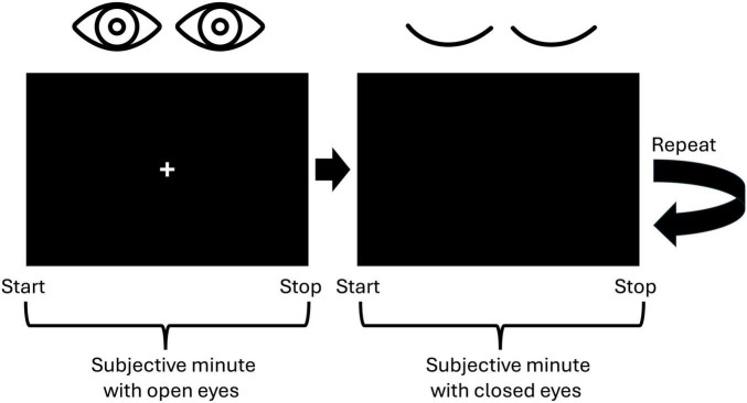
Methods: The study sample included 41 healthy volunteers, who were to produce subjective minutes with eyes closed and open by pressing the response button marking the beginning and end of this time interval. High-density EEG was recorded in parallel and the activity of cortical sources within the theta, alpha, and beta frequency bands was analyzed with standardized low-resolution brain electromagnetic tomography.
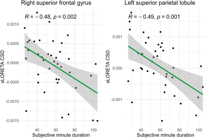
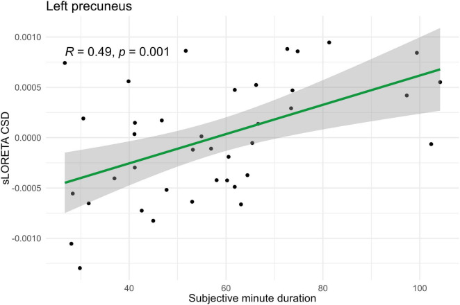
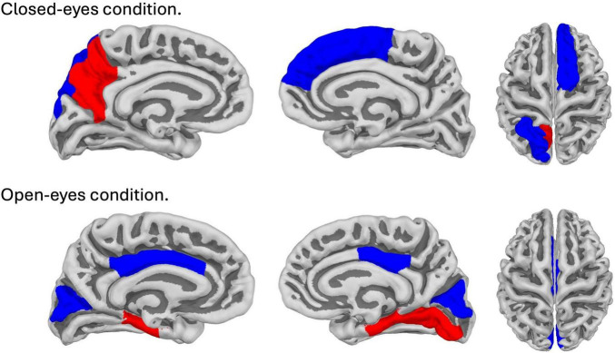
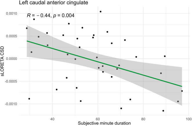
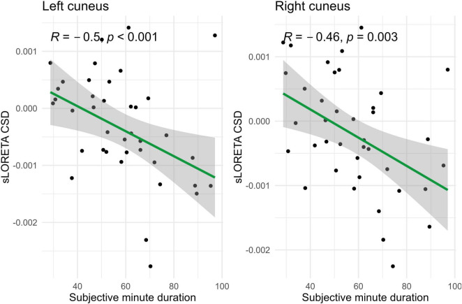
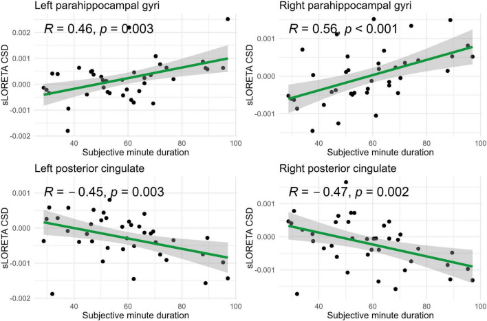
Results: The results revealed that activity of several cortical structures within the beta-band correlated with the duration of subjective minutes across participants, which highlights the role of the beta-rhythm in supra-second time perception. The sets of involved structures were different depending on eyes being open or closed, while the produced duration did not differ being around 58 s in both conditions. Individual minute correlated with beta power in the left precuneus, left superior parietal lobule, and right superior frontal gyrus (SFG) during eyes-closed sessions, and with that in the caudal anterior cingulate cortex, cuneus, posterior cingulate cortex, parahippocampal gyrus, and right lingual gyrus during the eyes-open condition. Noteworthy, some structures showed tendencies toward opposite correlations between conditions.
Discussion: Taken together, our findings bridge the gap between functional magnetic resonance imaging and EEG time perception studies and suggest reliance on different aspects of subjective experience when judging about time with eyes open or closed.
Keywords: EEG; current source density; duration production task; sLORETA; time perception.
Copyright © 2024 Proshina, Mitiureva and Sysoeva.
Conflict of interest statement
The authors declare that the research was conducted in the absence of any commercial or financial relationships that could be construed as a potential conflict of interest.
Figures
References
-
- Benjamini Y., Hochberg Y. (1995). Controlling the false discovery rate: a practical and powerful approach to multiple testing. J. R. Stat. Soc. Ser. B Stat. Methodol. 57 289–300. 10.1111/j.2517-6161.1995.tb02031.x - DOI
LinkOut - more resources
Full Text Sources
Miscellaneous