Review
. 2024 Dec 19:15:1453191.
doi: 10.3389/fgene.2024.1453191. eCollection 2024.

Dysregulation of ubiquitination modification in renal cell carcinoma

Affiliations
Review

Dysregulation of ubiquitination modification in renal cell carcinoma

Hongjie You et al. Front Genet. .

Abstract

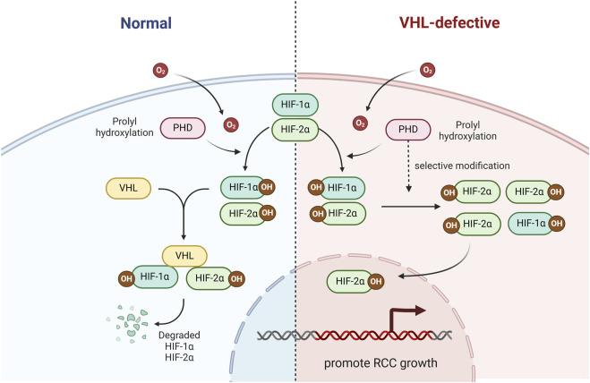
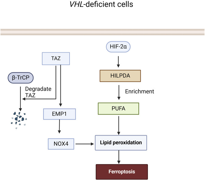
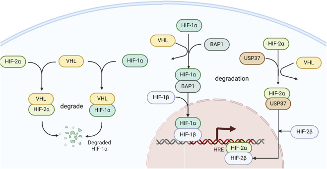
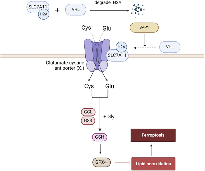
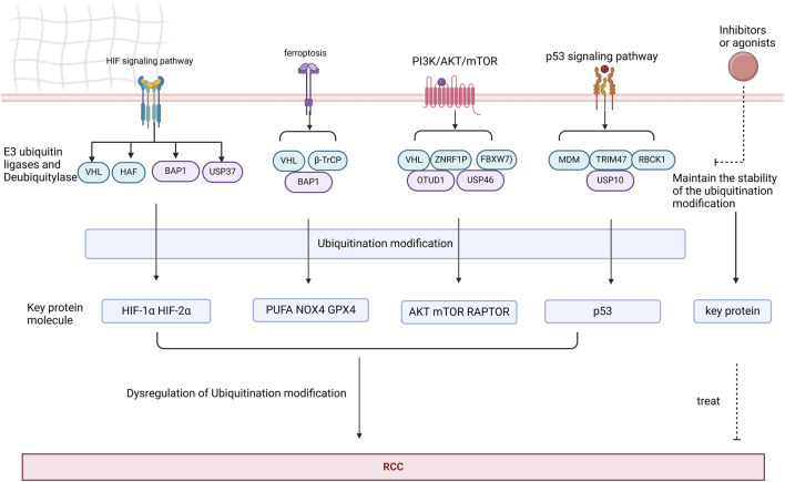
Renal cell carcinoma (RCC) is a malignant tumor of the renal tubular epithelial cells with a relatively high incidence rate worldwide. A large number of studies have indicated that dysregulation of the ubiquitination, including ubiquitination and dysregulation, is associated with the occurrence and development of RCC. This review focuses on several abnormal signaling pathways caused by E3 ligases and deubiquitinases. Additionally, we discuss research progress in RCC treatment by targeting key enzymes related to ubiquitination modifications.

Keywords: E3 ubiquitin ligases; PROTAC; deubiquitinase; immunotherapy; renal cell carcinoma (RCC); ubiquitination.


Conflict of interest statement

The authors declare that the research was conducted in the absence of any commercial or financial relationships that could be construed as a potential conflict of interest.

Figures

FIGURE 1
FIGURE 1
The role of VHL in the HIF signaling pathway. VHL ubiquitinate and degrade HIF-1α and HIF-2α.
FIGURE 2
FIGURE 2
The role of HAF in the HIF signaling pathway. HAF mediates HIF-1α polyubiquitination and degradation, binds and activates HIF-2α, and promotes the transcription of HIF-2α target genes including OCT-3/4, SOX2, and NANOG.
FIGURE 3
FIGURE 3
The role of BAP1 and USP37 in the HIF signaling pathway. BAP1 deubiquitinates and binds to stabilize HIF-1α, facilitating its association with HIF-1β to promote transcriptional activity in cells. USP37 deubiquitinates and binds to stabilize HIF-2α, thereby enhancing the transcription of its target genes.
FIGURE 4
FIGURE 4
The role of VHL and β-TrCP in the Ferroptosis signaling pathway. In VHL-deficient cells, HIF-2α is not ubiquitinated and degraded by VHL, leading to an increase in HILPDA expression and accumulation of PUFA, which in turn promotes the accumulation of lipid peroxides and triggers ferroptosis. TAZ can induce lipid peroxide accumulation and trigger ferroptosis through the EMP1-NOX4 pathway, while β-TrCP can ubiquitinate and degrade TAZ to terminate this process.
FIGURE 5
FIGURE 5
The role of BAP1 in the Ferroptosis signaling pathway. BAP1 promotes ferroptosis by ubiquitinating H2A on the SLC7A11 promoter, a component of the Xc-transporter, thereby reducing glutathione synthesis and GPX4 generation.
FIGURE 6
FIGURE 6
The role of Deubiquitylase in the PI3K/AKT/mTOR signaling pathway signaling pathway. VHL can ubiquitinate and degrade RAPTOR, an integral component of the mTORC1 complex, thereby inhibiting the AKT signaling pathway. VHL can also directly inhibit AKT activity to inhibit the AKT signaling pathway. In VHL-deficient cells, HIF-2α upregulates SLC7A5 amino acid carrier expression to enhance mTORC1 activity. ZNRF1 can polyubiquitinate and degrade AKT, and also interacts with LZTFL1 to inhibit AKT. Fbxw7 ubiquitinates and degrades mTOR. USP46 inhibits AKT levels. OTUD1 deubiquitinates and stabilizes PTEN, and regulates its inhibitory effect on AKT through deubiquitination.
FIGURE 7
FIGURE 7
The role of Deubiquitylase in the p53 signaling pathway signaling pathway. MDM2, TRIM47, RBCK1 can ubiquitinate and degrade p53, which affects the development of RCC cells. USP10 deubiquitinates p53 and stabilizes it, counteracting the degradation of p53 by MDM2.
FIGURE 8
FIGURE 8
Key proteins in the HIF, ferroptosis, PI3K/AKT/mTOR, and p53 signaling pathways are subjected to ubiquitination modifications by deubiquitinases and E3 ubiquitin ligases. Dysregulation of these ubiquitination modifications may contribute to the development of RCC. Targeting agonists or inhibitors of deubiquitinases and E3 ubiquitin ligases represents a potential therapeutic strategy for RCC.
FIGURE 9
FIGURE 9
Structural comparison of PROTACs and DUBTACs and mechanistic comparison of PROTAC-Mediated ubiquitination and DUBTAC-Mediated deubiquitination of POI.

Similar articles

References

    1. Adams C. M., Mitra R., Xiao Y., Michener P., Palazzo J., Chao A., et al. (2023). Targeted MDM2 degradation reveals a new vulnerability for p53-inactivated triple-negative breast cancer. Cancer Discov. 13 (5), 1210–1229. 10.1158/2159-8290.Cd-22-1131 - DOI - PMC - PubMed
    1. Adelaiye-Ogala R., Damayanti N. P., Orillion A. R., Arisa S., Chintala S., Titus M. A., et al. (2018). Therapeutic targeting of sunitinib-induced AR phosphorylation in renal cell carcinoma. Cancer Res. 78 (11), 2886–2896. 10.1158/0008-5472.Can-17-3386 - DOI - PMC - PubMed
    1. Akutsu M., Dikic I., Bremm A. (2016). Ubiquitin chain diversity at a glance. J. Cell Sci. 129 (5), 875–880. 10.1242/jcs.183954 - DOI - PubMed
    1. Alaseem A. M. (2023). Advancements in MDM2 inhibition: clinical and pre-clinical investigations of combination therapeutic regimens. Saudi Pharm. J. 31 (10), 101790. 10.1016/j.jsps.2023.101790 - DOI - PMC - PubMed
    1. Amendolare A., Marzano F., Petruzzella V., Vacca R. A., Guerrini L., Pesole G., et al. (2022). The underestimated role of the p53 pathway in renal cancer. Cancers (Basel) 14 (23), 5733. 10.3390/cancers14235733 - DOI - PMC - PubMed

LinkOut - more resources