

Research progress in Berger's space: An interesting retrolenticular space
- PMID: 39748980
- PMCID: PMC11693895
- DOI: 10.1016/j.heliyon.2024.e40432
Research progress in Berger's space: An interesting retrolenticular space
Abstract
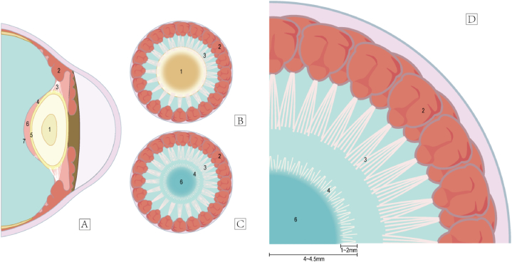
Purpose of the review: In recent years, Berger's space (BS), the potential space between the posterior lens capsule and the anterior hyaloid membrane, has received little attention from the ophthalmic clinical community. This is primarily due to the limited documentation, with only a few isolated case reports detailing foreign bodies in this area.
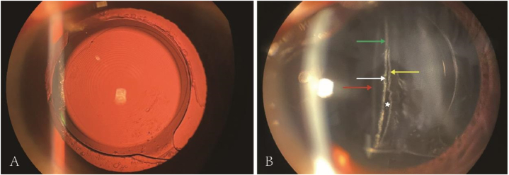
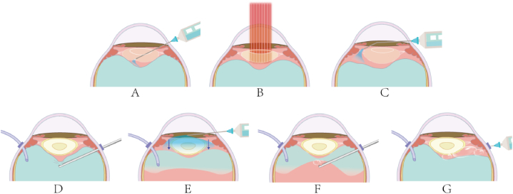
Recent findings: Recent advances in medical imaging technology have enabled the visualization of the BS under various circumstances. This progress has deepened clinicians' understanding of this elusive structure, allowing ophthalmic surgeons to utilize this space better and enhancing the safety and efficacy of relevant surgical procedures. This study explored the discovery, anatomical features, and clinical significance of the BS through anatomical and clinical studies.
Summary: This review emphasizes the observation of the BS via different imaging methods, the changes in the BS following the entry of abnormal substances, and its application in specific intraocular surgical procedures.
Keywords: Anterior segment; Berger's space; Ophthalmic surgical procedures; Ophthalmological imaging techniques.
© 2024 Published by Elsevier Ltd.
Conflict of interest statement
The authors declare the following financial interests/personal relationships which may be considered as potential competing interests: Xiaogang Wang reports a relationship with Elsevier Inc that includes: board membership. If there are other authors, they declare that they have no known competing financial interests or personal relationships that could have appeared to influence the work reported in this paper.
Figures


References
-
- Berger E. Beiträge zur Anatomie des Auges in normalem und pathologischem Zustande. 1887. Bergmann.
-
- Weidle E.G. Visualization of Berger's space in the living eye Ophthalmic Surgery. Lasers and Imaging Retina. 1985;16(11):733–734. - PubMed
-
- Bergua A., Kchle M. Visualization of the hyalo-capsular ligament in the living eye. J Graefe’s Arch Clin Exp Ophthalmol. 2002;240(6):503–505. - PubMed
Publication types
LinkOut - more resources
Full Text Sources
Research Materials
