

Lower Cranial Nerves in the Neck: An Anatomical Study
- PMID: 39749067
- PMCID: PMC11695066
- DOI: 10.7759/cureus.75049
Lower Cranial Nerves in the Neck: An Anatomical Study
Abstract
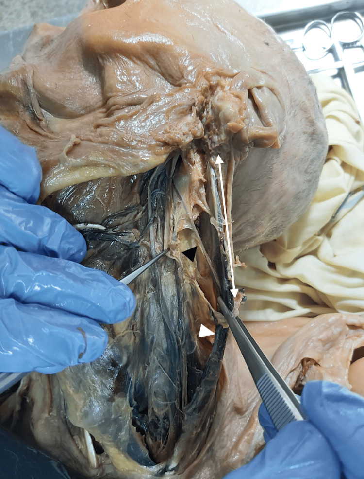
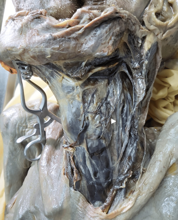
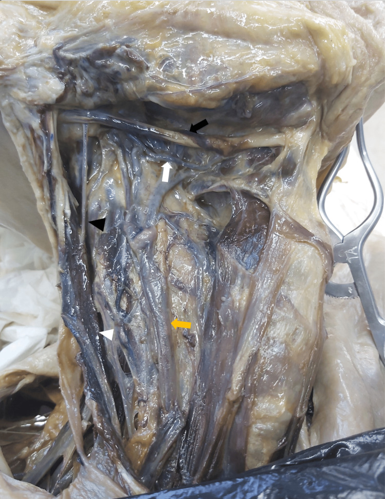
Objectives The aim of this anatomical study was to analyze distances and anatomical relations between the lower cranial nerves and important neck landmarks. Methods Anatomical study based on neck dissection in Thiel-embalmed cadavers. Anatomical relations and distances between the vagus (X), accessory (XI), and hypoglossal (XII) nerves and important neck landmarks were registered and compared. The relation between the emergence of the great auricular nerve and the posterior border of the sternocleidomastoid muscle and certain anatomical aspects of the ansa cervicalis were also studied. Results A total of 18 neck dissections (seven bilateral, four unilateral) were performed on 11 adult cadavers (mean (SD) age: 74.2 (12.9) years, four male, seven female). The X nerve was posterolateral to the common carotid artery and medial to the internal jugular vein (IJV) in 55.6% of the cases. In relation to the IJV, the XI nerve traveled mostly anteromedial (44.4%) at the level of the jugular foramen, posterior (50%) at the posterior belly of the digastric muscle, and posterolateral (77.8%) below the digastric muscle. The XII nerve was inferior (50%), medial (33.3%), and superior (16.7%) to the digastric tendon. The distance between the XII nerve and the carotid bifurcation was significantly superior in the male gender (mean (SD): 31.7 (6.8) mm vs. 18.5 (7.9) mm, p = .003). Also, the distance between the origin of the occipital artery and the point where it crosses the XII nerve was significantly higher in females (median (IQR): 7 (12.0) vs. 4 (4.0), p = .012). Conclusions There is a great variability in the anatomical position, course, and distances between the lower cranial nerves and traditional anatomical landmarks in the neck. The topography of the lower cranial nerves can vary even with the gender. Proper anatomical knowledge is crucial in neck surgery to prevent potential nerve injuries.
Keywords: accessory nerve; hypoglossal nerve; lower cranial nerves; neck surgery; vagus nerve.
Copyright © 2024, Pinto et al.
Conflict of interest statement
Human subjects: All authors have confirmed that this study did not involve human participants or tissue. Animal subjects: All authors have confirmed that this study did not involve animal subjects or tissue. Conflicts of interest: In compliance with the ICMJE uniform disclosure form, all authors declare the following: Payment/services info: All authors have declared that no financial support was received from any organization for the submitted work. Financial relationships: All authors have declared that they have no financial relationships at present or within the previous three years with any organizations that might have an interest in the submitted work. Other relationships: All authors have declared that there are no other relationships or activities that could appear to have influenced the submitted work.
Figures
References
-
- Complications after functional neck dissection in head and neck cancer patients: an observational, retrospective, single-centre study. Chiesa-Estomba CM, Soriano-Reixach M, Thomas-Arrizabalaga I, Sistiaga-Suarez JA, González-García JA, Larruscain E, Altuna X. ORL J Otorhinolaryngol Relat Spec. 2021;83:372–380. - PubMed
-
- A systematic review of 1143 parapharyngeal space tumors reported over 20 years. Riffat F, Dwivedi RC, Palme C, Fish B, Jani P. Oral Oncol. 2014;50:421–430. - PubMed
-
- Hypoglossal nerve palsy after anterior cervical spine surgery: a case report and literature review. Colón LF, Isaza J, Katsuura Y, Nuss D. JBJS Case Connect. 2020;10:0. - PubMed
-
- Cranial nerve injury after carotid endarterectomy: incidence, risk factors, and time trends. Kakisis JD, Antonopoulos CN, Mantas G, Moulakakis KG, Sfyroeras G, Geroulakos G. Eur J Vasc Endovasc Surg. 2017;53:320–335. - PubMed
LinkOut - more resources
Full Text Sources