. 2024 Dec 28:18:6389-6411.
doi: 10.2147/DDDT.S487476. eCollection 2024.

Qingre Huoxue Decoction Alleviates Atherosclerosis by Regulating Macrophage Polarization Through Exosomal miR-26a-5p

Affiliations

Qingre Huoxue Decoction Alleviates Atherosclerosis by Regulating Macrophage Polarization Through Exosomal miR-26a-5p

Weifeng He et al. Drug Des Devel Ther. .

Abstract

Background: Qingre Huoxue Decoction (QRHX) is a classical Chinese herbal prescription widely used in clinical practice for the treatment of atherosclerosis (AS). Our previous study demonstrated its efficacy in stabilizing plaque and improving prognosis, as well as its ability to regulate macrophage polarization. This study aimed to further investigate the effects of QRHX on AS and explore the underlying mechanisms.

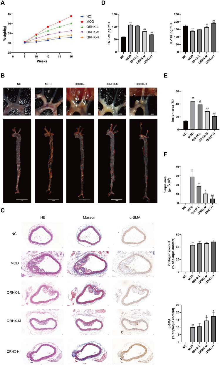
Methods: ApoE-/- mice were fed a high-fat diet (HFD) for 8 weeks in order to establish an AS model. Oil Red O, H&E, Masson, and IHC staining were employed to assess lipid accumulation, plaque development, collagen loss and target of the aortas tissue. ELISA was employed to measure the levels of TNF-α and IL-10 in serum. Dual luciferase reporter assay was conducted to ascertain the connection between miR-26a-5p and PTGS2 in vitro. Western blot and RT-qPCR assay were conducted to assess the NF-κB signaling pathway and macrophage polarization. The effects of miR-26a-5p were tested after transfecting miR-26a-5p over-expressive lentivirus.

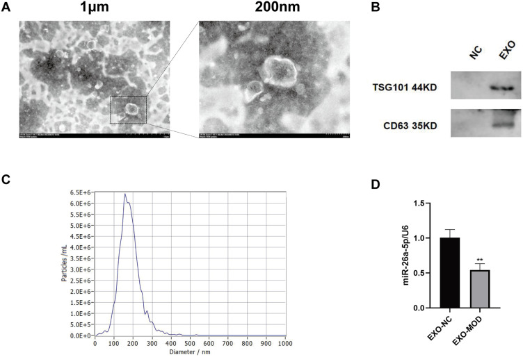
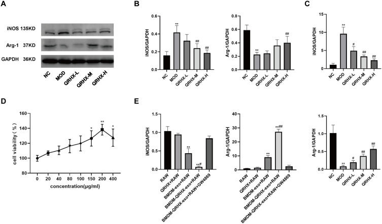
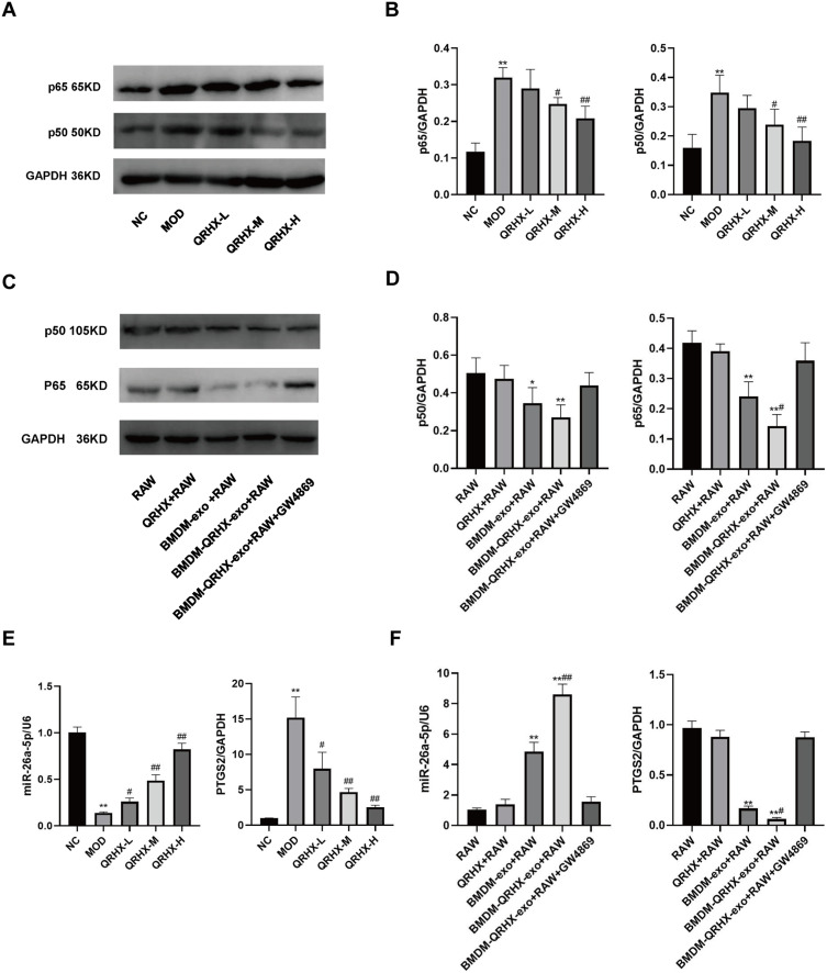
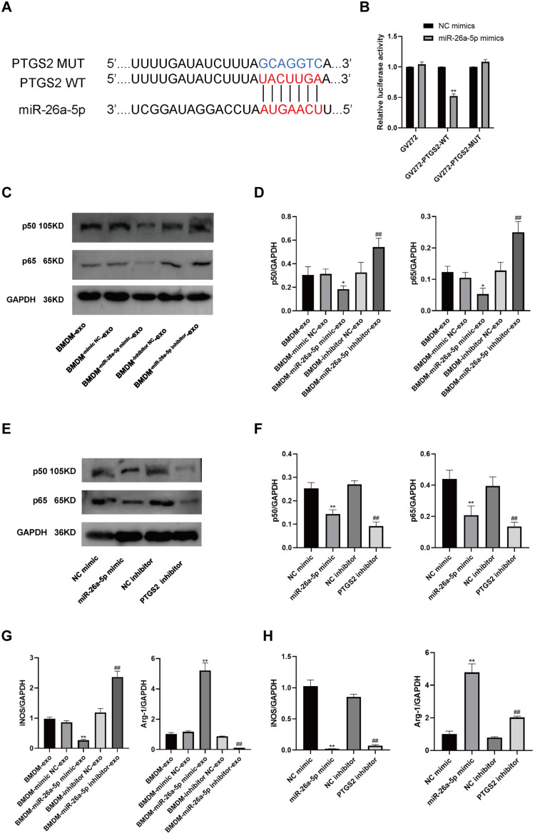
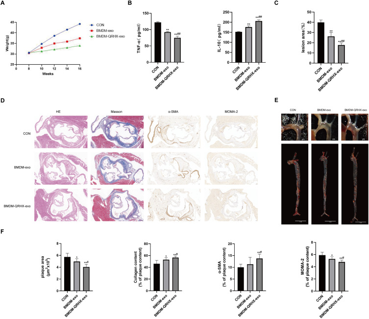
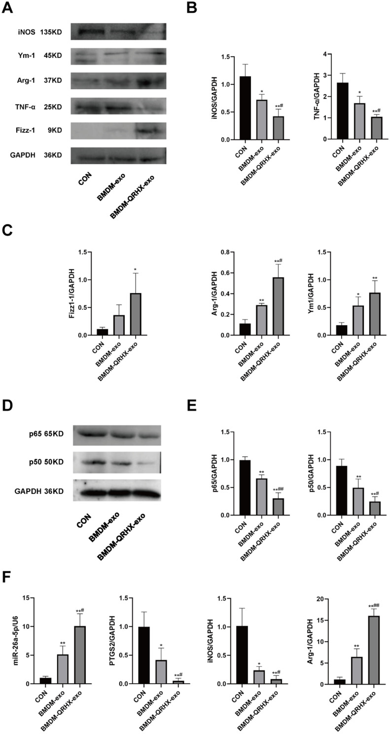
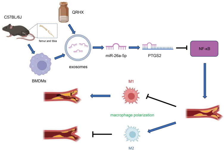
Results: QRHX attenuated HFD-induced plaque progression and inflammation of AS model mice. BMDM-derived exosomes (BMDM-exo) increased miR-26a-5p and decreased PTGS2 expressions, inhibited the NF-κB signaling pathway and regulated macrophage polarization in vivo. These effects of BMDM-exo were further enhanced after QRHX intervention. Dual luciferase reporter assay results showed that miR-26a-5p directly binds to the 3'-UTR of PTGS2 mRNA and regulates the expression of PTGS2. The miR-26a-5p of BMDM-exo played a key role in macrophage polarization. After overexpression of miR-26a-5p, the NF-κB signaling pathway was inhibited and macrophages were converted from M1 to M2 in vitro.

Conclusion: QRHX can exert anti-inflammatory and plaque-stabilizing effects through exosomal miR-26a-5p via inhibiting the PTGS2/NF-κB signaling pathway and regulating macrophage phenotype from M1 to M2 polarization in AS.

Keywords: Qingre Huoxue decoction; atherosclerosis; exosomes; macrophage polarization; miR-26a-5p.


Conflict of interest statement

The author reports no conflicts of interest in this work.

Figures

Figure 1
Figure 1
Characterization and miR-26a-5p expression of BMDM-derived exosomes. (A) Representative images of BMDM-derived exosomes. Scale bar: 1µm (left) and 200nm (right). (B) Exosomes were validated by assessing exosomal marker proteins CD63 and TSG101 (n=5). (C) Size distribution of BMDM-derived exosomes by Nanoparticle Tracking Analysis. (D) Expression of miR-26a-5p in exosomes isolated from different sources of BMDMs (n=5). NC: dying cell debris from BMDM cells supernatant; EXO: BMDM-derived exosomes; EXO-NC: BMDM-exo of C57BL/6J mice; EXO-MOD: BMDM-exo of AS model mice. Data were presented as mean ± standard deviation (x ± s) in an Independent Samples t-test. **p<0.01 vs EXO-NC group.
Figure 2
Figure 2
QRHX attenuated atherosclerosis in HFD-fed ApoE−/− mice. (A) Body weights of ApoE−/− mice (n=10). 0 to 8 weeks: HFD induction; 9 to 16 weeks: HFD induction, QRHX intervention. (B and E) The oil red O staining on entire aorta of ApoE−/− mice. The ratio is the relative area of lipid deposition staining area to the total aortic area (n=5). (C and F) H&E, Masson, and α-SMA staining of ApoE−/− mice. For Masson and α-SMA staining, the ratio is the relative area of the stained area within the plaque to the total area of the plaque (n=5). (D) ELISA for serum levels of TNF-α and IL-10 (n=10). NC: normal diet; Model: high-fat diet; QRHX-L: QRHX low-dose group (HFD + 7.5 g/kg/d QRHX); QRHX-M: QRHX medium-dose group (HFD + 15 g/kg/d QRHX); QRHX-H: QRHX high-dose group (HFD + 30 g/kg/d QRHX). Data were presented as mean ± standard deviation (x ± s) in one-way ANOVA. **p<0.01 vs NC group; #p<0.05, ##p<0.01 vs MOD group.
Figure 3
Figure 3
Effect of QRHX on macrophage polarization in ApoE−/− mice and miR-26a-5p of BMDM-derived exosomes in RAW264.7. (A and B) Western blot analysis was conducted to evaluate the expression of iNOS and Arg-1 in the aortas of ApoE−/− mice (n=5). (C and E) RT-PCR was performed to assess the gene expression levels of iNOS and Arg-1 in the aortas of ApoE−/− mice (n=5) and RAW264.7 cells (n=5). (D) The BMDMs were evaluated following treatment with varying concentrations of QRHX, utilizing the Cell Counting Kit-8 assay (n=6). NC: normal diet; Model: high-fat diet; QRHX-L: QRHX low-dose group (HFD + 7.5 g/kg/d QRHX); QRHX-M: QRHX medium-dose group (HFD + 15 g/kg/d QRHX); QRHX-H: QRHX high-dose group (HFD + 30 g/kg/d QRHX). RAW: RAW264.7 cells; QRHX+RAW: RAW264.7 cells, QRHX intervention; BMDM-exo: RAW264.7 cells, BMDM-exo intervention; BMDM-QRHX-exo+RAW: RAW264.7 cells, BMDM-exo and QRHX intervention; BMDM-QRHX-exo+RAW+GW4869: RAW264.7 cells, BMDM-exo, QRHX, and GW4869 intervention. Data were presented as mean ± standard deviation (x ± s) in one-way ANOVA. *p<0.05, **p<0.01 vs NC group; #p<0.05, #p<0.01 vs MOD group. *p<0.05, **p<0.01 vs RAW group; #p<0.05, ##p<0.01 vs BMDM-exo group.
Figure 4
Figure 4
Effect of QRHX on miR-26a-5p/PTGS2/NF-κB signaling pathway in ApoE−/− mice and miR-26a-5p of BMDM-derived exosomes in RAW264.7. (AD) Western blot analysis was conducted to assess the expression levels of p65 and p50 proteins in the aortas of ApoE−/− mice and RAW264.7 cells across each group (n=5). (E and F) RT-qPCR was employed to evaluate the expression levels of miR-26a-5p and PTGS2 genes in the aortas of ApoE−/− mice and RAW264.7 cells within each group. NC: normal diet, Model: high-fat diet, QRHX-L: QRHX low-dose group (HFD + 7.5 g/kg/d QRHX), QRHX-M: QRHX medium-dose group (HFD + 15 g/kg/d QRHX), QRHX-H: QRHX high-dose group (HFD + 30 g/kg/d QRHX). RAW: RAW264.7 cells; QRHX+RAW: RAW264.7 cells, QRHX intervention; BMDM-exo: RAW264.7 cells, BMDM-exo intervention; BMDM-QRHX-exo+RAW: RAW264.7 cells, BMDM-exo and QRHX intervention; BMDM-QRHX-exo+RAW+GW4869: RAW264.7 cells, BMDM-exo, QRHX, and GW4869 intervention. Data were presented as mean ± standard deviation (x ± s) in one-way ANOVA. *p<0.05, **p<0.01 vs NC group; #p<0.05, #p<0.01 vs MOD group. *p<0.05, **p<0.01 vs RAW group; #p<0.05, ##p<0.01 vs BMDM-exo group.
Figure 5
Figure 5
PTGS2 is a target gene of miR-26a-5p. miR-26a-5p regulation of PTGS2 affects the NF-κB signaling pathway and macrophage polarization. (A) The binding site prediction maps for miR-26a-5p and both wild-type (WT) and mutant (MUT) PTGS2 are presented. (B) Results from the dual luciferase reporter assay are shown (n=6). (CF) Western blot analyses were conducted to assess the protein expression levels of p65 and p50 in RAW264.7 cells (n=5). (G and H) RT-qPCR was performed to measure the expression levels of the iNOS and Arg-1 genes in RAW264.7 cells. BMDM-exo: RAW264.7 cells, BMDM-exo; BMDM-mimic NC-exo: RAW264.7 cells, BMDM-exo (mimic vector); BMDM-miR-26a-5p mimic-exo: RAW264.7 cells, BMDM-exo (miR-26a-5p mimic); BMDM-inhibitor NC-exo: RAW264.7 cells, BMDM-exo (inhibitor vector); BMDM-miR-26a-5p inhibitor-exo: RAW264.7 cells, BMDM-exo (miR-26a-5p inhibitor). NC mimic: RAW264.7 cells (mimic vector); miR-26a-5p mimic: RAW264.7 cells (miR-26a-5p mimic); NC inhibitor: RAW264.7 cells (inhibitor vector); PTGS2 inhibitor: RAW264.7 cells (PTGS2 inhibitor). Data were presented as mean ± standard deviation (x ± s) in one-way ANOVA. *p<0.05, **p<0.01 vs NC mimics group. *p<0.05, **p<0.01 vs BMDM-mimic NC-exo group; #p<0.05, ##p<0.01 vs BMDM-inhibitor NC-exo group. *p<0.05, **p<0.01 vs NC mimic group; #p<0.05, ##p<0.01 vs NC inhibitor group.
Figure 6
Figure 6
QRHX and miR-26a-5p of BMDM-derived exosomes attenuate atherosclerosis in HFD-fed ApoE−/− mice. (A) Body weight of ApoE−/− mice (n=10). 0~8 weeks: HFD induction; 9~16 weeks: HFD induction, QRHX intervention. (B and D) H&E, Masson, α-SMA, MOMA-2 staining. For Masson, α-SMA, and MOMA-2 staining, the ratio is the relative area of the stained area within the plaque to the total area of the plaque (n=5). Scale bar: 100 µm. (C and F) The oil red O staining on the entire aorta of ApoE−/− mice. The ratio is the relative area of stained area for lipid deposits to the relative area of total aortic area (n=5). (E) ELISA for serum levels of TNF-α and IL-10 (n=10). CON: high-fat diet, 200 μL PBS injected in the tail vein; BMDM-exo: high-fat diet, BMDM-derived exosomes (1010/each, dissolution in 200 μL PBS injected in the tail vein); BMDM-QRHX-exo: high-fat diet, BMDM-derived exosomes of QRHX intervention (1010/each, dissolution in 200 μL PBS injected in the tail vein). Data were presented as mean ± standard deviation (x ± s) in one-way ANOVA. *p<0.05, **p<0.01 vs CON group; #p<0.05, ##p<0.01 vs BMDM-exo group.
Figure 7
Figure 7
Effects of QRHX and miR-26a-5p of BMDM-derived exosomes on miR-26a-5p/PTGS2/NF-κB signaling pathway and macrophage polarization in ApoE−/− mice. (AE) Western blot to detect the protein expression of iNOS, TNF-α, Arg-1, Ym-1, Fizz-1, p65, and p50 in aortas of ApoE−/− mice in each group (n=5). (F) RT-qPCR was performed to detect miR-26a-5p, PTGS2, iNOS, and Arg-1 gene expression levels in the aorta of ApoE−/− mice in each group. CON: high-fat diet, 200 μL PBS injected in the tail vein; BMDM-exo: high-fat diet, BMDM-derived exosomes (1010/each, dissolution in 200 μL PBS injected in the tail vein); BMDM-QRHX-exo: high-fat diet, BMDM-derived exosomes of QRHX intervention (1010/each, dissolution in 200 μL PBS injected in the tail vein). Data were presented as mean ± standard deviation (x ± s) in one-way ANOVA. *p<0.05, **p<0.01 vs CON group; #p<0.05, ##p<0.01 vs BMDM-exo group.
Figure 8
Figure 8
Graphical mechanism by which QRHX and miR-26a-5p of BMDM-derived exosomes attenuate HFD-induce atherosclerosis. QRHX can play an anti-inflammatory and plaque-stabilizing role by mediating the exosomal miR-26a-5p via inhibiting PTGS2/NF-κB signaling pathway and regulating the macrophage phenotype in AS plaques from M1 to M2 polarization.

Similar articles

Cited by

References

    1. Hiwasa T, Zhang XM, Kimura R, et al. Elevated adiponectin antibody levels in sera of patients with atherosclerosis-related coronary artery disease, cerebral infarction and diabetes mellitus. J Circ Biomark. 2016;5:8. doi:10.5772/63218 - DOI - PMC - PubMed
    1. Hou P, Fang J, Liu Z, et al. Macrophage polarization and metabolism in atherosclerosis. Cell Death Dis. 2023;14(10):691. doi:10.1038/s41419-023-06206-z - DOI - PMC - PubMed
    1. Stoger JL, Gijbels MJ, van der Velden S, et al. Distribution of macrophage polarization markers in human atherosclerosis. Atherosclerosis. 2012;225(2):461–468. doi:10.1016/j.atherosclerosis.2012.09.013 - DOI - PubMed
    1. Otsuka F, Sakakura K, Yahagi K, Joner M, Virmani R. Has our understanding of calcification in human coronary atherosclerosis progressed? Arterioscler Thromb Vasc Biol. 2014;34(4):724–736. doi:10.1161/ATVBAHA.113.302642 - DOI - PMC - PubMed
    1. Davis FM, Gallagher KA. Epigenetic mechanisms in monocytes/macrophages regulate inflammation in cardiometabolic and vascular disease. Arterioscler Thromb Vasc Biol. 2019;39(4):623–634. doi:10.1161/ATVBAHA.118.312135 - DOI - PMC - PubMed