

Engineering extracellular vesicles for targeted therapeutics in cardiovascular disease
- PMID: 39749310
- PMCID: PMC11693616
- DOI: 10.3389/fcvm.2024.1503830
Engineering extracellular vesicles for targeted therapeutics in cardiovascular disease
Abstract
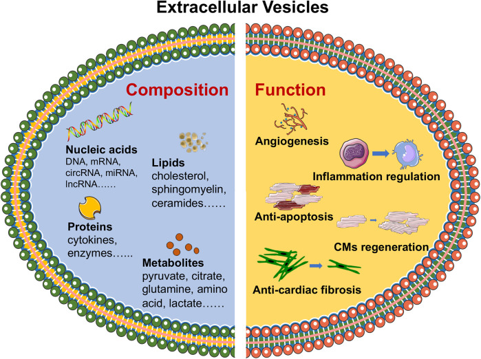
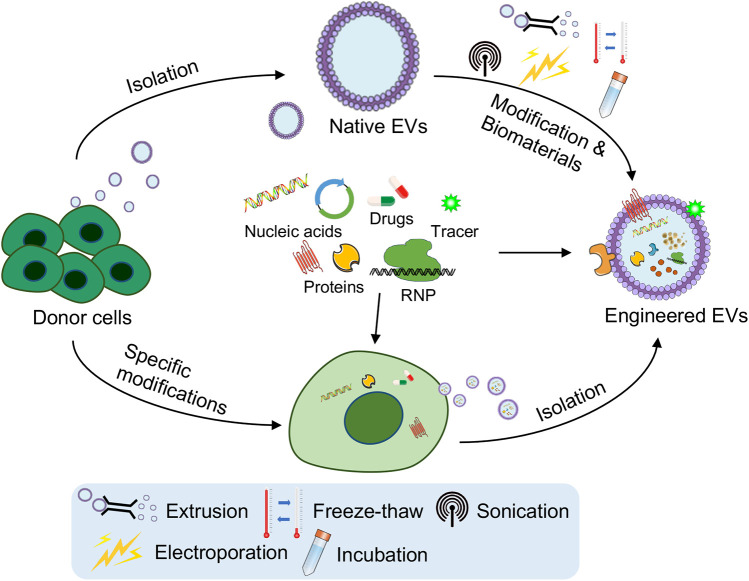
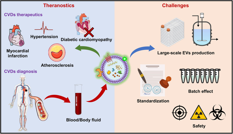
Extracellular vesicles (EVs) are nanosized particles secreted by cells that play crucial roles in intercellular communication, especially in the context of cardiovascular diseases (CVDs). These vesicles carry complex cargo, including proteins, lipids, and nucleic acids, that reflects the physiological or pathological state of their cells of origin. Multiomics analysis of cell-derived EVs has provided valuable insights into the molecular mechanisms underlying CVDs by identifying specific proteins and EV-bound targets involved in disease progression. Recent studies have demonstrated that engineered EVs, which are designed to carry specific therapeutic molecules or modified to enhance their targeting capabilities, hold promise for treating CVDs. Analysis of the EV proteome has been instrumental in identifying key proteins that can be targeted or modulated within these engineered vesicles. For example, proteins involved in inflammation, thrombosis, and cardiac remodeling have been identified as potential therapeutic targets. Furthermore, the engineering of EVs to increase their delivery to specific tissues, such as the myocardium, or to modulate their immunogenicity and therapeutic efficacy is an emerging area of research. By leveraging the insights gained from multiomics analyses, researchers are developing EV-based therapies that can selectively target pathological processes in CVDs, offering a novel and potentially more effective treatment strategy. This review integrates the core findings from EV multiomics analysis in the context of CVDs and highlights the potential of engineered EVs in therapeutic applications.
Keywords: cardiovascular diseases (CVDs); engineering; extracellular vesicles (EVs); multiomics analysis; targeted therapeutics.
© 2024 Fu, Pan and Li.
Conflict of interest statement
The authors declare that the research was conducted in the absence of any commercial or financial relationships that could be construed as a potential conflict of interest.
Figures
Similar articles
-
Engineered extracellular vesicles and their mimics in cardiovascular diseases.J Control Release. 2022 Jul;347:27-43. doi: 10.1016/j.jconrel.2022.04.046. Epub 2022 May 4. J Control Release. 2022. PMID: 35508222 Review.
-
Targeted therapy using engineered extracellular vesicles: principles and strategies for membrane modification.J Nanobiotechnology. 2023 Sep 16;21(1):334. doi: 10.1186/s12951-023-02081-0. J Nanobiotechnology. 2023. PMID: 37717008 Free PMC article. Review.
-
Harnessing engineered extracellular vesicles for enhanced therapeutic efficacy: advancements in cancer immunotherapy.J Exp Clin Cancer Res. 2025 May 2;44(1):138. doi: 10.1186/s13046-025-03403-w. J Exp Clin Cancer Res. 2025. PMID: 40317075 Free PMC article. Review.
-
Multi-omics analysis identified extracellular vesicles as biomarkers for cardiovascular diseases.Talanta. 2024 Dec 1;280:126710. doi: 10.1016/j.talanta.2024.126710. Epub 2024 Aug 16. Talanta. 2024. PMID: 39213888 Review.
-
Recent advances in the roles of extracellular vesicles in cardiovascular diseases: pathophysiological mechanisms, biomarkers, and cell-free therapeutic strategy.Mol Med. 2025 May 5;31(1):169. doi: 10.1186/s10020-025-01200-x. Mol Med. 2025. PMID: 40325357 Free PMC article. Review.
Cited by
-
Multi-omics analysis of two rat models reveals potential role of vesicle transport and autophagy in right ventricular remodeling.Sci Rep. 2025 Apr 18;15(1):13401. doi: 10.1038/s41598-025-98347-8. Sci Rep. 2025. PMID: 40251385 Free PMC article.
-
Beyond boundaries: exploring the role of extracellular vesicles in organ-specific metastasis in solid tumors.Front Immunol. 2025 Jun 12;16:1593834. doi: 10.3389/fimmu.2025.1593834. eCollection 2025. Front Immunol. 2025. PMID: 40574836 Free PMC article. Review.
-
Pharmacological Agent GW4869 Inhibits Tick-Borne Langat Virus Replication to Affect Extracellular Vesicles Secretion.Viruses. 2025 Jul 10;17(7):969. doi: 10.3390/v17070969. Viruses. 2025. PMID: 40733586 Free PMC article.
References
Publication types
LinkOut - more resources
Full Text Sources