Review
. 2025 Feb;12(6):e2408653.
doi: 10.1002/advs.202408653. Epub 2025 Jan 3.

Emerging Wearable Acoustic Sensing Technologies

Affiliations
Review

Emerging Wearable Acoustic Sensing Technologies

Tao Liu et al. Adv Sci (Weinh). 2025 Feb.

Abstract

Sound signals not only serve as the primary communication medium but also find application in fields such as medical diagnosis and fault detection. With public healthcare resources increasingly under pressure, and challenges faced by disabled individuals on a daily basis, solutions that facilitate low-cost private healthcare hold considerable promise. Acoustic methods have been widely studied because of their lower technical complexity compared to other medical solutions, as well as the high safety threshold of the human body to acoustic energy. Furthermore, with the recent development of artificial intelligence technology applied to speech recognition, speech recognition devices, and systems capable of assisting disabled individuals in interacting with scenes are constantly being updated. This review meticulously summarizes the sensing mechanisms, materials, structural design, and multidisciplinary applications of wearable acoustic devices applied to human health and human-computer interaction. Further, the advantages and disadvantages of the different approaches used in flexible acoustic devices in various fields are examined. Finally, the current challenges and a roadmap for future research are analyzed based on existing research progress to achieve more comprehensive and personalized healthcare.

Keywords: acoustic sensor; human‐machine interface; ultrasonic healthcare; wearable and implantable.


Conflict of interest statement

The authors declare no conflict of interest.

Figures

Figure 1
Figure 1
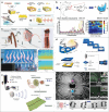
Application of flexible wearable acoustic sensors targeting different parts of the human body. (Reproduced with permission.[ 22 ] Copyright 2023, Springer Nature. Reproduced with permission.[ 23 ] Copyright 2016, American Association for the Advancement of Science. Reproduced with permission.[ 24 ] Copyright 2015, Wiley. Reproduced with permission.[ 25 ] Copyright 2020, Springer Nature. Reproduced with permission.[ 26 ] Copyright 2019, American Chemical Society. Reproduced with permission.[ 27 ] Copyright 2024, Springer Nature. Reproduced with permission.[ 28 ] Copyright 2022, American Association for the Advancement of Science. Reproduced with permission.[ 29 ] Copyright 2021, American Association for the Advancement of Science. Reproduced with permission.[ 30 ] Copyright 2023, Springer Nature. Reproduced with permission.[ 31 ] Copyright 2022, Elsevier. Reproduced with permission.[ 32 ] Copyright 2024, Springer Nature. Reproduced with permission.[ 33 ] Copyright 2023, Wiley. Reproduced with permission.[ 34 ] Copyright 2021, Springer Nature. Reproduced with permission.[ 35 ] Copyright 2022, American Association for the Advancement of Science. Reproduced with permission.[ 36 ] Copyright 2021, Wiley. Reproduced with permission.[ 37 ] Copyright 2022, American Chemical Society. Reproduced with permission.[ 38 ] Copyright 2022, Elsevier. Reproduced with permission.[ 39 ] Copyright 2023, Wiley. Reproduced with permission.[ 40 ] Copyright 2024, Wiley. Reproduced with permission.[ 41 ] Copyright 2024, Springer Nature. Reproduced with permission.[ 42 ] Copyright 2019, American Chemical Society. Reproduced with permission.[ 43 ] Copyright 2023, Springer Nature. Reproduced with permission.[ 44 ] Copyright 2023, Springer Nature. Reproduced with permission.[ 45 ] Copyright 2022, Springer Nature. Reproduced with permission.[ 46 ] Copyright 2024, American Association for the Advancement of Science.
Figure 2
Figure 2
Different kinds of acoustic sensing mechanisms: a) Piezoelectric sensing mechanism. b) Capacitive sensing mechanism. c) Piezoresistive sensing mechanism. d) Triboelectric sensing mechanism. e) Electromagnetic sensing mechanism. f) Photoacoustic sensing mechanism.
Figure 3
Figure 3
Wearable sensor for mechano‐acoustic signal monitoring. a) Fully portable MEMS stethoscope can be worn on the chest‐back. Reproduced with permission.[ 28 ] Copyright 2022, American Association for the Advancement of Science. b) Wireless human mechano‐acoustic sensing network for monitoring heart, lung, and bowel sounds. Reproduced with permission.[ 152 ] Copyright 2023, Springer Nature. c) Flexible sensing system based on folded double‐layer piezoelectric sensor for integrated detection of human acoustic signals. Reproduced under the terms of the CC‐BY license.[ 156 ] Copyright 2023, Liuyang Han et al., Published by Wiley. d) Thin elastic conductive nanocomposites based on LIG micropatterning. Reproduced with permission.[ 66 ] Copyright 2024, Springer Nature.
Figure 4
Figure 4
Gesture recognition through ultrasound method. a) Stretchable ultrasound array for contactless gesture recognition with small distance between each PZT composite. Reproduced under the terms of the CC‐BY license.[ 40 ] Copyright 2024, Ankan Dutta et al., Published by Wiley. b) A fully integrated wearable ultrasound echomyography system that can be attached to the skin for long‐term accurate wireless monitoring of muscles. Reproduced with permission.[ 186 ] Copyright 2024, Springer Nature. c) Monitoring change of muscle thickness during different movements through A‐mode ultrasound to estimate the torque of joint. Reproduced under terms of the CC‐BY license.[ 41 ] Copyright 2024, Yichu Jin et al., Springer Nature.
Figure 5
Figure 5
Wearable auditory sensors for speech recognition. a) Self‐powered eardrum‐inspired membrane sensor for arterial pulse wave monitoring and laryngeal microphones. Reproduced with permission.[ 24 ] Copyright 2015, Wiley. b) New speech recognition platform developed with highly sensitive MXene/MoS2 flexible vibration sensor. Reproduced with permission.[ 199 ] Copyright 2022, Springer. c) Highly sensitive self‐powered triboelectric acoustic sensors for robotic electronic hearing systems. Reproduced with permission.[ 125 ] Copyright 2018, American Association for the Advancement of Science. d) A paper‐based pressure sensor prepared from MXene/bacterial cellulose film with a three‐dimensional barrier layer structure. Reproduced with permission.[ 197 ] Copyright 2022, American Chemical Society.
Figure 6
Figure 6
Artificial throat acoustic sensor. a) Standalone stretchable wearable platform that measures laryngeal vibration and muscle activity. Reproduced under the terms of CC‐BY license.[ 208 ] Copyright 2023, Hongcheng Xu et al., published by Springer Nature. b) Flexible wireless device for monitoring physiological activity in the larynx such as breathing and swallowing. Reproduced under the terms of CC‐BY license.[ 203 ] Copyright 2022, Youn J. Kang et al., published by Springer Nature. c) Attachable speech recognition system based on reduced graphene oxide/PDMS composite film with biologically inspired microcracks and layered surface textures. Reproduced with permission.[ 42 ] Copyright 2019, American Chemical Society. d) Self‐powered artificial throat for the deaf population in a smart home scenario and recognizing process of common words. Reproduced with permission.[ 204 ] Copyright 2019, Elsevier.
Figure 7
Figure 7
Silent speech recognition system. a) Self‐powered flexible triboelectric lip recognition system based on trained dilated recurrent neural network model for prototype learning. Reproduced under the terms of CC‐BY license.[ 219 ] Copyright 2022, Yijia Lu et al., published by Springer Nature. b) Serpentine mesh tattooed strain transducer network fitting around the lips. Reproduced under the terms of CC‐BY license.[ 221 ] Copyright 2022, Taemin Kim et al., published by Springer Nature. c) Real‐time micro‐movement of masticatory muscles into HMI inspired by the croaking behavior of frogs. Reproduced under the terms of CC‐BY license.[ 230 ] Copyright 2021, Hong Zhou et al., published by Wiley. d) Personalized Skin‐Integrated Facial Interface (PSiFI) for simultaneous detection and integration of facial expression and speech. Reproduced under the terms of CC‐BY license.[ 232 ] Copyright 2024, Jin Pyo Lee et al., published by Springer Nature. e) All‐weather silent speech recognition interface that transmits biological data from tattoo‐like electrodes to machine learning in real time. Reproduced under the terms of CC‐BY license.[ 222 ] Copyright 2021, Youhua Wang et al,. published by Springer Nature.
Figure 8
Figure 8
Bio‐inspired acoustic sensor. a) Underwater bionic ultra‐thin flexible sensors inspired by shark skin. Reproduced with permission.[ 236 ] Copyright 2024, Wiley. b) Metamaterials with bionic nautilus structure for multi‐information fusion compressive sensing acoustic imaging device. Reproduced with permission.[ 242 ] Copyright 2023, Elsevier. c) Bionic hearing system consisting of soft elastic metamaterials and piezoelectric transducer diaphragms. Reproduced under the terms of CC‐BY license.[ 245 ] Copyright 2023, Hanchuan Tang et al., published by Wiley. d) Fiber membrane composed of piezoelectric nanofibers to sense acoustic vibration signals in different frequency bands by adjusting the diameter of the membrane. Reproduced under the terms of CC‐BY license.[ 246 ] Copyright 2020, Giuseppe Viola et al., published by American Chemical Society. e) Sensitive elements using BTO/P (VDF‐TrFE) composites as piezoelectric fibers that can be woven into high elastic modulus fabric media for bionic eardrums and wearable acoustic transmitters and receivers. Reproduced with permission.[ 252 ] Copyright 2022, Springer Nature. f) A micro‐robot with ciliated bands that mimics the surface of a starfish larva, with acoustic waves driving the cilia to vibrate. Reproduced under the terms of CC‐BY license.[ 253 ] Copyright 2021, Cornel Dillinger et al., published by Springer Nature.
Figure 9
Figure 9
Ultrasound energy‐harvester for implantable device in vivo. a) Ultrasonic energy harvester arrays integrating a large number of piezoelectric active devices with multilayer flexible electrodes that produce continuous voltage and current outputs on planar and curved surfaces. Reproduced with permission.[ 265 ] Copyright 2019, Elsevier. b) Multilayer piezoelectric with strain enhancement for in vivo energy harvesting, with parallel stomatal arrays introduced to improve energy efficiency. Reproduced with permission.[ 266 ] Copyright 2022, Wiley. c) Implantable vibratory TENG with ultrasound‐induced micrometer‐scale displacement of polymer films to generate electrical energy through contacts. Reproduced with permission.[ 74 ] Copyright 2019, American Association for the Advancement of Science. d) Integration into a flexible printed circuit board triboelectric nanogenerator using electrodes with optimized structural parameters improved output power by 66% and can provide a stable 1.8 V DC voltages. Reproduced with permission.[ 31 ] Copyright 2022, Elsevier.
Figure 10
Figure 10
Ultrasonic transducer for in vivo communication. a) A novel design and implementation of a hybrid induced energy transfer strategy using photoacoustic (PA) and piezo‐ultrasound (PU) technology in a 3D twining wireless implant. Reproduced with permission.[ 273 ] Copyright 2021, Royal Society of Chemistry. b) An implantable acoustic energy transmission and communication device (AECD) that maintains good ultrasound function after bending and provides intelligent monitoring and emergency treatment of the heart. Reproduced under the terms of CC‐BY license.[ 275 ] Copyright 2021, Peng Jin et al., published by American Association for the Advancement of Science. c) Micro‐structured triboelectric ultrasonic device (µTUD), using a vacuum chamber to eliminate ambient effects. Reproduced under the terms of CC‐BY license.[ 25 ] Copyright 2020, Chen Chen et al., published by Springer Nature. d) Lithium niobate piezoelectric micromachined ultrasonic transducers for high data‐rate intrabody communication. Reproduced under the terms of CC‐BY license.[ 276 ] Copyright 2022, Flavius Pop et al., published by Springer Nature.
Figure 11
Figure 11
Implantable neural therapy and recording device stimulating through the ultrasonic method. a) Flexible piezoelectric energy harvester for deep brain electrical stimulation based on SM‐PMN‐PT single crystals converts collected acoustic energy into electrical stimulation. Reproduced with permission.[ 35 ] Copyright 2022, American Association for the Advancement of Science. b) Ultrasound‐driven battery‐free neurostimulator based on a high‐performance PVDF/TMCM‐MnCl3 hybrid piezo‐triboelectric nanogenerator. Reproduced with permission.[ 277 ] Copyright 2023, American Chemical Society. c) A wireless, leadless, battery‐free millimeter‐scale implantable neurostimulator for recording and outward transmission of neuroelectric signals. Reproduced with permission.[ 279 ] Copyright 2022, Springer Nature. d) Schematic of a retinal stimulation prosthesis induced by ultrasound, which can be properly programmed and wirelessly transmitted via an external ultrasound transducer to an ultrasound field having a spatial acoustic power distribution. Reproduced under the terms of CC‐BY license.[ 284 ] Copyright 2022, Laiming Jiang et al., published by Springer Nature.
Figure 12
Figure 12
Ultrasound–assisted bandages and patch for wound therapy. a) Schematic of the therapeutic mechanism of novel electroactive composite device consisting of lithium‐doped ZnO/PLLA microfibres coated with antioxidant 4OI. Reproduced with permission.[ 293 ] Copyright 2023, Elsevier. b) Schematic of composite patch treatment consisting of recombinant human collagen (RHC) hydrogel, near‐field electrospinning (NFES) microfibers, and gold nanoparticle‐decorated barium tetragonal titanate (BTO@Au) used for the treatment of keratitis, where ultrasound irradiation generates reactive oxygen species and thus destroys bacteria in the wound. Reproduced with permission.[ 104 ] Copyright 2024, Wiley. c) Flexible ultrasound patch for the treatment of chronic wounds that accelerates wound healing by stimulating Rac1 in the dermis and epidermis with ultrasound signals. Reproduced with permission.[ 36 ] Copyright 2021, Wiley. d) Bio‐adhesive triboelectric nanogenerators (BA‐TENG) for wound sealing and accelerated wound healing with ultrasound. Reproduced with permission.[ 296 ] Copyright 2021, Wiley.
Figure 13
Figure 13
Ultrasound–assisted drug delivery. a) Non‐invasive topical drug delivery by acoustic droplet vaporization (ADV), which delivers the drug to the dermis and adjusts the amount of penetration by adjusting the applied ADV force. Reproduced with permission.[ 297 ] Copyright 2018, Wiley. b) Schematic of acne treatment by ultrasound‐activated sodium hyaluronate needle patch and comparison of the results of different treatment strategies. Reproduced with permission.[ 298 ] Copyright 2023, American Association for the Advancement of Science. c) Wearable, sensing‐controlled, ultrasound‐based microneedle smart system for diabetes management. Reproduced with permission.[ 299 ] Copyright 2023, American Chemical Society. d) Conformal ultrasound patches for transdermal transport of nicotinamide by acoustic cavitation induced by intermediate frequency sonophoresis. Reproduced under the terms of the CC‐BY License.[ 303 ] Copyright 2023, Chia‐Chen Yu, et al., published by Wiley.
Figure 14
Figure 14
Hemodynamic monitoring by ultrasound methods. a) Schematic diagram of the structure of a wearable ultrasound transducer patch and the mechanism for monitoring arterial venous blood pressure waveforms. Reproduced with permission.[ 304 ] Copyright 2018, Springer Nature. b) Schematic diagram of an epidermal patch integrating an ultrasound transducer and an electrochemical transducer for obtaining hemodynamic and sweat parameters in humans. Reproduced with permission.[ 305 ] Copyright 2021, Springer Nature. c) A flexible continuous wave Doppler ultrasound device for real‐time, continuous monitoring of absolute blood flow velocity. Reproduced under the terms of CC‐BY license.[ 29 ] Copyright 2021, Fengle Wang, et al., published by American Association for the Advancement of Science. d) Stretchable ultrasonic phased array for obtaining ventricular blood flow during cardiac diastole and systole by Doppler methods. Reproduced with permission.[ 309 ] Copyright 2021, Springer Nature.
Figure 15
Figure 15
Continuous organ imaging with wearable ultrasonic transducer. a) Wearable bio‐adhesive ultrasound elastography (BAUS‐E) generates acoustic radiation force impulses (ARFI) to induce shear waves for elastography measurements, which can be used to observe stiffness changes in the human liver. Reproduced with permission.[ 314 ] Copyright 2024, American Association for the Advancement of Science. b) Schematic of wearable ultrasound cardiac imager and ultrasound image of four ventricles. Reproduced under the terms of CC‐BY license.[ 22 ] Copyright 2023, Hongjie Hu et al., published by Springer Nature. c) Conformable ultrasound breast patch (cUSBr‐Patch) to allow large area, deep scanning, and multi‐angle breast imaging. Reproduced with permission.[ 315 ] Copyright 2023, American Association for the Advancement of Science. d) Conformable ultrasound bladder patch for bladder volume assessment. Reproduced with permission.[ 30 ] Copyright 2024, Springer Nature.
Figure 16
Figure 16
Ultrasonic imaging for deep tissue. a) Noninvasive elasticity measurements of tissues up to 4 cm subcutaneously by stretched ultrasound arrays and comparative validation with MRI. Reproduced with permission.[ 316 ] Copyright 2023, Springer Nature. b) Simultaneous improvement of bio‐adhesion and ultrasound imaging quality with coupling agents made of soft, tough, anti‐dehydrating and bio‐adhesive hydrogel‐elastomer blends. Reproduced with permission.[ 313 ] Copyright 2022, American Association for the Advancement of Science. c) Wearable ultrasound system for continuous imaging of human arteries and veins and machine learning methods to track moving targets. Reproduced with permission.[ 318 ] Copyright 2023, Springer Nature. d) Flexible large‐area ultrasound array using column P (VDF‐TrFE) as acoustic transducer element for carotid artery imaging detection. Reproduced under the terms of CC‐BY license.[ 319 ] Copyright 2024, Paul L. M. J. van Neer et al., published by Springer Nature.
Figure 17
Figure 17
Wearable photoacoustic imaging. a) A flexible patch integrating an ultrasound piezoelectric unit and a vertical cavity surface emitting laser (VCSEL) allows the reconstruction of a three‐dimensional map of hemoglobin with sub‐millimeter resolution. Reproduced under the terms of CC‐BY license.[ 45 ] Copyright 2022, Xiaoxiang Gao et al., published by Springer Nature. b) Flexible photoacoustic blood “stethoscope” for non‐invasive, multi‐parameter, and continuous cardiovascular monitoring with integrated micro‐focused lens array capable of generating a larger effective illumination area. Reproduced under the terms of CC‐BY license.[ 321 ] Copyright 2023, Haoran Jin et al., published by Springer Nature. c) Ultrasonic sensing chronic cranial window (CCW) fabricated using soft nanoimprint lithography, transparent microwave resonator integrated into CCW to detect vascular structures in the brain. Reproduced under the terms of CC‐BY license.[ 325 ] Copyright 2019, Hao Li et al., published by Springer Nature. d) Head‐mounted photoacoustic fiberscope for freely behaving mice. Reproduced under the terms of CC‐BY license.[ 326 ] Copyright 2024, Xiaoxuan Zhong et al., published by Springer Nature.
Figure 18
Figure 18
Other frontier research in acoustic field. a) Schematic of acoustic tweezers realized using bacteria capable of producing large numbers of submicron microbubbles. Reproduced under the terms of CC‐BY license.[ 43 ] Copyright 2023, Ye Yang et al., published by Springer Nature. b) Schematic of closed‐loop ultrasound brain‐machine interface (BMI) system using focused ultrasound for use in rhesus monkeys. Reproduced under the terms of CC‐BY license.[ 328 ] Copyright 2023, Whitney S. Griggs et al., published by Springer Nature. c) Autonomous intravascular aggregation and propulsion of microbubble‐containing microrobots using an ultrasound‐activated approach to vascularization. Reproduced under the terms of CC‐BY license.[ 44 ] Copyright 2023, Alexia Del Campo Fonseca et al., published by Springer Nature. d) In vivo ultrasound 3D printing using focused ultrasound. Reproduced with permission.[ 330 ] Copyright 2023, American Association for the Advancement of Science.

Similar articles

Cited by

References

    1. Senan E. M., Jadhav M. E., Rassem T. H., Aljaloud A. S., Mohammed B. A., Al‐Mekhlafi Z. G., Comput. Math. Method Med. 2022, 2022, 8330833. - PMC - PubMed
    1. Hunter B., Hindocha S., Lee R. W., Cancers 2022, 14, 1524. - PMC - PubMed
    1. Rezai A. R., Ranjan M., D'Haese P.‐F., Haut M. W., Carpenter J., Najib U., Mehta R. I., Chazen J. L., Zibly Z., Yates J. R., Hodder S. L., Kaplitt M., Proc. Natl. Acad. Sci. USA 2020, 117, 9180. - PMC - PubMed
    1. Shin J., Jeong B., Kim J., Nam V. B., Yoon Y., Jung J., Hong S., Lee H., Eom H., Yeo J., Choi J., Lee D., Ko S. H., Adv. Mater. 2020, 32, 1905527. - PubMed
    1. Li F., Xue H., Lin X., Zhao H., Zhang T., ACS Appl. Mater. Interfaces 2022, 14, 43844. - PubMed

LinkOut - more resources