. 2025 Mar;55(3):41.
doi: 10.3892/ijmm.2025.5482. Epub 2025 Jan 3.

Aquaporin‑1 regulates microglial polarization and inflammatory response in traumatic brain injury

Affiliations

Aquaporin‑1 regulates microglial polarization and inflammatory response in traumatic brain injury

Yixuan Ma et al. Int J Mol Med. 2025 Mar.

Abstract

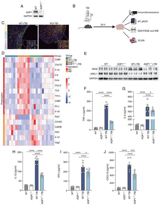
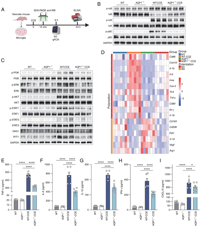
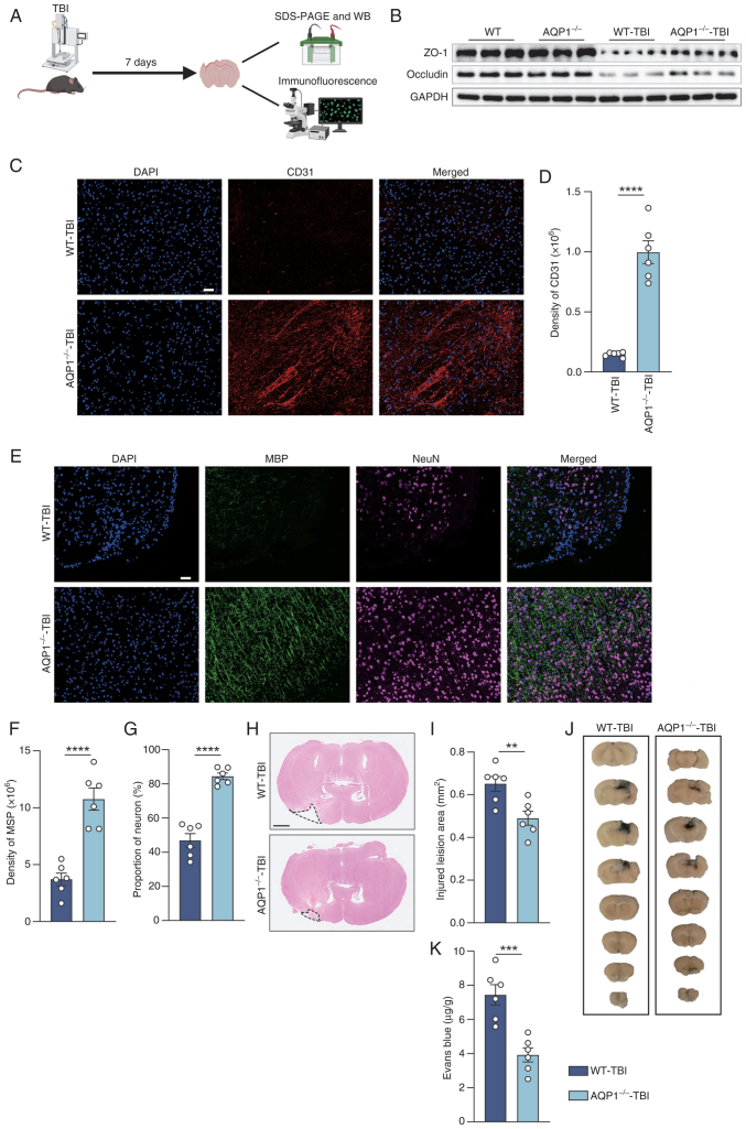
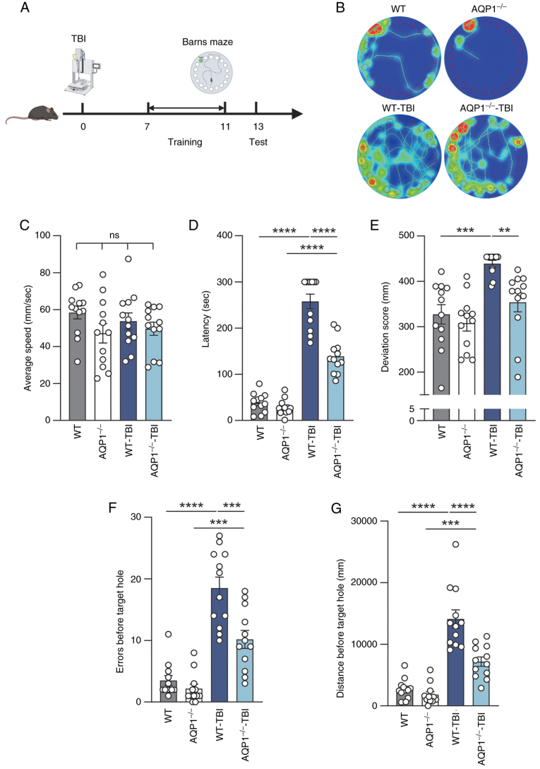
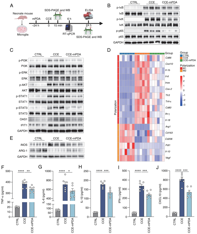
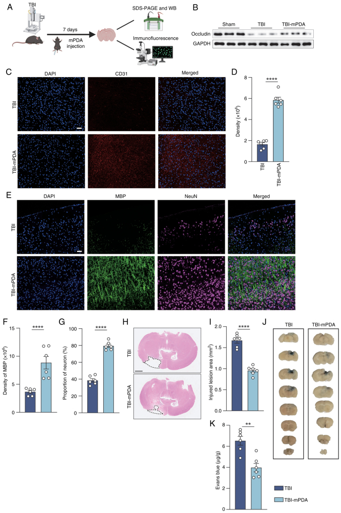
The present study investigated the mechanisms by which aquaporin 1 (AQP1) influences microglial polarization and neuroinflammatory processes in traumatic brain injury (TBI). A model of TBI was generated in AQP1‑knockout mice to assess the impact of AQP1 deletion on inflammatory cytokine release, neuronal damage and cognitive function. Immunofluorescence, reverse transcription‑quantitative PCR, western blotting and enzyme‑linked immunosorbent assay were employed to evaluate pro‑inflammatory and anti‑inflammatory markers. Behavioral assessments, including the Barnes maze, were performed to determine cognitive outcomes. Moreover, AQP1 knockout inhibited the activation of inflammation‑related signaling pathways, including nuclear factor‑κB, Janus kinase/signal transducer and activator of transcription, phosphoinositide 3‑kinase/protein kinase B and extracellular signal‑regulated kinase/mitogen‑activated protein kinase pathways. Further studies indicated that the AQP1 inhibitor m‑phenylenediacrylic acid demonstrated significant neuroprotective effects in a mouse model of TBI. These findings suggested that AQP1 may be essential in post‑TBI inflammatory responses and neuronal injury, establishing a theoretical foundation for future therapies aimed at AQP1.

Keywords: AQP1; TBI; inflammation; microglia.


Conflict of interest statement

The authors declare that they have no competing interests.

Figures

Figure 1
Figure 1
Knockout of AQP1 reduces inflammatory factors in the lesion area after TBI. (A) Verification of the knockout effect in AQP1−/− mice. (B) Schematic diagram of the experimental procedure (created using BioRender.com). Mice were sacrificed and their brains collected 24 h after establishing the TBI model for subsequent experiments. (C) Triple-marker four-color immunofluorescence labeling was conducted to evaluate the proportions of pro-inflammatory and anti-inflammatory microglia surrounding the lesion in AQP1−/− and WT-TBI mice following TBI induction (n=6). Scale bar, 50 μm. (D) mRNA levels of pro-inflammatory and anti-inflammatory markers in the lesion were quantified using RT-qPCR (n=6). (E) SDS-PAGE and western blotting was performed to detect pro-inflammatory and anti-inflammatory markers in microglia (n=3). Inflammatory factor levels in the lesion were assessed using ELISA (n=6), including (F) TNF-α, (G) IL-6, (H) IL-1β, (I) IFN-γ and (J) CXCL10. Data are presented as the mean ± SE. *P<0.05, ***P<0.001, ****P<0.0001. AQP1, aquaporin 1; WT, wild-type; TBI, traumatic brain injury; ELISA, enzyme-linked immunosorbent assay; RT-qPCR, reverse transcription-quantitative PCR, IBA1, ionized calcium binding adaptor protein 1; CXCL10, C-X-C motif chemokine ligand 10; TNF-α, tumor necrosis factor-α; IL, interleukin-; IFN-γ, interferon-γ; iNOS, inducible nitric oxide synthase; ARG1, arginase 1.
Figure 2
Figure 2
Knockout of AQP1 inhibits pro-inflammatory polarization of microglia after addition of CCE by blocking the inflammation-related signaling pathways. (A) Schematic diagram of the experimental procedure (created using BioRender.com). Primary microglia were isolated from the brains of neonatal WT and AQP1−/− mice, and were stimulated with 5 μg/ml CCE. (B) Activation of the NF-κB pathway was assessed 15 min after CCE stimulation (n=3 independent experiments). (C) Activation analysis of other inflammation-related signaling pathways (n=3 independent experiments). (D) A total of 6 h after stimulation, RT-qPCR was performed to detect mRNA expression levels of pro-inflammatory and anti-inflammatory markers in microglia (n=6 independent experiments). (E-I) A total of 24 h after stimulation, the levels of inflammatory factors in the cell culture supernatant were measured by ELISA (n=6 independent experiments). Data are presented as the mean ± SE. *P<0.05, ****P<0.0001. AQP1, aquaporin 1; WT, wild-type; CCE, cortical crude extracts; ELISA, enzyme-linked immunosorbent assay; RT-qPCR, reverse transcription-quantitative PCR, p-, phosphorylated; PI3K, phosphoinositide 3-kinase; ERK, extracellular signal-regulated kinase; AKT, protein kinase B; STAT, signal transducer and activator of transcription; OAS1, 2′,5′-oligoadenylate synthetase 1; IFIT1, interferon-inducible protein 1; CXCL10, C-X-C motif chemokine ligand 10; IL, interleukin; IFN-γ, interferon-γ; TNF-α, tumor necrosis factor-α.
Figure 3
Figure 3
Knockout of AQP1 alleviates neuronal and vascular injury after TBI. (A) Schematic diagram of the experimental procedure. Mice were sacrificed and their brains collected 7 days after TBI induction for subsequent experiments (created using BioRender.com). (B) Detection of tight junction proteins in the injured area (n=3). (C and D) Assessment of vascular endothelial cell density. (C) Representative images of immunofluorescence used to detect and evaluate angiogenesis around the lesion. (D) Quantification of the density of CD31. Scale bar, 50 μm (n=6). (E-G) Immunofluorescence analysis of neuronal and myelin densities in the area surrounding the injury. (E) Representative images used to assess neuronal and myelin densities around the lesion. (F) Quantitative analysis of MBP density. (G) Quantitative analysis of neuronal density. Scale bar, 50 μm (n=6). (H and I) Assessment of lesion size by H&E staining. (H) Images of the largest lesion cross-section. (I) Quantification of the lesion area. Scale bar, 500 μm (n=6). (J and K) Evaluation of blood-brain barrier integrity, evaluated using Evans Blue (n=6). (J) Images of various brain cross-sections in mice. (K) Quantitative analysis of Evans Blue. Data are presented as the mean ± SE. **P<0.01, ***P<0.001, ****P<0.0001. AQP1, aquaporin 1; WT, wild-type; TBI, traumatic brain injury; ZO-1, zonula occludens-1; MBP, myelin basic protein, NeuN, neuronal nuclei.
Figure 4
Figure 4
Knockout of AQP1 reduces cognitive dysfunction in mice with TBI. (A) Schematic diagram of the experimental procedure (created using BioRender. com). A total of 7 days after TBI induction, mice underwent Barnes maze training for 5 consecutive days, followed by testing 1 day after the completion of training (n=12). (B) Movement trajectories and heat maps of mice. (C) Analysis of the average speed of mice. Behavioral analysis: (D) Latency to find the escape hole, (E) deviation score from the target hole, (F) number of errors before finding the escape hole and (G) distance moved in the four groups of mice. Data are presented as the mean ± SE. **P<0.01, ***P<0.001, ****P<0.0001. AQP1, aquaporin 1; WT, wild-type; TBI, traumatic brain injury; ns, not significant.
Figure 5
Figure 5
AQP1 inhibitor mPDA inhibits pro-inflammatory polarization of microglia after the addition of CCE. (A) Schematic diagram of the experimental procedure (created using BioRender.com). Primary microglia isolated from neonatal WT mice were treated with 8 μM mPDA for 24 h, then stimulated with 5 μg/ml CCE. (B) Activation status of the nuclear factor-κB signaling pathway was assessed 15 min after CCE stimulation (n=3 independent experiments). (C) Analysis of activation of inflammation-related signaling pathways n=3 independent experiments). (D) A total of 6 h after CCE stimulation, mRNA expression levels of pro-inflammatory and anti-inflammatory phenotype markers in microglia were detected by RT-qPCR (n=6 independent experiments). (E) A total of 24 h after CCE stimulation, western blotting was performed to detect pro-inflammatory and anti-inflammatory markers in microglia (n=3 independent experiments). A total of 24 h after CCE stimulation, levels of inflammatory factors in the cell culture supernatant were measured by ELISA (n=6 independent experiments), including (F) TNF-α, (G) IL-6, (H) IL-1β, (I) IFN-γ and (J) CXCL10. Data are presented as the mean ± SE. *P<0.05, **P<0.01, ***P<0.001, ****P<0.0001. CTRL, control; mPDA, m-phenylenediacrylic acid, CCE, cortical crude extract, ELISA, enzyme-linked immunosorbent assay; RT-qPCR: reverse transcription-quantitative PCR; p-, phosphorylated; PI3K, phosphoinositide 3-kinase; ERK, extracellular signal-regulated kinase; AKT, protein kinase B; STAT, signal transducer and activator of transcription; OAS1, 2′,5′-oligoadenylate synthetase 1; IFIT1, interferon-inducible protein 1; CXCL10, C-X-C motif chemokine ligand 10; IL, interleukin; IFN-γ, interferon-γ; TNF-α, tumor necrosis factor-α; iNOS, inducible nitric oxide synthase; ARG1, arginase 1.
Figure 6
Figure 6
AQP1 inhibitor mPDA alleviates neurovascular injury after TBI. (A) Schematic diagram of the experimental procedure (created using BioRender. com). Mice with TBI were intraperitoneally injected with 0.17 mg/kg mPDA daily for 7 consecutive days and sacrificed on day 7 for further analysis. (B) Detection of tight junction proteins in the injured area (n=3, male). (C and D) Assessment of vascular endothelial cell density. (C) Representative images of immunofluorescence used to detect and evaluate angiogenesis around the lesion. (D) Quantification of the density of CD31. Scale bar, 50 μm (n=6). (E-G) Immunofluorescence analysis of neuronal and myelin densities in the area surrounding the injury. (E) Representative images used to assess neuronal and myelin densities around the lesion. (F) Quantitative analysis of MBP density. (G) Quantitative analysis of neuronal density. Scale bar, 50 μm (n=6). (H and I) Assessment of lesion size by H&E staining. (H) Images of the largest lesion cross-section. (I) Quantification of the lesion area. Scale bar, 500 μm (n=6). (J and K) Evaluation of blood-brain barrier integrity (n=6) using Evans Blue. (J) Images of various brain cross-sections in mice. (K) Quantitative analysis of Evans Blue. Data are presented as the mean ± SE. **P<0.01, ****P<0.0001. AQP1, aquaporin 1; WT, wild-type; TBI, traumatic brain injury; mPDA, m-phenylenediacrylic acid; MBP, myelin basic protein; NeuN, neuronal nuclei.
Figure 7
Figure 7
mPDA reduces cognitive dysfunction in mice with TBI. (A) Schematic diagram of the experimental procedure (created using BioRender.com). After TBI induction, mice were intraperitoneally injected daily with 0.17 mg/kg mPDA. Following 7 days of treatment, a 5-day Barnes maze training was conducted, and behavioral testing was performed 1 day after the completion of training (n=6). (B) Movement trajectories and heat maps of mice in the Barnes maze. (C) Comparison of the average movement speed of mice in the maze. Behavioral analysis: (D) Latency to find the escape hole, (E) number of errors made before finding the escape hole and (F) total distance moved before finding the escape hole among the groups of mice. Data are presented as the mean ± SE. *P<0.05, **P<0.01, ***P<0.001, ****P<0.0001. TBI, traumatic brain injury; mPDA, m-phenylenediacrylic acid; ns, not significant.

Similar articles

References

    1. Ditthapron A, Lammert AC, Agu EO. Continuous TBI monitoring from spontaneous speech using parametrized sinc filters and a cascading GRU. IEEE J Biomed Health Inform. 2022;26:3517–3528. doi: 10.1109/JBHI.2022.3158840. - DOI - PubMed
    1. Gardner RC, Byers AL, Barnes DE, Li Y, Boscardin J, Yaffe K. Mild TBI and risk of parkinson disease: A chronic effects of neurotrauma consortium study. Neurology. 2018;90:e1771–e1779. doi: 10.1212/WNL.0000000000005522. - DOI - PMC - PubMed
    1. Willis EF, MacDonald KPA, Nguyen QH, Garrido AL, Gillespie ER, Harley SBR, Bartlett PF, Schroder WA, Yates AG, Anthony DC, et al. Repopulating microglia promote brain repair in an IL-6-dependent manner. Cell. 2020;180:833–846.e16. doi: 10.1016/j.cell.2020.02.013. - DOI - PubMed
    1. Loane DJ, Kumar A. Microglia in the TBI brain: The good, the bad, and the dysregulated. Exp Neurol. 2016;275:316–327. doi: 10.1016/j.expneurol.2015.08.018. - DOI - PMC - PubMed
    1. Karve IP, Taylor JM, Crack PJ. The contribution of astrocytes and microglia to traumatic brain injury. Br J Pharmacol. 2016;173:692–702. doi: 10.1111/bph.13125. - DOI - PMC - PubMed

LinkOut - more resources